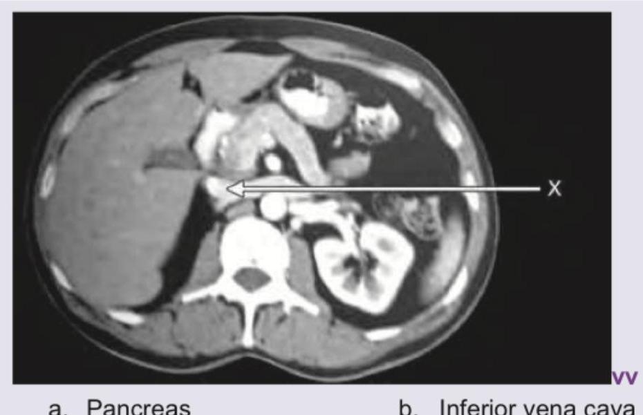
Name the structure marked as $X$ in the CT abdomen shown below: (Recent NEET Pattern 2016-17)

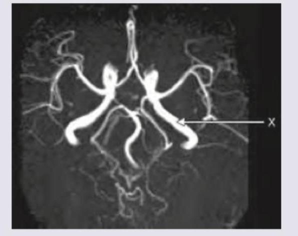
The blood vessel marked as $X$ in the CT angiography image is:

What is the correct diagnosis for the image shown?

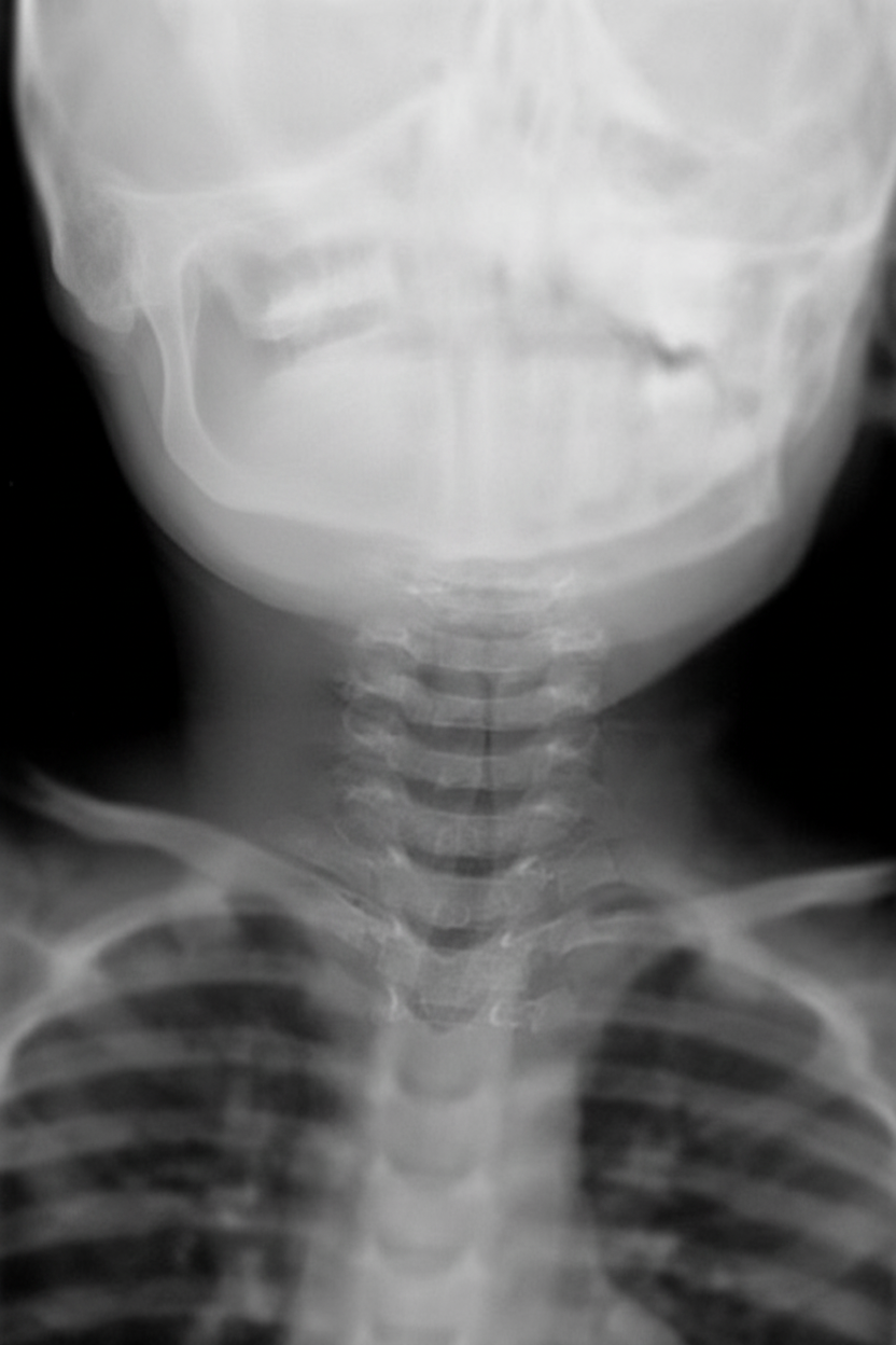
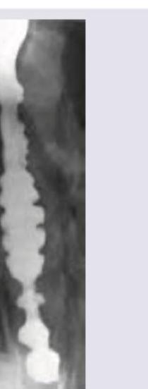
The neck X-ray of a patient shows:

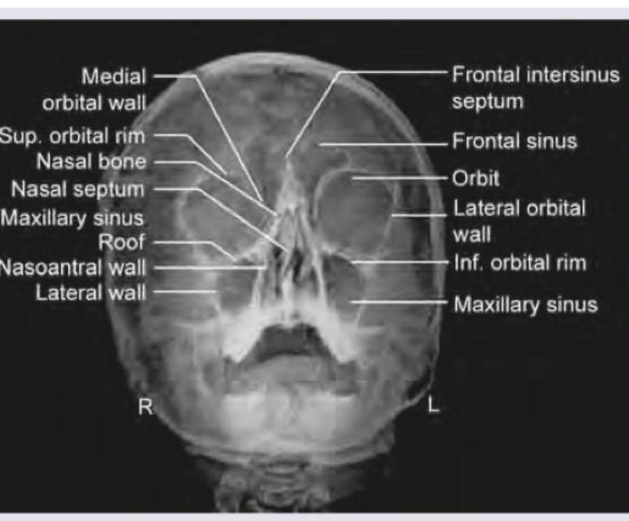
The given X-ray of paranasal sinuses shows which view?

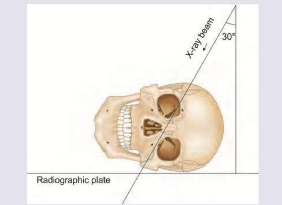
A radiographic projection with X-ray beam angled at 30 degrees to the radiographic plate, with the skull positioned laterally (as shown in the image), is used to visualize which of the following structures?

The following X-ray is used to evaluate \qquad sinus?

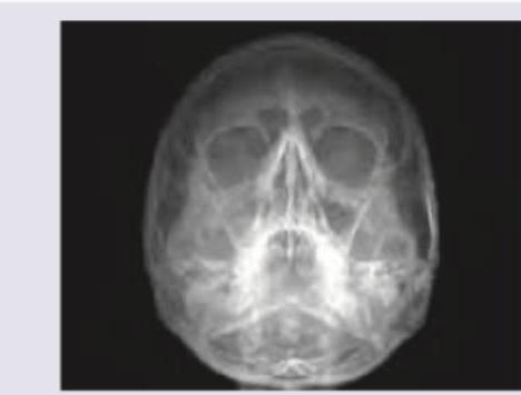
The images show the presence of:

All of the following can be used to describe the radiological image shown below EXCEPT:

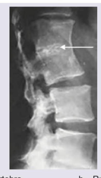
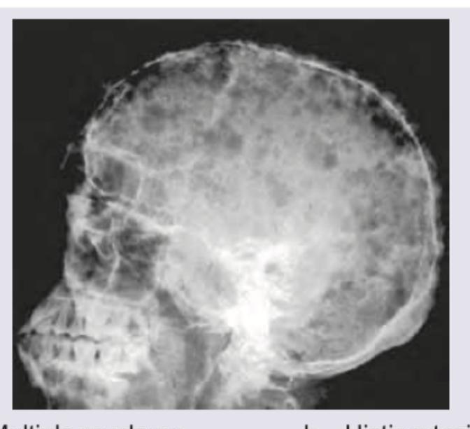
The findings in the following skull X-ray are most characteristic of:

Explanation: ***Inferior vena cava*** - The structure marked as X is located to the **right of the aorta** and is typically seen as a large, relatively **thin-walled vessel**. - Its position anterior and to the right of the vertebral body, draining into the heart, is consistent with the **inferior vena cava (IVC)**. *Pancreas* - The pancreas is typically located more anteriorly and superiorly in this cross-section, usually nestled between the duodenum and spleen. - It would appear as a **glandular organ** with a different texture and position on a CT scan. *Aorta* - The aorta is typically the **larger, thick-walled, pulsatile vessel** located to the **left of the vertebral body** and to the left of the structure marked X. - It usually appears circular in cross-section and is often seen with higher attenuation due to arterial contrast. *Left renal vein* - The left renal vein typically arises from the left kidney and crosses the midline to drain into the IVC, passing **anterior to the aorta**. - The structure marked X is the IVC itself, which receives the left renal vein, rather than the left renal vein.
Explanation: ***P2 PCA*** - The image displays a CT angiography of the **Circle of Willis**. The vessel marked 'X' is a segment of the **posterior cerebral artery** (PCA). - The PCA is conventionally divided into four segments: P1 (pre-communicating), P2 (peduncular), P3 (quadrigeminal), and P4 (cortical). The 'X' points to the segment of the PCA that is distal to the posterior communicating artery, indicating the **P2 segment**. *P1 PCA* - The **P1 segment** of the PCA is the initial part, located between the basilar artery bifurcation and the posterior communicating artery. - The vessel marked 'X' is clearly **distal to the connection point** where the posterior communicating artery would typically join, thus it is not the P1 segment. *Internal carotid artery* - The **internal carotid arteries** typically ascend higher and give rise to the anterior and middle cerebral arteries, forming the anterior circulation. - The vessel marked 'X' is part of the **posterior circulation**, originating from the basilar artery system, not the internal carotid artery. *M1, Middle cerebral artery* - The **M1 (main trunk) segment of the middle cerebral artery** extends laterally from the internal carotid artery. - The vessel marked 'X' is situated more posteriorly and medially, clearly identifying it as part of the **posterior cerebral artery**, and not the middle cerebral artery.
Explanation: ***Fish vertebra*** - The image indicates **biconcave vertebral bodies** with a central depression, mimicking the appearance of a **fish vertebra**. This shape results from bone softening conditions, leading to compression of the vertebral body by the turgid intervertebral discs. - This finding is classically associated with **osteoporosis** or **osteomalacia**, where the bone mineral density is reduced making the vertebrae more susceptible to this type of deformation. *Hemivertebra* - A hemivertebra is a **congenital anomaly** where only half of a vertebral body forms, causing a wedge-shaped vertebra and often leading to **scoliosis**. - The image does not show a malformed, wedge-shaped vertebral body; instead, it shows a **biconcave deformity** of a fully formed vertebra. *Rugger jersey spine* - Rugger jersey spine refers to the appearance of alternating lucent and sclerotic bands on the superior and inferior endplates of vertebral bodies, resembling the stripes on a rugby jersey. - This finding is characteristic of **renal osteodystrophy** due to secondary hyperparathyroidism, which is not depicted in the given image. *Block vertebra* - A block vertebra is a **congenital or acquired fusion** of two or more adjacent vertebral bodies, resulting in a single, enlarged vertebral segment with rudimentary or absent intervertebral discs. - The image clearly shows distinct vertebral bodies with intervening discs, albeit distorted, and no evidence of fusion.
Explanation: ***Steeple sign*** - The image shows a **narrowing of the subglottic trachea**, which resembles a "steeple" or a church spire on a frontal neck X-ray. - This sign is highly suggestive of **croup** (laryngotracheobronchitis), caused by inflammation and edema of the subglottic region. *Zenker's diverticulum* - This is an **outpouching of the posterior pharyngeal wall** through Killian's triangle, typically seen as a fluid or air-filled sac posterior to the esophagus. - It would appear inferior to the larynx and esophagus, not as a tracheal narrowing within the airway. *Laryngocele* - A laryngocele is an **abnormal air-filled dilation of the laryngeal saccule**, usually appearing as a radiolucent mass in the neck, often extending into the soft tissues. - It would be located more superiorly and laterally to the trachea and does not present as a classic subglottic narrowing. *Thumb sign* - The **"thumb sign"** refers to the appearance of an **enlarged, edematous epiglottis** on a lateral neck X-ray, resembling a thumb. - This sign is characteristic of **epiglottitis**, a different condition affecting the supraglottic region, not the subglottic trachea.
Explanation: ***Waters' view*** - This view, also known as the **occipitomental view**, is primarily used to visualize the **maxillary sinuses**. - Key features include the **maxillary sinuses** being projected above the petrous ridges, allowing clear visualization of their floors and the inferolateral orbital walls. - The patient's mouth is open in this view, and the **petrous pyramids are projected below the maxillary sinuses**. *Caldwell view* - Also known as the **occipitofrontal view**, it primarily visualizes the **frontal sinuses** and anterior ethmoid air cells. - In a Caldwell view, the **petrous ridges** obscure the lower third of the orbits. *Orthopantomogram* - An **Orthopantomogram (OPG)** is a panoramic dental X-ray that shows all the teeth and surrounding bones in a single image. - It provides a broad view of the **mandible** and **maxilla**, which is not depicted in the image. *Luc's View* - Luc's view is an uncommon projection, sometimes referring to a **lateral view of the maxillary sinus** or a **basal/submentovertex view**. - It is used to visualize the **sphenoid sinus** and **posterior structures**, not the maxillary sinuses as prominently shown in this image.
Explanation: ***Sinus tympani*** - The image shows a **radiographic projection** designed to visualize structures of the **temporal bone**. Specifically, an X-ray beam angled at 30 degrees to the radiographic plate, with the skull positioned laterally, is characteristic of specific views for the **middle ear structures**, such as the sinus tympani. - The **sinus tympani** is a small, deep recess in the posterior wall of the middle ear, and its detailed visualization often requires specialized **oblique radiographic projections**. *Recess of fourth ventricle* - The **fourth ventricle** is located within the brain stem, and its recesses are deep brain structures. - Visualization of the fourth ventricle and its recesses typically requires advanced imaging modalities like **CT or MRI**, not standard X-ray projections of the skull. *Round window* - The **round window** is a structure of the inner ear, specifically located at the cochlea. - While it is part of the temporal bone, standard skull X-rays are not optimal for its detailed visualization, and a specialized view for the **hypotympanum** would be needed to see it clearly, which is not depicted by this projection angle. *Lateral ventricle* - The **lateral ventricles** are located within the cerebral hemispheres of the brain. - Similar to the fourth ventricle, these structures are best visualized with **CT or MRI** and are not effectively seen with basic skull X-ray projections, especially not with an angled view designed for the temporal bone.
Explanation: ***Maxillary*** - The image provided is a **Waters' view** (occipitomental view) X-ray of the paranasal sinuses, which is primarily used to visualize the **maxillary sinuses**. - In a Waters' view, the **petrous ridges** (dense bone at the base of the skull) are projected below the maxillary sinuses, allowing for a clear view of these sinuses. *Frontal* - While the **frontal sinuses** are visible in a Waters' view, they are generally better visualized in a **Caldwell view** (occipitofrontal view) or lateral view. - In this projection, their visualization can be obscured by other bony structures, and they are not the primary focus. *Ethmoidal* - The **ethmoidal sinuses** are typically comprised of multiple small air cells located between the orbits and are best seen on a **Caldwell view** or specialized oblique views. - In a Waters' view, their evaluation is limited due to superimposition of other facial bones. *Sphenoidal* - The **sphenoidal sinuses** are located deep within the skull, inferior to the sella turcica, and are quite difficult to visualize on standard plain radiographs like the Waters' view. - They are best assessed using a **lateral view of the skull** or advanced imaging like **CT scans**.
Explanation: ***A= Ventriculoperitoneal shunt, B= Ventriculo-atrial shunt*** - Image A shows a catheter extending from the cerebral ventricle and terminating in the **peritoneal cavity** (abdomen), characteristic of a **ventriculoperitoneal shunt**. - Image B depicts a catheter extending from the ventricle to the **right atrium** of the heart, indicating a **ventriculo-atrial shunt**. *A= Ventriculo-atrial shunt, B= Ventriculo-peritoneal shunt* - This option incorrectly reverses the identification of the shunts shown in the images. - The catheter termination points are mismatched; image A terminates in the abdomen (VP shunt), not the heart (VA shunt). *A= Denver shunt, B= Leveen shunt* - **Denver shunts** and **Leveen shunts** are **peritoneovenous shunts** used for refractory **ascites**, not hydrocephalus. - These shunts drain ascitic fluid from the peritoneal cavity to systemic circulation, completely different from ventricular shunts for CSF drainage. *B= Leveen shunt, A= Denver shunt* - This option also misidentifies the shunts as peritoneovenous shunts used for **ascites management**. - Both Denver and Leveen shunts are unrelated to **hydrocephalus treatment** and ventricular CSF drainage systems.
Explanation: ***Cobble stone*** - The image shows a barium swallow with a **"corkscrew" or "rosary bead" appearance**, characteristic of **distal esophageal spasm**. - **Cobblestoning** is typically seen in the colon, associated with severe **Crohn's disease**, and refers to ulcerations and edema creating a nodular appearance, which is not depicted here. *Corkscrew appearance* - This term accurately describes the radiological image, specifically identifying **distal esophageal spasm** where uncoordinated contractions present as multiple irregular indentations. - The **barium column** takes on a spiral or corkscrew shape due to simultaneous, non-peristaltic contractions. *Rosary bead esophagus* - This term is a synonym for the **corkscrew esophagus** appearance and is also used to describe the irregular, segmentally constricted appearance seen in diffuse esophageal spasm. - The contractions create multiple small, bead-like segments, resembling a rosary. *Pseudodiverticula* - These are small outpouchings that can be present in the esophagus when there is severe luminal narrowing from spastic contractions. - The image shows these constrictions and bulging segments that can resemble pseudodiverticula in the context of esophageal spasm.
Explanation: ***Multiple myeloma*** - The X-ray shows multiple well-defined, lytic, **punched-out lesions** throughout the skull, which is the classic presentation of multiple myeloma. - These lesions represent areas of bone destruction due to proliferation of **plasma cells** within the bone marrow. *Histiocytosis-X* - Histiocytosis X (Langerhans cell histiocytosis) can cause lytic skull lesions, but they are often described as **"beveled edge"** or **"hole within a hole"** appearance, which is not clearly depicted here. - While it can cause bony lesions, the widespread, uniformly punched-out appearance is more characteristic of multiple myeloma. *Hyperparathyroidism* - Hyperparathyroidism typically causes **generalized demineralization** of the skull, leading to a **"salt-and-pepper"** appearance, and possibly **subperiosteal bone resorption**. - It does not typically present with discrete, punched-out lytic lesions like those seen in the image. *Sickle cell anemia* - In sickle cell anemia, the skull X-ray may show **widening of the diploic spaces** and a **"hair-on-end"** or **"crew-cut"** appearance, especially in severe cases, due to marrow hyperplasia. - This is distinct from the multiple punched-out lesions observed in the provided image.
Radiographic Anatomy of Skull and Face
Practice Questions
Radiographic Anatomy of Spine
Practice Questions
Radiographic Anatomy of Chest
Practice Questions
Radiographic Anatomy of Abdomen
Practice Questions
Radiographic Anatomy of Extremities
Practice Questions
Cross-sectional Anatomy: Brain and Head
Practice Questions
Cross-sectional Anatomy: Neck
Practice Questions
Cross-sectional Anatomy: Thorax
Practice Questions
Cross-sectional Anatomy: Abdomen and Pelvis
Practice Questions
Vascular Anatomy
Practice Questions
Developmental Anatomy Variations
Practice Questions
Anatomic Landmarks for Interventional Procedures
Practice Questions
Get full access to all questions, explanations, and performance tracking.
Start For Free