Multiple radioopacities are seen in which of the following conditions?
A 56-year old man presented with bony pain. What is the most likely diagnosis given an X-ray skull lateral view showing findings consistent with hyperostosis frontalis interna?
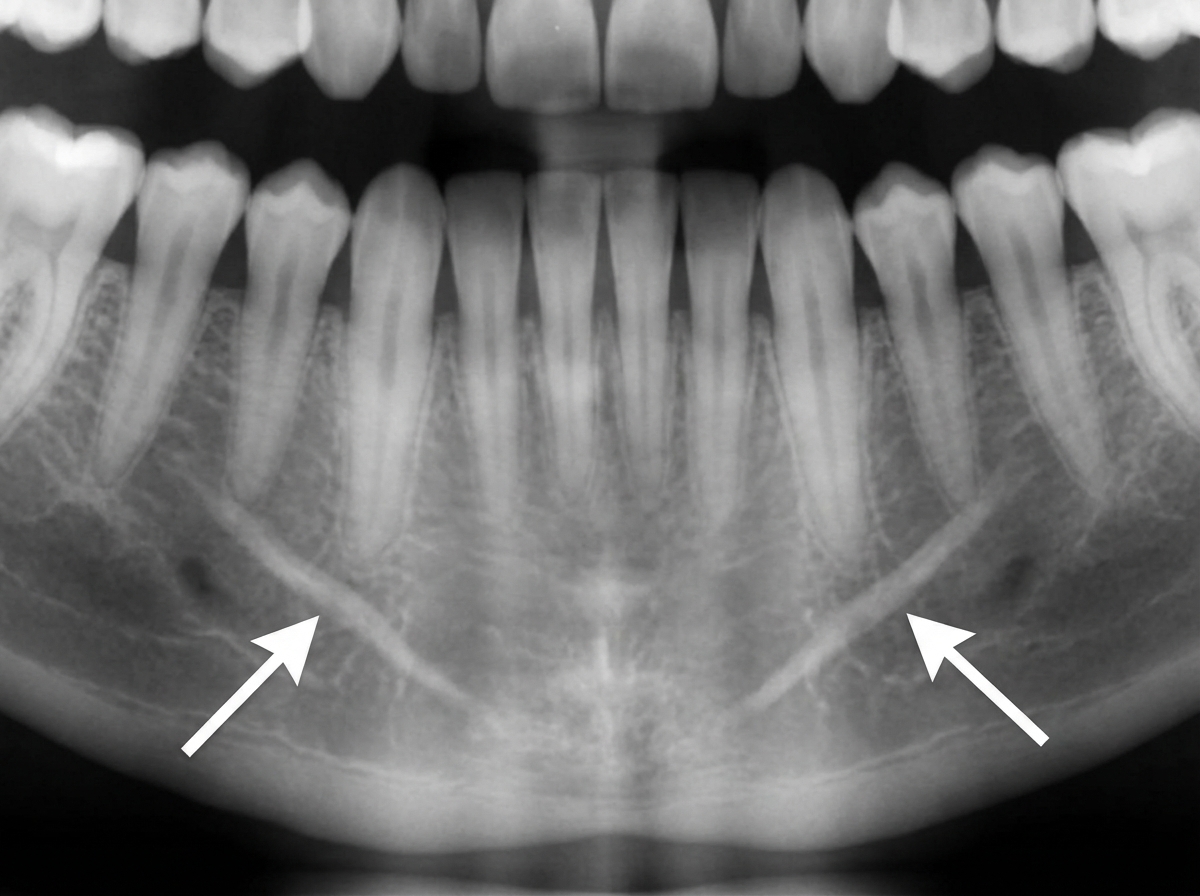
The symmetrical radiopacities marked with arrows are most likely?

Which of the following will appear radiopaque in an X-ray except?
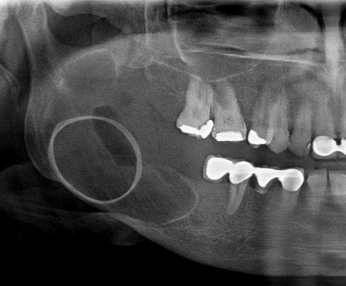
A radiograph of a 32-year-old patient reveals an asymptomatic lesion that was an accidental finding. What is the most likely diagnosis?

What is the best investigation to detect calcification?
A patient with a history of trauma presents with hearing loss. A High-Resolution Computed Tomography (HRCT) scan was performed. Which of the following structures is not typically visualized on HRCT?
Identify the rib highlighted in the X-ray.
In the given barium swallow image, which of the following shows the left atrium impression on the esophagus?
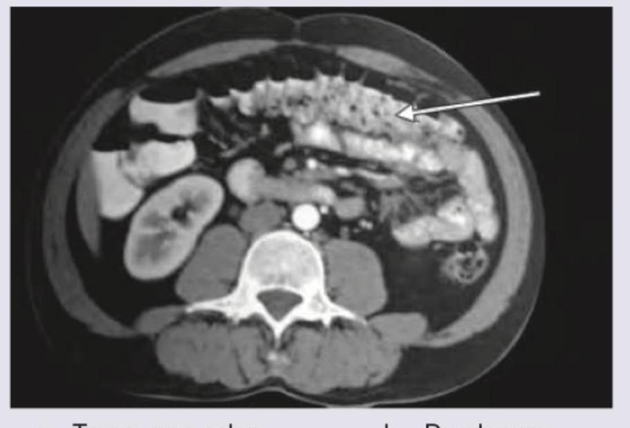
The structure marked by arrow in CT abdomen:

Explanation: **Explanation:** **Odontomas** are the most common odontogenic tumors and are considered hamartomas rather than true neoplasms. They are composed of dental tissues (enamel, dentin, cementum, and pulp). On a radiograph, they appear as **multiple radioopacities** surrounded by a narrow radiolucent halo. Specifically, **Compound Odontomas** present as a cluster of small, tooth-like structures (denticles), typically in the anterior maxilla, which accounts for the multiple radioopaque shadows. **Analysis of Incorrect Options:** * **Multiple Myeloma:** Characteristically presents as multiple **radiolucent** (dark) "punched-out" osteolytic lesions, not radioopacities. * **Cherubism:** A genetic disorder characterized by bilateral, symmetrical **multilocular radiolucent** (soap-bubble appearance) lesions in the mandible and maxilla. * **Osteopetrosis:** Known as "Marble Bone Disease," it presents as a **diffuse, generalized increase in bone density** (sclerosis) rather than discrete multiple radioopacities. **High-Yield Clinical Pearls for NEET-PG:** * **Complex Odontoma:** Presents as an amorphous, irregular mass of calcified tissue, usually in the posterior mandible. * **Gardner Syndrome:** If multiple odontomas are associated with osteomas and intestinal polyps, suspect this syndrome. * **Radiopacity vs. Radiolucency:** Always remember that "opaque" is white (dense) and "lucent" is black (destruction/void) on X-rays. * **Sunray Appearance:** Characteristic of Osteosarcoma. * **Cotton Wool Appearance:** Characteristic of Paget’s disease (late stage).
Explanation: ### Explanation **Hyperostosis Frontalis Interna (HFI)** is a benign condition characterized by the thickening of the inner table of the frontal bone. On a lateral skull X-ray, it appears as a dense, undulating, or nodular bony overgrowth. **Why the correct answer is right:** The hallmark of HFI is that the thickening is strictly limited to the **inner table** of the frontal bone, typically sparing the midline (where the superior sagittal sinus lies) and the outer table. It is most commonly an incidental finding in postmenopausal women and is often associated with metabolic or endocrine disturbances (e.g., Morgagni-Stewart-Morel syndrome). **Why the incorrect options are wrong:** * **Paget’s Disease:** Characterized by "Cotton Wool" spots on the skull. It involves both the inner and outer tables, leading to generalized skull thickening and enlargement (diploe expansion), unlike the localized inner-table involvement of HFI. * **Fibrous Dysplasia:** Typically presents with a "Ground Glass" appearance. While it can affect the frontal bone, it causes expansion of the bone and distortion of the normal architecture, often involving the facial bones (leontiasis ossea). * **Osteopetrosis:** A generalized increase in bone density ("Marble Bone Disease"). It affects the entire skeleton and the entire skull base/vault uniformly, rather than being localized to the internal frontal bone. **High-Yield Clinical Pearls for NEET-PG:** * **Demographics:** Most common in elderly females (incidental finding). * **Radiological Sign:** Sparing of the midline/sagittal suture is a classic feature of HFI. * **Morgagni-Stewart-Morel Syndrome:** A triad of HFI, obesity, and virilism/hirsutism. * **Differential Diagnosis:** Must be distinguished from a meningioma, which usually shows associated soft tissue changes or a sunburst periosteal reaction.
Explanation: ***Mental ridges*** - **Bilateral symmetrical radiopacities** on the anterior mandible below the canine-premolar region are characteristic of **mental ridges**, which are bony prominences that provide muscle attachment sites. - They appear as **paired radiopaque lines** running horizontally on either side of the mandibular symphysis and are normal anatomical landmarks. *Genial tubercle* - Appears as a **radiopaque ring or button-like structure** at the **midline symphysis**, not as bilateral symmetrical radiopacities. - Located at the **lingual aspect** of the mandibular symphysis and serves as attachment for **geniohyoid and genioglossus muscles**. *Mylohyoid ridges* - Appear as **oblique radiopaque lines** running posteriorly from the **premolar to molar region**, not as anterior symmetrical structures. - Located on the **lingual surface** of the mandible and provide attachment for the **mylohyoid muscle**. *Both genial tubercle and mental ridges* - **Genial tubercles** appear as a single midline radiopaque structure, not bilateral symmetrical radiopacities as described. - This combination would show different radiographic patterns - a **central ring-like opacity** (genial tubercle) plus **bilateral horizontal ridges** (mental ridges).
Explanation: **Explanation:** In dental radiology, the appearance of a material on an X-ray (radiopaque vs. radiolucent) depends on its **atomic number** and **density**. Materials with high atomic numbers absorb more X-ray photons and appear white (radiopaque), while those with lower density appear dark (radiolucent). **Why Zinc Oxide Eugenol (ZOE) is the correct answer (in the context of this specific question):** While ZOE is technically radiopaque due to the presence of Zinc (Atomic No. 30), it is often **less radiopaque** than modern restorative materials like Composite or Calcium Hydroxide liners. However, there is a common academic distinction in dental materials: **Acrylic resin** is the only material in this list that is inherently **radiolucent** (appears dark). *Note: There appears to be a discrepancy in the provided key. In standard radiology, Acrylic resin is radiolucent, while ZOE, Calcium Hydroxide, and Composites are radiopaque. If the question asks which is NOT radiopaque, **Acrylic Resin (A)** is the most accurate clinical answer.* **Analysis of Options:** * **Acrylic Resin (A):** Composed of organic polymers with low atomic numbers (C, H, O). It is **radiolucent** and can be difficult to distinguish from caries or oral tissues unless radiopaque fillers are added. * **Calcium Hydroxide (B):** Used as a liner; it is manufactured with radiopaque fillers (like Barium sulfate) to ensure it is visible on X-rays. * **Zinc Oxide Eugenol (C):** Contains Zinc, making it inherently **radiopaque**. It is used as a base or temporary filling. * **Composite (D):** Modern composites contain glass fillers (Barium, Strontium, or Zirconium) specifically to make them **radiopaque** so they aren't confused with recurrent decay. **NEET-PG High-Yield Pearls:** 1. **Most Radiopaque:** Amalgam, Gold crowns, and Gutta-percha. 2. **Radiolucent:** Acrylic resin, older composites, and dental caries. 3. **Rule of Thumb:** Any material containing heavy metals (Zinc, Barium, Strontium) will appear radiopaque. 4. **Clinical Significance:** Restorative materials must be more radiopaque than dentin to allow for the detection of secondary caries.
Explanation: ***Stafne bone cavity*** - Characteristically an **asymptomatic incidental radiographic finding** below the **inferior alveolar canal** in the posterior mandible, typically between the premolars and angle. - Represents a **developmental depression** in the lingual cortex filled with salivary gland tissue, appearing as a well-defined radiolucency with **sclerotic borders**. *Radicular cyst* - Associated with **non-vital teeth** and typically presents with symptoms like pain, swelling, or **periapical pathology**. - Located at the **apex of a tooth** rather than below the inferior alveolar canal, and usually has a history of dental trauma or caries. *Dentigerous cyst* - Surrounds the **crown of an unerupted tooth**, most commonly **impacted third molars** or canines. - Presents as a **pericoronal radiolucency** and may cause **displacement** of the associated tooth, not as an isolated mandibular depression. *Lateral periodontal cyst* - Occurs along the **lateral root surface** of vital teeth, typically in the **anterior mandible** or premolar region. - Often presents with **localized gingival swelling** and is associated with vital teeth, unlike the asymptomatic nature described.
Explanation: **Explanation:** **CT Scan (Correct Answer):** Computed Tomography (CT) is the "Gold Standard" for detecting calcification. It possesses superior **high-contrast resolution**, allowing it to detect even minute, speck-like calcifications (e.g., in the pineal gland, granulomas, or early vascular calcification) that are invisible on other modalities. On CT, calcium appears as high-attenuation (hyperdense) areas with Hounsfield Units (HU) typically ranging from **+100 to over +1000**. **Why other options are incorrect:** * **MRI Scan:** MRI is generally poor at detecting calcification because calcium lacks mobile protons, often appearing as a "signal void" (dark). It can be easily confused with flowing blood or air. While specialized sequences like **SWI (Susceptibility Weighted Imaging)** can detect it, CT remains the primary choice. * **X-ray:** While X-rays can show gross calcifications (like kidney stones or bone tumors), they lack the sensitivity to detect small or faint calcifications due to the superposition of overlying tissues. * **Ultrasound (USG):** USG can detect calcification (appearing as hyperechoic structures with posterior acoustic shadowing), but it is operator-dependent and cannot visualize calcifications deep within the lungs or behind bony structures. **High-Yield Clinical Pearls for NEET-PG:** * **Hounsfield Unit (HU) for Bone/Calcification:** > +400 to +1000. * **Psammomatous calcification:** Classically seen in Papillary Thyroid Carcinoma, Meningioma, and Serous Cystadenocarcinoma of the Ovary. * **Eggshell calcification:** Characteristic of Silicosis and Sarcoidosis (hilar lymph nodes). * **Popcorn calcification:** Pathognomonic for Pulmonary Hamartoma and involuting Breast Fibroadenoma.
Explanation: ***Organ of Corti***- The **Organ of Corti** is a delicate, purely **soft-tissue structure** located within the cochlear duct (scala media).- **HRCT** primarily excels at visualizing **bone and air interfaces**; the soft-tissue resolution is insufficient to distinctly delineate this microscopic sensorineural structure.*Cochlea*- The bony shell of the **cochlea**, forming the **bony labyrinth** wall, is clearly visible on HRCT due to its high **density and calcification**.- HRCT is essential for evaluating the internal auditory canal and **cochlear anatomy**, especially for surgical planning or trauma assessment.*Vestibule*- The **vestibule** is part of the **bony labyrinth** housing the utricle and saccule, and its dense bony walls are clearly depicted by HRCT.- Visualization of the vestibule is crucial for assessing **temporal bone fractures** and identifying developmental anomalies.*Semicircular canal*- The three **semicircular canals** (anterior, posterior, lateral) are osseous structures easily resolved by the fine spatial detail of **HRCT**.- HRCT accurately assesses pathology such as **semicircular canal dehiscence** or traumatic disruption of these structures.
Explanation: ***3rd rib posterior part*** - On a posteroanterior (PA) chest X-ray, the **posterior ribs** are more prominent and have a more horizontal orientation as they articulate with the thoracic vertebrae, which matches the highlighted structure. - When counting from the top, the first rib is the highest and most curved. The highlighted rib is clearly the third one down from the apex of the thorax, confirming it as the **3rd posterior rib**. *3rd rib anterior part* - The **anterior parts** of the ribs are more difficult to visualize and course downwards and medially towards the sternum. The highlighted rib is oriented horizontally. - Anterior ribs connect to the sternum via **costal cartilage**, which is not as radiopaque as bone and thus appears less distinct on an X-ray than the highlighted structure. *4th rib anterior part* - This option is incorrect as the highlighted structure is a **posterior rib**, not an anterior one, based on its orientation. - Additionally, counting reveals the rib to be the third, not the fourth. The **fourth rib** would be located inferior to the highlighted one. *1st rib anterior part* - The **1st rib** is the most superior and has a very sharp curvature, often partially obscured by the clavicle. The highlighted rib is located below the first and second ribs. - This is also incorrect because the image highlights the **posterior aspect** of a rib, not the anterior aspect.
Explanation: ***Correct: Option 3*** - This pointer correctly identifies the impression on the esophagus caused by the **left atrium**, which is the most posterior chamber of the heart. - An enlarged left atrium, often seen in conditions like **mitral stenosis**, can cause a prominent posterior indentation on the esophagus at this level. - The left atrium impression is typically seen at the level of T4-T6 vertebrae on barium swallow studies. *Incorrect: None of them* - This option is incorrect as the image clearly shows normal anatomical impressions on the esophagus, and pointer 3 correctly identifies the left atrial impression. - A barium swallow is a standard radiological procedure to visualize these impressions from adjacent cardiovascular structures. *Incorrect: Option 1* - This pointer indicates the impression of the **aortic arch**, which is the first and most superior indentation seen on a lateral or oblique view of a barium swallow. - The aortic arch crosses the esophagus on its left side at approximately T4 level, creating a distinct notch as it arches posteriorly. *Incorrect: Option 2* - This pointer shows the impression made by the **left main bronchus** as it crosses anterior to the esophagus. - This indentation is located inferior to the aortic arch impression at approximately T5 level and is typically less pronounced than the aortic or left atrial impressions.
Explanation: ***Correct Answer: Duodenum*** - The arrow points to a C-shaped structure located in the **retroperitoneum**, anterior to the right kidney and vertebral body, consistent with the location and appearance of the **duodenum** on axial CT - The characteristic **thickened, enhancing wall** and mucosal folds further distinguish the duodenum - The duodenum is a retroperitoneal structure that wraps around the head of the pancreas in a C-shaped configuration *Incorrect: Transverse colon* - The transverse colon is typically located more anteriorly in the abdomen and often contains **feces** and **intraluminal gas**, which are not seen in the indicated structure - It has a larger caliber and generally a different mucosal pattern than the structure pointed to by the arrow - The transverse colon is an intraperitoneal structure, not retroperitoneal *Incorrect: Superior mesenteric vein* - The superior mesenteric vein is a **vascular structure** located in the mesentery, typically seen anterior to the aorta and to the right of the superior mesenteric artery - It appears as a **round, enhancing vessel filled with contrast media**, which is distinct from the C-shaped, bowel-like structure marked by the arrow - Would not show mucosal folds or bowel wall characteristics *Incorrect: IVC* - The IVC (Inferior Vena Cava) is a large **retroperitoneal vein** situated to the right of the aorta and posterior to the head of the pancreas and duodenum - It would appear as a **large, circular or oval contrast-filled vessel**, much larger than the structure indicated by the arrow and in a more posterior location - The IVC does not have the C-shaped configuration or mucosal characteristics of the marked structure
Radiographic Anatomy of Skull and Face
Practice Questions
Radiographic Anatomy of Spine
Practice Questions
Radiographic Anatomy of Chest
Practice Questions
Radiographic Anatomy of Abdomen
Practice Questions
Radiographic Anatomy of Extremities
Practice Questions
Cross-sectional Anatomy: Brain and Head
Practice Questions
Cross-sectional Anatomy: Neck
Practice Questions
Cross-sectional Anatomy: Thorax
Practice Questions
Cross-sectional Anatomy: Abdomen and Pelvis
Practice Questions
Vascular Anatomy
Practice Questions
Developmental Anatomy Variations
Practice Questions
Anatomic Landmarks for Interventional Procedures
Practice Questions
Get full access to all questions, explanations, and performance tracking.
Start For Free