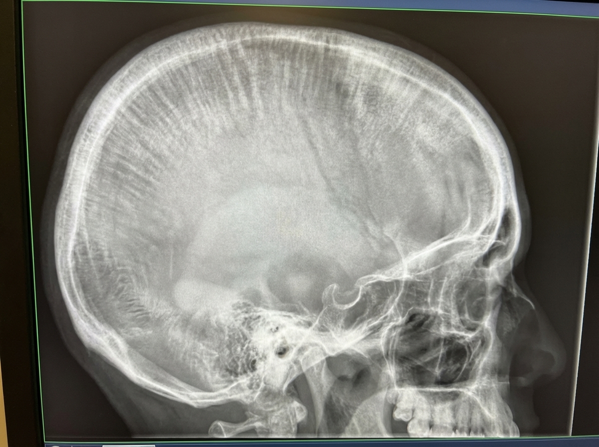
Which of the following conditions can manifest with the shown X-ray findings in the skull?

Which region in the crown of a tooth is often mistaken for caries in an intraoral periapical radiograph?
A 15-year-old male patient presents with pain in a lower back tooth and a history of frequent snacking. Intra-oral examination reveals occlusal caries on teeth 16 and 36, proximal caries between teeth 26 and 27, and a grossly carious tooth 46. Which of the following must be used when taking a radiograph for this patient?
Bitewing radiographs are best suited to monitor:
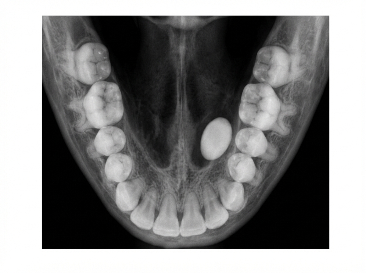
The following occlusal radiograph shows:

A 50-year-old male patient complains of reduced mouth opening. The patient has a history of a Road Traffic Accident (RTA) one week prior. A submentovertex view x-ray was taken which revealed a zygomatic arch fracture. Which of the following is true regarding the central beam in this x-ray?
Insal-Salvati index is used for evaluation of which bone?
Which bones ossify at birth?
Which of the following structures is not visualized on a coronal CT scan of the paranasal sinuses?
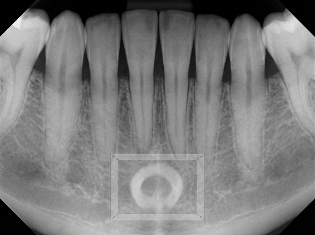
The radiopaque structure shown in the box is most likely:

Explanation: ***Thalassemia*** - Causes **"hair-on-end"** or **"crew-cut"** appearance on skull X-ray due to **diploic widening** from compensatory **extramedullary hematopoiesis**. - The chronic hemolytic anemia leads to **bone marrow expansion** and characteristic **perpendicular trabeculae** extending outward from the skull. *Acute Myeloid Leukemia* - Primarily causes **lytic bone lesions** and **osteopenia** rather than the specific hair-on-end pattern. - X-ray findings typically show **punched-out lesions** and **pathological fractures**, not diploic expansion. *Iron deficiency anemia* - Does not cause significant **bone marrow expansion** as it's primarily a **microcytic hypochromic** anemia. - X-ray findings are typically **normal** or may show **osteopenia** but never the hair-on-end appearance. *Megaloblastic Anemia* - Results from **vitamin B12** or **folate deficiency** causing **ineffective erythropoiesis**. - Does not produce the **compensatory extramedullary hematopoiesis** necessary for diploic widening and hair-on-end pattern.
Explanation: ### Explanation The correct answer is **Cementoenamel junction (CEJ)**. This phenomenon is known as **Cervical Burnout**. **1. Why the Cementoenamel Junction (CEJ) is correct:** Cervical burnout is a radiolucent artifact seen at the neck of the tooth (the CEJ). It occurs because the neck of the tooth has a smaller cross-sectional area compared to the enamel-covered crown above and the bone-covered root below. Consequently, fewer X-ray photons are absorbed in this region, resulting in a localized radiolucency. On an intraoral periapical radiograph, this can mimic the appearance of **cervical caries** or root caries. It is distinguished from true caries by its diffuse borders and the fact that it disappears when the X-ray angulation is changed. **2. Why the other options are incorrect:** * **Dentin-enamel junction (DEJ):** This is the interface between enamel and dentin. While a "Mach band" effect can sometimes occur here, it is not the classic site for mistaking anatomy for caries. * **Pulp horn:** These are extensions of the pulp chamber. While radiolucent, their anatomical position deep within the tooth makes them easily distinguishable from peripheral decay. * **Secondary dentin:** This is dentin formed after root formation is complete. It is **more radiopaque** (whiter) than primary dentin and actually reduces the size of the pulp chamber; it would not be mistaken for a radiolucent cavity. **3. High-Yield Clinical Pearls for NEET-PG:** * **Cervical Burnout vs. Caries:** Caries usually show a "saucer-shaped" defect with loss of the outer tooth surface, whereas cervical burnout maintains the integrity of the outer tooth edge. * **Mach Band Effect:** An optical illusion where the eye perceives exaggerated contrast at boundaries (e.g., seeing a dark line under a very radiopaque enamel cap), often confused with DEJ caries. * **Adumbration:** Another term sometimes used for the shadowing effect seen in cervical burnout.
Explanation: **Explanation:** The primary objective in dental radiography, especially in a young patient (15 years old), is to minimize radiation exposure while maintaining diagnostic quality. This is achieved through the principle of **ALARA (As Low As Reasonably Achievable)**. **Why Rectangular Collimator is Correct:** A **rectangular collimator** is the single most effective way to reduce radiation dose in intraoral radiography. It restricts the X-ray beam to a size that just covers the dimensions of the intraoral film or sensor (approx. 3x4 cm). Compared to a standard round collimator, a rectangular collimator reduces the patient’s radiation exposure by **60-70%** and improves image contrast by reducing "scatter radiation." **Analysis of Incorrect Options:** * **A. Round Collimator:** While commonly used, it produces a beam diameter of 7 cm (2.75 inches) at the skin. This is significantly larger than the film, exposing the patient to unnecessary "excess" radiation. * **C. Short Pointed Aiming Tubes:** These are strictly contraindicated in modern practice. They cause high levels of secondary scatter radiation to the patient's face and thyroid. Modern units use open-ended, lead-lined cylinders. * **D. Short Source-to-Skin Distance:** A short distance (e.g., 8 inches) increases beam divergence and skin dose. A **long source-to-skin distance** (12-16 inches) is preferred as it produces a more parallel beam, reducing the skin dose and improving image sharpness (less penumbra). **Clinical Pearls for NEET-PG:** * **Collimation:** Restricting the size and shape of the X-ray beam. * **Filtration:** Removing low-energy (long wavelength) photons that do not contribute to the image but increase patient dose. Aluminum is the standard filter. * **Fastest Film Speed:** Using F-speed film or digital sensors significantly reduces exposure time compared to D-speed film. * **Thyroid Shield:** Crucial for pediatric/adolescent patients due to the high sensitivity of the thyroid gland to radiation.
Explanation: **Explanation:** **Bitewing radiographs** are a specific type of intraoral imaging where the patient bites on a tab, allowing the film/sensor to capture the crowns of both maxillary and mandibular teeth simultaneously. **Why Crestal Bone Levels is Correct:** The primary clinical indication for bitewing radiographs is the detection of **interproximal caries** and the evaluation of **crestal bone levels**. Because the X-ray beam is directed perpendicular to the long axis of the teeth, it provides an undistorted view of the alveolar crest. This makes it the "gold standard" for monitoring early changes in periodontal disease and bone loss, as periapical views often suffer from vertical angulation distortion. **Analysis of Incorrect Options:** * **A. Unerupted teeth:** These are best evaluated using **Periapical (PA) radiographs** or **Orthopantomograms (OPG)**, as bitewings do not capture the full root length or the surrounding periapical bone. * **C. Crown preparations:** While bitewings show the crown, the detailed assessment of a preparation’s margins and fit is typically done via clinical examination and periapical films for apical health. * **D. Endodontic procedures:** These require a full view of the root apex and pulp canal. **Periapical radiographs** are mandatory here to visualize the entire root length and the periapical lamina dura. **High-Yield Pearls for NEET-PG:** * **Ideal Angulation:** For bitewings, a vertical angulation of **+5° to +10°** is used to compensate for the slight palatal tilt of maxillary teeth. * **Vertical Bitewings:** Used specifically when there is extensive bone loss to visualize more of the alveolar process. * **Rule of Thumb:** Bitewings = Caries & Bone levels; Periapicals = Root & Apex; OPG = General survey/Trauma.
Explanation: ***Submandibular calculus*** - **Occlusal radiographs** are the gold standard for detecting **submandibular calculi** because they provide optimal visualization of **Wharton's duct** running along the floor of the mouth. - The **anatomical location** of the submandibular gland and its duct makes them clearly visible on occlusal projections, appearing as **radiopaque masses** in the expected pathway. *Parotid calculus* - **Parotid calculi** are located in **Stensen's duct**, which runs along the buccal surface and is **not optimally visualized** on occlusal radiographs. - **Lateral oblique** or **panoramic radiographs** are preferred for parotid gland pathology, not occlusal views. *Sublingual calculus* - **Sublingual calculi** are extremely rare compared to submandibular calculi and typically occur in **Bartholin's duct**. - The **sublingual glands** have **multiple small ducts** rather than one major duct, making calculus formation uncommon in this location. *None of the above* - The **occlusal radiograph** is specifically designed to show structures in the **floor of the mouth**, making submandibular calculi the most likely finding. - Given the clinical context and radiographic technique, **submandibular calculus** is the most appropriate diagnosis for this imaging modality.
Explanation: The **Submentovertex (SMV) view**, also known as the **Jug-handle view**, is a specialized radiographic projection used primarily to visualize the zygomatic arches, the base of the skull (sphenoid sinus, foramen ovale, and spinosum), and the mandible. ### **Explanation of the Correct Option** In the SMV view, the patient’s head is hyperextended until the infraorbitomeatal line (IOML) is parallel to the image receptor. The **central beam** is directed perpendicular to the IOML. Anatomically, the beam enters through the midline of the neck (submental region) and is **centered approximately 2 cm (¾ inch) anterior to a line connecting the right and left mandibular condyles** (or midway between the angles of the mandible). This positioning ensures that the zygomatic arches are projected clear of the mandible, appearing like "jug handles." ### **Why Other Options are Incorrect** * **Option A:** The central beam is **perpendicular** to the image receptor, not parallel. * **Option B:** The beam is directed from the **mandible (submental) toward the skull (vertex)**, hence the name "Submentovertex." * **Option D:** The centering point is related to the **condyles**, not the coronoid processes. Centering posterior to the condyles would result in suboptimal visualization of the zygomatic arches and skull base. ### **High-Yield Clinical Pearls for NEET-PG** * **Best View for Zygomatic Arch:** Submentovertex (SMV) view. * **Best View for Maxillary Sinus:** Waters’ view (Occipitomental). * **Best View for Frontal/Ethmoid Sinus:** Caldwell’s view (Occipitofrontal). * **Clinical Correlation:** Reduced mouth opening (trismus) after trauma often indicates a zygomatic arch fracture impinging on the coronoid process of the mandible. * **Technical Note:** To visualize the zygomatic arches specifically, the exposure factors (kVp) are usually reduced compared to a standard skull base SMV.
Explanation: **Explanation:** The **Insall-Salvati index** is a radiographic measurement used to assess the position of the **patella** (Option B) relative to the tibia. It is calculated on a lateral knee X-ray (ideally flexed at 30°) by dividing the length of the **patellar tendon (LT)** by the **greatest diagonal length of the patella (LP)**. * **Normal Index:** 0.8 – 1.2 * **Patella Alta (High-riding patella):** Index **> 1.2**. Associated with patellar tendon rupture or recurrent patellar subluxation. * **Patella Baja (Low-riding patella):** Index **< 0.8**. Associated with quadriceps tendon rupture, neuromuscular disorders, or post-surgical changes. **Analysis of Incorrect Options:** * **Olecranon (A):** Evaluated for fractures or bursitis, but no specific "Insall-Salvati" index applies here. * **Talus (B):** Assessed using angles like the Boehler’s angle or Gissane’s angle (primarily for the calcaneus). * **Scaphoid (D):** Evaluated for fractures or AVN (Preiser’s disease) using specific views like the "Scaphoid view" (Ulnar deviation), but not this index. **High-Yield Clinical Pearls for NEET-PG:** * **Modified Insall-Salvati Index:** Uses the length of the posterior articular surface of the patella instead of the whole bone; it is considered more accurate in cases of an elongated inferior pole (Cyrano patella). * **Other Knee Indices:** Blackburne-Peel and Caton-Deschamps indices are alternative methods to measure patellar height. * **Radiology Fact:** The lateral view at 30° flexion is crucial because it puts the patellar tendon under tension, ensuring an accurate measurement.
Explanation: **Explanation:** The presence of specific ossification centers at birth is a critical concept in pediatric radiology and forensic medicine for determining gestational age and fetal maturity. **1. Why "All of the Above" is correct:** At full-term birth (approx. 40 weeks), several primary and secondary ossification centers are expected to be radiologically visible. These serve as markers of skeletal maturity: * **Lower end of Femur:** This is the most reliable indicator of a full-term fetus. It typically appears between **36–40 weeks** of intrauterine life. * **Upper end of Tibia:** This center usually appears just before birth, around **38–40 weeks**. * **Calcaneum:** This is the first tarsal bone to ossify, appearing much earlier (around the **5th–6th month** of intrauterine life). **2. Analysis of Options:** * **Lower end of Femur & Upper end of Tibia:** These are secondary ossification centers (epiphyses). Their presence on an X-ray of a newborn's knee confirms the baby is full-term. * **Calcaneum & Talus:** These are primary ossification centers of the tarsal bones present at birth. The **Talus** appears around the 7th month. * **Cuboid:** Another high-yield bone that may ossify just before or at birth (often used as a sign of maturity). **3. Clinical Pearls for NEET-PG:** * **Rule of Thumb:** If you see the distal femoral epiphysis, the fetus is >36 weeks; if you see the proximal tibial epiphysis, the fetus is >38 weeks. * **First bone to ossify:** Clavicle (5th–6th week of IUL). * **First carpal bone to ossify:** Capitate (at 1–3 months post-birth). **No carpal bones are ossified at birth.** * **Forensic Significance:** These centers are used in the "Modified Rule of Haase" and other methods to estimate the age of a newborn in medico-legal cases.
Explanation: ### Explanation The **coronal plane** is the gold standard for evaluating the paranasal sinuses (PNS) because it best mimics the surgical orientation used in Functional Endoscopic Sinus Surgery (FESS) and clearly demonstrates the drainage pathways. **1. Why "Frontoethmoid Recess" is the correct answer:** The **frontoethmoid recess** (or frontal recess) is the narrow drainage pathway connecting the frontal sinus to the middle meatus. While it can be seen on coronal sections, it is **best visualized and evaluated in the sagittal plane**. The sagittal view is superior for understanding the anterior-to-posterior relationship of the frontal recess, the attachment of the uncinate process, and the boundaries formed by the ethmoid bulla. In many clinical contexts and board exams, if a structure is "not well visualized" or "better seen elsewhere" compared to the coronal plane, the frontal recess/outflow tract is the primary candidate. **2. Why the other options are incorrect:** * **Ostiomeatal Complex (OMC):** This is the functional unit comprising the maxillary ostium, infundibulum, and middle meatus. The coronal plane is the **best** view to visualize the OMC. * **Orbit:** The floor, roof, and medial walls of the orbit are clearly demarcated on coronal CT, which is essential for assessing orbital complications of sinusitis or blow-out fractures. * **Sinus Cavities:** The air-filled maxillary, ethmoid, and frontal sinuses are the most prominent features of a coronal PNS scan. ### Clinical Pearls for NEET-PG: * **Best view for OMC:** Coronal CT. * **Best view for Cribriform plate:** Coronal CT (to assess the Keros classification for risk of CSF leak). * **Best view for Frontal Recess:** Sagittal CT. * **Haller Cells:** Infraorbital ethmoid cells seen on coronal views; they can narrow the maxillary ostium. * **Onodi Cells:** Sphenoethmoid cells (posterior-most ethmoid cells) that lie lateral or superior to the sphenoid sinus; important because they are adjacent to the optic nerve.
Explanation: ***Genial tubercle*** - The **genial tubercle** appears as a **radiopaque structure** on dental radiographs due to its bony composition and serves as attachment point for **genioglossus** and **geniohyoid muscles**. - Located at the **lingual surface** of the mandibular symphysis, it creates a characteristic **dense radiopaque shadow** on periapical and panoramic radiographs. *Lingual foramen* - The **lingual foramen** appears as a **radiolucent (dark) area**, not radiopaque, as it represents a **canal or opening** in the bone. - Located **below the genial tubercle**, it allows passage of the **lingual artery** and appears as a small circular radiolucency. *PCD* - **Periapical cemento-osseous dysplasia (PCD)** typically affects the **periapical regions** of anterior teeth, not the symphyseal area. - It shows **mixed radiolucent-radiopaque patterns** around tooth roots, not a single radiopaque mass in the symphysis. *None of the above* - This option is incorrect as the **genial tubercle** is the most appropriate answer for a radiopaque structure in this anatomical location. - The radiographic appearance and location are **pathognomonic** for genial tubercle on mandibular radiographs.
Radiographic Anatomy of Skull and Face
Practice Questions
Radiographic Anatomy of Spine
Practice Questions
Radiographic Anatomy of Chest
Practice Questions
Radiographic Anatomy of Abdomen
Practice Questions
Radiographic Anatomy of Extremities
Practice Questions
Cross-sectional Anatomy: Brain and Head
Practice Questions
Cross-sectional Anatomy: Neck
Practice Questions
Cross-sectional Anatomy: Thorax
Practice Questions
Cross-sectional Anatomy: Abdomen and Pelvis
Practice Questions
Vascular Anatomy
Practice Questions
Developmental Anatomy Variations
Practice Questions
Anatomic Landmarks for Interventional Procedures
Practice Questions
Get full access to all questions, explanations, and performance tracking.
Start For Free