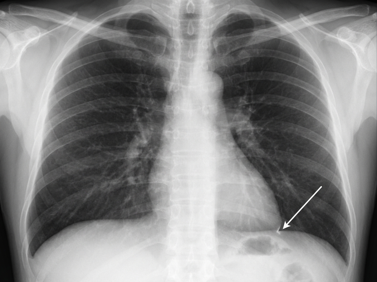
All of the following are true in a chest X-ray PA view EXCEPT:
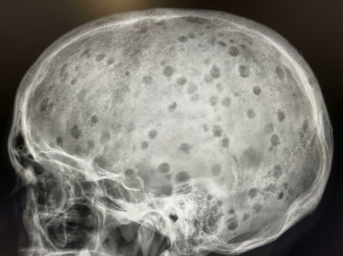
What is shown in the X-ray skull?

Identify the structure indicated in the given chest X-ray.

At 1 year of age, how many carpal bones are typically visible on a skiagram of the hand?
'Towne's view' in X-ray is used for visualizing which of the following structures?
The radiological feature of Pindborg's tumour is:
The 'hair on end' appearance on a skull X-ray is characteristic of which condition?
A low kilovoltage technique is most advantageous in demonstrating which of the following?
Shenton line is a radiological landmark seen in which joint on X-ray?
Wide diploic space of the skull with a brush border (hair on end) appearance is characteristic of which condition?
Explanation: In a Chest X-ray (PA view), the cardiac silhouette is formed by specific chambers and vessels. Understanding this anatomy is crucial for identifying chamber enlargement. **1. Why Option A is the Correct Answer (The "Except" Statement):** The **Right Ventricle (RV)** is the most anterior chamber of the heart. In a standard PA view, it sits directly behind the sternum and **does not form any part of the heart border**. It only contributes to the cardiac silhouette in a Lateral view (forming the anterior border). Therefore, the statement that the RV forms the right heart border is false. **2. Analysis of Other Options:** * **Option B (Left ventricle forms the left heart border):** This is **true**. The left heart border is formed by the aortic arch (knuckle), pulmonary artery segment, left atrial appendage, and primarily the **Left Ventricle**. * **Option C (Cardiothoracic ratio is 50% or less):** This is **true**. The CTR is the ratio of the maximum horizontal cardiac diameter to the maximum inner thoracic diameter. A ratio >0.5 (50%) in adults suggests cardiomegaly. * **Option D (Right atrium forms the right heart border):** This is **true**. The right heart border is formed by two main structures: the **Superior Vena Cava (SVC)** superiorly and the **Right Atrium (RA)** inferiorly. **High-Yield Clinical Pearls for NEET-PG:** * **Right Ventricular Enlargement (RVE):** On a PA view, RVE causes the **apex to be lifted upwards** (boot-shaped heart/Coeur en sabot), as seen in Tetralogy of Fallot. * **Left Atrial Enlargement (LAE):** Look for the "Double atrial shadow," splaying of the carina, and straightening of the left heart border. * **AP vs. PA View:** In an AP view (often portable), the heart appears artificially magnified, making the CTR unreliable for diagnosing cardiomegaly.
Explanation: ***Pepper pot skull*** - Characterized by **multiple small punched-out lytic lesions** throughout the skull, giving a characteristic "salt and pepper" appearance on X-ray. - Commonly associated with **hyperparathyroidism** and **multiple myeloma**, where bone destruction creates numerous small radiolucent areas. *Artifacts* - Result from **technical errors** during X-ray acquisition, such as patient movement, equipment malfunction, or processing issues. - Present as **irregular patterns** or distortions that don't follow anatomical structures and can be reproduced with repeat imaging. *Silver-beaten appearance* - Shows **generalized thinning** of the inner table of the skull with a beaten metal-like appearance on X-ray. - Associated with **raised intracranial pressure** in children, causing chronic pressure effects on the developing skull. *Mug shot with pellets* - Refers to **retained metallic foreign bodies** (like shotgun pellets) visible as **radiopaque densities** on skull X-ray. - Appears as **discrete, well-defined metallic objects** with characteristic shape and density, not the diffuse lytic pattern seen here.
Explanation: ***Left-Cardiophrenic angle*** - The **left-cardiophrenic angle** is formed by the junction of the **left heart border** and the **left hemidiaphragm**, creating a sharp, acute angle. - It is located more **medially** compared to the costophrenic angle and represents the **cardiac silhouette's** interface with the diaphragm. *Left-Costophrenic angle* - The **left-costophrenic angle** is formed by the junction of the **left hemidiaphragm** and the **lateral chest wall** (ribs), not involving the heart border. - It is located more **laterally** and represents the **pleural space** where fluid collections (pleural effusions) are first detected. *Right-Costophrenic angle* - The **right-costophrenic angle** is located on the **opposite side** of the chest from the indicated structure. - It is formed by the junction of the **right hemidiaphragm** and the **right lateral chest wall**, appearing as a sharp angle laterally. *Right-Cardiophrenic angle* - The **right-cardiophrenic angle** is located on the **opposite side** of the chest from the indicated structure. - It is formed by the junction of the **right heart border** and the **right hemidiaphragm**, positioned medially on the right side.
Explanation: **Explanation:** The appearance of carpal bones on a radiograph (skiagram) is a classic high-yield topic for assessing skeletal maturity and bone age in pediatric radiology. At birth, the carpal bones are entirely cartilaginous and therefore not visible on X-ray. They ossify in a predictable, clockwise sequence starting from the center. **Why Two is Correct:** By the end of the **first year (12 months)**, typically **two** carpal bones have begun to ossify and are visible: 1. **Capitate:** The first to appear (usually at 1–3 months). 2. **Hamate:** The second to appear (usually at 2–4 months). **Analysis of Incorrect Options:** * **A. None:** Incorrect, as the Capitate and Hamate ossify well before the first birthday. * **B. One:** Incorrect, as both the Capitate and Hamate are typically present by 6 months of age. * **D. Three:** Incorrect, as the third bone (**Triquetrum**) usually appears around **3 years** of age. **High-Yield Clinical Pearls for NEET-PG:** * **Order of Ossification (Mnemonic: "Go Catch Her, The Tiger Takes Laps"):** 1. **C**apitate (1–3 months) 2. **H**amate (2–4 months) 3. **T**riquetrum (3 years) 4. **L**unate (4 years) 5. **S**caphoid (5 years) 6. **T**rapezium (6 years) 7. **T**rapezoid (6 years) 8. **P**isiform (9–12 years; the last to ossify). * **Rule of Thumb:** The number of carpal bones visible on an X-ray is roughly equal to the **Age in Years + 1** (valid up to age 8). For a 1-year-old: 1 + 1 = 2 bones. * **Standard for Bone Age:** The **Greulich and Pyle Atlas** (using the left hand and wrist) is the gold standard for comparing these ossification centers to determine skeletal age.
Explanation: **Explanation:** **Towne’s View (Anteroposterior Axial Projection)** is a specialized radiographic view primarily used to visualize the **occipital bone** and the **petrous portions of the temporal bones**. **Why the correct answer is 'All of the above':** In Towne’s view, the X-ray beam is angled 30° caudally to the orbitomeatal line. This projection displaces the dense bones of the skull base, allowing for a clear, symmetrical visualization of the internal structures of the petrous pyramids. Specifically: * **Cochlea:** Located in the anterior part of the bony labyrinth, it is well-projected in this view. * **Arcuate Eminence:** This is the rounded prominence on the anterior surface of the petrous temporal bone (caused by the superior semicircular canal), which is clearly profiled. * **Superior Semicircular Canal:** Along with the posterior canal and the vestibule, these components of the inner ear are visible within the petrous ridge. **Analysis of Options:** Since all three structures (Cochlea, Arcuate eminence, and Superior semicircular canal) are anatomical components of the **inner ear/petrous temporal bone**, they are all effectively visualized in a well-executed Towne’s projection. **High-Yield Clinical Pearls for NEET-PG:** * **Primary Use:** Best view for the **occipital bone**, foramen magnum, and posterior clinoid processes. * **Clinical Indication:** Often used to evaluate **acoustic neuroma** (widening of the internal auditory canal) and fractures of the condylar neck of the mandible. * **Comparison:** While **Stenver’s view** is the "gold standard" for the petrous apex, Towne’s view provides an excellent bilateral comparison of the petrous ridges. * **Angle:** Remember the **30° caudal angle** to the orbitomeatal line (or 37° to the infraorbitomeatal line).
Explanation: ### Explanation **Pindborg’s Tumour**, medically known as **Calcifying Epithelial Odontogenic Tumour (CEOT)**, is a rare, benign but locally aggressive odontogenic neoplasm. **1. Why "Driven Snow Appearance" is Correct:** The characteristic radiological feature of CEOT is a multilocular (or occasionally unilocular) radiolucency containing varying amounts of radiopaque foci. These calcifications represent the mineralization of amyloid-like material within the tumour. When these scattered, dense white flecks are seen within the radiolucent area, they resemble **"driven snow."** This feature is most prominent when the tumour is associated with an impacted tooth (usually a mandibular molar). **2. Analysis of Incorrect Options:** * **Onion-peel appearance:** This refers to concentric layers of new periosteal bone formation. It is classically seen in **Ewing’s Sarcoma** and Garre’s Osteomyelitis. * **Sun burst appearance:** This represents divergent spicules of bone formation radiating from the periosteum. It is a hallmark of **Osteosarcoma**. * **Cherry blossom appearance:** This is a classic sialographic finding (punctate sialectasis) seen in **Sjögren’s syndrome**. **3. NEET-PG High-Yield Pearls:** * **Location:** Most common in the **posterior mandible** (molar-ramus area). * **Histology:** Look for **Liesegang rings** (concentric calcifications) and polyhedral epithelial cells with prominent intercellular bridges. * **Association:** Frequently associated with an **impacted tooth** (60% of cases). * **Differential Diagnosis:** If a lesion is pericoronal with radiopacities, consider CEOT, Adenomatoid Odontogenic Tumour (AOT), or Calcifying Odontogenic Cyst (COC).
Explanation: ### Explanation The **'hair-on-end'** (or crew-cut) appearance is a classic radiological sign seen on a lateral skull X-ray. It occurs due to **compensatory extramedullary hematopoiesis** in response to chronic hemolytic anemia. **1. Why Thalassemia is Correct:** In conditions like **Thalassemia Major** and **Sickle Cell Anemia**, the body attempts to compensate for chronic hypoxia and ineffective erythropoiesis by expanding the bone marrow. This causes: * **Widening of the diploic space** of the skull. * **Thinning of the outer table.** * The formation of new bone trabeculae perpendicular to the inner table, which creates the characteristic "sunburst" or "hair-on-end" vertical striations. **2. Why Other Options are Incorrect:** * **Hydrocephalus:** This typically presents with "beaten silver" or **"copper beaten"** skull appearance due to increased intracranial pressure causing gyral impressions on the inner table of the skull. * **Chronic Malaria:** While malaria causes hemolysis, it rarely leads to the degree of marrow hyperplasia required to produce the hair-on-end sign. * **All of the above:** Incorrect, as the sign is specific to marrow-expanding disorders. **3. High-Yield Clinical Pearls for NEET-PG:** * **Other conditions with 'hair-on-end':** Sickle cell anemia, Hereditary spherocytosis, and occasionally Iron deficiency anemia (rarely in children). * **Facial changes:** Marrow expansion in Thalassemia also involves the maxilla, leading to **"Chipmunk facies"** (prominent cheekbones and malocclusion). * **Note:** The **frontal bone** is most commonly involved, but the **occipital bone** is usually spared because it lacks significant hematopoietic marrow in adults.
Explanation: **Explanation:** The correct answer is **C. Incipient caries.** In dental radiography, the choice of kilovoltage (kVp) determines the **contrast** of the image. A **low kilovoltage technique (65-70 kVp)** produces a "short-scale" contrast, meaning there is a high degree of difference between black and white areas with fewer shades of gray. **Incipient caries** are early-stage lesions characterized by a very slight decrease in mineral density (demineralization) of the enamel. To detect these subtle changes, high contrast is essential to distinguish the radiolucent (dark) demineralized area from the surrounding radiopaque (white) healthy enamel. Therefore, low kVp is the gold standard for detecting early decay. **Analysis of Incorrect Options:** * **A & B (Alveolar crest resorption and Periapical lesions):** These conditions involve bone changes. Evaluating bone requires a "long-scale" contrast (high kVp) to visualize subtle variations in bone trabeculation and density. High kVp provides better latitude, allowing the clinician to see through varying thicknesses of bone. * **D (Nutrient canals):** These are small anatomical landmarks within the bone. Like other bony structures, they are better visualized using standard or higher kVp settings that provide a wider range of gray scales to differentiate delicate anatomical details. **High-Yield Facts for NEET-PG:** * **Low kVp (<70):** High contrast (Short-scale); best for **caries detection**. * **High kVp (>70-90):** Low contrast (Long-scale); best for **periodontal disease** and **periapical assessment** as it allows for better visualization of bone density changes. * **Rule of Thumb:** Increasing kVp increases the energy/penetration of the beam but decreases image contrast. * **Exposure:** Low kVp techniques generally result in a higher skin dose to the patient compared to high kVp techniques for the same film density.
Explanation: **Explanation:** **Shenton’s Line** is a fundamental radiological landmark used to assess the integrity of the **Hip joint** on an Anteroposterior (AP) X-ray of the pelvis. 1. **Why Hip is Correct:** Shenton’s line is an imaginary continuous arc formed by the **inferior border of the superior pubic ramus** and the **medial margin of the femoral neck**. In a normal, healthy hip, this line is smooth and unbroken. A disruption or "step-off" in this line is a critical diagnostic sign indicating pathology such as a femoral neck fracture, developmental dysplasia of the hip (DDH), or slipped capital femoral epiphysis (SCFE). 2. **Why Other Options are Incorrect:** * **Shoulder:** Landmarks here include the *Moloney’s line* (scapulohumeral arch), which assesses for dislocations. * **Elbow:** Key lines include the *Anterior Humeral Line* and the *Radiocapitellar Line*, used primarily to detect supracondylar fractures and radial head dislocations. * **Knee:** Radiological assessment focuses on the *Blumensaat’s line* (intercondylar notch) or the *Insall-Salvati ratio* for patellar height. **Clinical Pearls for NEET-PG:** * **DDH:** Disruption of Shenton’s line is a classic finding in Developmental Dysplasia of the Hip, along with an increased acetabular angle and lateral displacement of the femoral head. * **Positioning:** The line can occasionally appear broken if the hip is significantly externally rotated, so proper positioning is vital for accurate interpretation. * **Other Hip Lines:** Remember **Ward’s Triangle** (area of low bone density in the femoral neck) and **Skinner’s Line** as other high-yield hip landmarks.
Explanation: ### Explanation **Correct Answer: A. Congenital hemolytic anemia** The "hair-on-end" appearance (also known as the crew-cut sign) is a classic radiological finding caused by **compensatory extramedullary hematopoiesis**. In conditions like **Thalassemia major** and **Sickle cell anemia**, chronic hemolysis leads to a massive demand for red blood cell production. This results in: 1. **Marrow Hyperplasia:** The diploic space of the skull expands to accommodate the proliferating bone marrow. 2. **Trabecular Reorientation:** The outer table of the skull becomes thinned, and new bone trabeculae are laid down perpendicular to the inner table to provide structural support, creating the characteristic "brush border" or "hair-on-end" spikes on a lateral X-ray. --- ### Why the other options are incorrect: * **B. Multiple Myeloma:** Typically presents with **"punched-out" lytic lesions** (well-circumscribed, non-sclerotic margins) due to plasma cell infiltration. It does not cause diploic expansion or vertical trabeculations. * **C. Raised Intracranial Tension:** In children, this leads to **sutural diastasis** (widening of sutures) and a **"beaten silver" or "copper beaten" appearance** of the skull vault due to chronic pressure from gyri. * **D. Meningioma:** While it can cause localized hyperostosis (bone thickening) of the overlying skull, it does not produce a generalized hair-on-end appearance across the diploic space. --- ### High-Yield NEET-PG Pearls: * **Thalassemia:** The most common cause of the hair-on-end appearance. It also causes **"Chipmunk facies"** due to maxillary marrow expansion and a "Salt and Pepper" appearance of the skull. * **Sickle Cell Anemia:** Shows similar skull changes but may also feature **H-shaped vertebrae** (Reynold’s sign) due to endplate infarction. * **Note:** The **facial bones** are often spared in Sickle Cell Anemia (due to early marrow conversion to fat) but are heavily involved in Thalassemia, leading to the characteristic malocclusion.
Radiographic Anatomy of Skull and Face
Practice Questions
Radiographic Anatomy of Spine
Practice Questions
Radiographic Anatomy of Chest
Practice Questions
Radiographic Anatomy of Abdomen
Practice Questions
Radiographic Anatomy of Extremities
Practice Questions
Cross-sectional Anatomy: Brain and Head
Practice Questions
Cross-sectional Anatomy: Neck
Practice Questions
Cross-sectional Anatomy: Thorax
Practice Questions
Cross-sectional Anatomy: Abdomen and Pelvis
Practice Questions
Vascular Anatomy
Practice Questions
Developmental Anatomy Variations
Practice Questions
Anatomic Landmarks for Interventional Procedures
Practice Questions
Get full access to all questions, explanations, and performance tracking.
Start For Free