What is the threshold radiation dose for the hematological syndrome?
Which of the following is a late complication of radiotherapy?
Which tissue is most radiosensitive?
Which of the following is NOT a radioprotector?
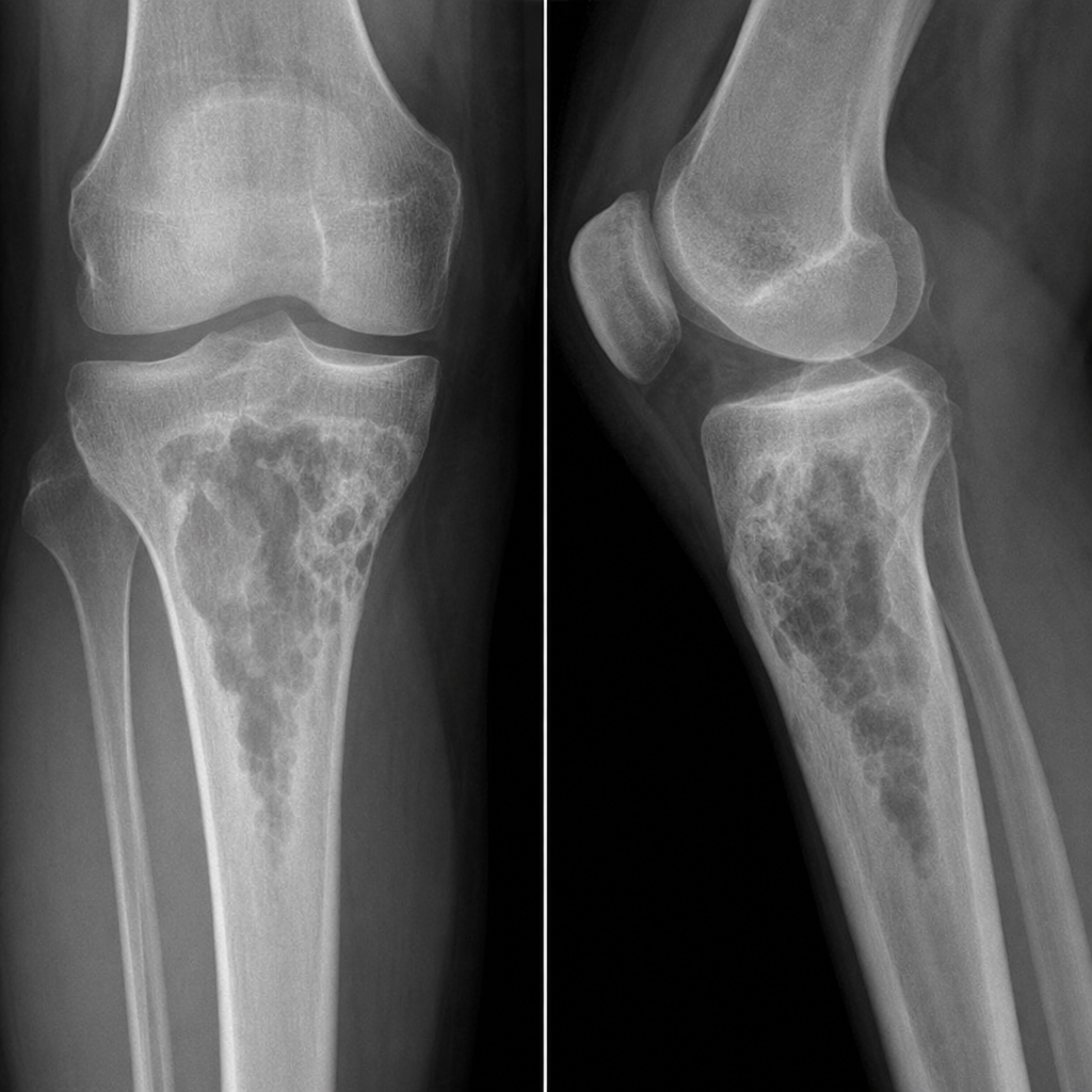
Which of the following statements about Linear Energy Transfer (LET) is true?
Acute radiation hepatic damage is most marked between which time period?
Which organ is most radiosensitive?
Radiation therapy to hypoxic tissues may be potentiated by treatment with which of the following?
Radiation caries is caused by irradiation of which structure?
What is the most important lesion produced in chromosomal DNA by exposure to ionizing radiation?
Explanation: ### Explanation **Acute Radiation Syndrome (ARS)** occurs after whole-body exposure to high doses of ionizing radiation. It is categorized into three distinct sub-syndromes based on the dose received and the organ system affected. **1. Why Option A (2 Gy) is Correct:** The **Hematological (Bone Marrow) Syndrome** occurs at doses between **2 and 10 Gy**. At this threshold, the radiation destroys the highly mitotic precursor cells in the bone marrow, leading to pancytopenia (depletion of white blood cells, platelets, and red blood cells). Death, if it occurs, is usually due to infection or hemorrhage within 3–6 weeks. **2. Why the Other Options are Incorrect:** * **Option B (6 Gy):** While 6 Gy falls within the range of hematological syndrome, it is above the *threshold* (starting point). At doses above 6–10 Gy, the Gastrointestinal syndrome begins to overlap and dominate. * **Option C (15 Gy):** This dose triggers the **Gastrointestinal (GI) Syndrome** (threshold: **6–10 Gy**). It involves the destruction of intestinal crypt cells, leading to severe diarrhea, dehydration, and electrolyte imbalance. Death typically occurs within 5–10 days. * **Option D (50 Gy):** This dose triggers the **Cerebrovascular (CNS) Syndrome** (threshold: **>20–50 Gy**). It results in immediate neurological symptoms, seizures, and coma, with death occurring within 24–48 hours. **High-Yield Clinical Pearls for NEET-PG:** * **LD 50/60:** The lethal dose required to kill 50% of the population in 60 days is approximately **3–4 Gy** (without medical intervention). * **Prodromal Phase:** The initial stage of ARS characterized by nausea, vomiting, and anorexia (NVA). * **Radiosensitivity:** According to the **Law of Bergonie and Tribondeau**, cells with high mitotic activity and low differentiation (like hematopoietic stem cells) are the most radiosensitive.
Explanation: In radiobiology, complications of radiotherapy are classified based on the timing of their appearance relative to the treatment course. **Correct Answer: C. Mucositis** Mucositis is traditionally categorized as an **acute complication** of radiotherapy. It occurs due to the rapid depletion of the basal cell layer of the oral or gastrointestinal mucosa, which has a high mitotic index. However, in the context of this specific question (often seen in previous medical exams), it is frequently contrasted against immediate systemic reactions. *Note for NEET-PG:* There is a common academic debate regarding this question. While mucositis is biologically "acute," it often persists longer than immediate reactions like nausea. However, if the question asks for a **late** complication (occurring months to years later), typical examples include **fibrosis, necrosis, and secondary malignancies**. If "Mucositis" is marked as the key, it is often due to its peak occurring toward the end of a 6-week treatment cycle compared to immediate "early" symptoms. **Analysis of Incorrect Options:** * **A. Nausea:** This is an **immediate/early** side effect, often part of "radiation sickness," occurring within hours of exposure. * **B. Thrombocytopenia:** This is an **acute** effect on the hematopoietic system. Bone marrow suppression occurs rapidly due to the high radiosensitivity of precursor cells. * **D. Erythema:** This is the classic **acute** skin reaction (resembling a sunburn) that occurs within days to weeks of starting therapy. **High-Yield Clinical Pearls for NEET-PG:** * **Acute Effects:** Occur in rapidly dividing tissues (Skin, Mucosa, Bone Marrow). * **Late Effects:** Occur in slowly dividing tissues (Lung, Kidney, Heart, CNS). The hallmark of late injury is **vascular damage and fibrosis**. * **Radiosensitivity:** The most sensitive phase of the cell cycle is **M (Mitosis)**, followed by G2. The most resistant phase is **S (Synthesis)**. * **Law of Bergonie and Tribondeau:** Radiosensitivity is directly proportional to the reproductive rate and inversely proportional to the degree of differentiation.
Explanation: The radiosensitivity of a tissue is governed by the **Law of Bergonié and Tribondeau**, which states that cells are most sensitive to radiation when they have a **high mitotic rate**, a **long mitotic future**, and are **undifferentiated (primitive)**. **1. Why Bone Marrow is Correct:** Bone marrow contains hematopoietic stem cells that are rapidly dividing and undifferentiated. Among all tissues listed, the hematopoietic system (specifically the erythroblasts and myeloblasts within the bone marrow) exhibits the highest rate of cell turnover. Therefore, it is the most radiosensitive organ in the body. **2. Analysis of Incorrect Options:** * **Gonads (Option A):** While highly radiosensitive (especially spermatogonia), they are generally ranked slightly below the hematopoietic system in terms of immediate sensitivity to cell death. * **Red Blood Cells (Option C):** Mature RBCs are highly **radioresistant** because they are non-dividing, highly differentiated, and lack a nucleus. * **Gastrointestinal Tract Epithelium (Option D):** The lining of the small intestine is highly radiosensitive due to rapid cell renewal in the crypts of Lieberkühn, but it ranks below the bone marrow in the hierarchy of sensitivity. **3. High-Yield Clinical Pearls for NEET-PG:** * **Most Radiosensitive Cell:** Lymphocyte (Exception to the law: it is sensitive despite being non-dividing). * **Most Radioresistant Cell:** Nerve cell / Myocyte. * **Order of Sensitivity (High to Low):** Bone Marrow > Gonads > GI Epithelium > Skin > Endothelium > Growing Bone/Cartilage > Muscle > Nerve. * **Cell Cycle Phase:** Cells are most sensitive in **M (Mitosis)** and **G2** phases; most resistant in **S (Synthesis)** phase.
Explanation: **Explanation:** In radiobiology, substances are classified based on how they modify the cellular response to ionizing radiation. The distinction between **radioprotectors** and **radiosensitizers** is a high-yield topic for NEET-PG. **Why BUDR is the correct answer:** **BUDR (5-Bromo-2'-deoxyuridine)** is a **radiosensitizer**, not a radioprotector. It is a halogenated pyrimidine analog that incorporates into the DNA of rapidly dividing cells in place of thymidine. This substitution makes the DNA chain more fragile and susceptible to radiation-induced strand breaks, thereby increasing the lethality of a given dose of radiation. **Analysis of incorrect options (Radioprotectors):** * **Amifostine (WR-2721):** This is the most potent and well-known radioprotector. It is a sulfhydryl compound that acts as a free radical scavenger. It is FDA-approved to reduce xerostomia in patients undergoing radiotherapy for head and neck cancers. * **IL-1 (Interleukin-1):** Cytokines like IL-1 act as biological response modifiers. They protect hematopoietic stem cells and promote recovery of the bone marrow after radiation exposure. * **GM-CSF (Granulocyte-Macrophage Colony-Stimulating Factor):** This is a growth factor that stimulates the proliferation of white blood cells. It is used clinically to mitigate hematologic toxicity (bone marrow syndrome) following radiation. **Clinical Pearls for NEET-PG:** * **Oxygen Effect:** Oxygen is the most potent naturally occurring radiosensitizer. * **Sulfhydryl Compounds:** Most radioprotectors work by scavenging free radicals (produced by indirect action of radiation) or by donating hydrogen atoms to repair DNA lesions. * **Radiosensitizers list:** BUDR, IUDR, Metronidazole, Misonidazole, and Cisplatin. * **Radioprotectors list:** Amifostine, Cysteine, Cysteamine, Vitamin E, and certain cytokines (IL-1, TNF-alpha).
Explanation: **Explanation:** **1. Why the Correct Answer is Right:** Linear Energy Transfer (LET) is the rate at which energy is deposited as an ionizing particle travels through matter (keV/µm). **High LET radiations** (e.g., alpha particles, neutrons) cause dense, direct ionization of DNA, leading to irreparable double-strand breaks. Because this damage is direct and physical, it does not rely on the presence of oxygen to "fix" the damage (the Oxygen Fixation Hypothesis). Therefore, High LET radiations have a **low Oxygen Enhancement Ratio (OER)**, meaning they are nearly as effective in hypoxic conditions as they are in oxygenated ones. **2. Why the Incorrect Options are Wrong:** * **Option A:** Low LET radiations (e.g., X-rays, Gamma rays) are **less lethal** per unit dose because they cause sparse ionizations and rely heavily on indirect action (free radical formation), which cells can often repair. * **Option B:** LET is a **property of the radiation** (the projectile), not the tissue. It depends on the charge and velocity of the ionizing particle. * **Option C:** LET and Relative Biological Effectiveness (RBE) are related but **not the same**. RBE is a ratio comparing the biological effect of a test radiation to a standard (250 kVp X-rays). As LET increases, RBE generally increases (up to a point of "overkill" at 100 keV/µm). **3. NEET-PG High-Yield Clinical Pearls:** * **OER Formula:** Dose required under hypoxic conditions / Dose required under aerobic conditions. * **OER Values:** For X-rays (Low LET), OER is ~2.5–3.0. For Alpha particles (High LET), OER is ~1.0. * **The "Overkill" Effect:** RBE peaks at an LET of **100 keV/µm**. Beyond this, energy is wasted because the DNA is already "over-killed," and RBE actually decreases. * **Direct vs. Indirect Action:** High LET = Direct action (dominant); Low LET = Indirect action (dominant, mediated by OH• radicals).
Explanation: **Explanation:** **Radiation-Induced Liver Disease (RILD)**, often referred to as radiation hepatitis, is a subacute clinical syndrome. Unlike the immediate "radiation sickness" seen in the GI tract, the liver's response to radiation is delayed due to the slow turnover rate of hepatocytes and vascular endothelial cells. 1. **Why 40–100 days is correct:** The underlying pathophysiology of acute radiation hepatic damage is **Veno-Occlusive Disease (VOD)**. Following radiotherapy, there is a progressive obstruction of the central veins of the liver lobules due to fibrin deposition and endothelial swelling. This process takes time to manifest clinically. Symptoms typically peak between **2 to 6 weeks (subacute phase)** but the most marked clinical and pathological damage is classically observed between **40 and 100 days** post-exposure. 2. **Why other options are incorrect:** * **1–7 days:** This period corresponds to the *Prodromal phase* of radiation syndrome (nausea, vomiting), not specific organ parenchymal damage. * **7–21 days:** This is the *Latent period* for the liver. While hematopoietic damage (bone marrow) manifests here, the liver remains clinically asymptomatic. * **21–40 days:** This is the early onset period. While damage begins, it has not yet reached its "most marked" or peak clinical presentation. **High-Yield Clinical Pearls for NEET-PG:** * **Pathological Hallmark:** Central venous congestion and "Veno-occlusive disease" (VOD). * **Clinical Triad:** Hepatomegaly, ascites, and elevated alkaline phosphatase (disproportionate to bilirubin). * **Tolerance Dose (TD 5/5):** The whole-liver tolerance dose is approximately **30–35 Gy**. Exceeding this significantly increases the risk of RILD. * **Radiological Sign:** On CT, acute radiation hepatitis appears as a well-defined area of low attenuation corresponding exactly to the radiation portal.
Explanation: **Explanation:** The radiosensitivity of a tissue is primarily governed by the **Law of Bergonie and Tribondeau**. This law states that cells are most sensitive to radiation when they have a **high mitotic rate**, a **long mitotic future** (many future divisions), and are **undifferentiated** (primitive). 1. **Why Bone Marrow is Correct:** Bone marrow contains hematopoietic stem cells that are rapidly dividing and undifferentiated. Among all body tissues, the hematopoietic system (specifically erythroblasts and myeloblasts) is considered the most radiosensitive. Exposure to even low doses of radiation can lead to a significant drop in blood cell counts (pancytopenia). 2. **Why the others are Incorrect:** * **Fat (Adipose Tissue):** These are specialized, differentiated cells with a very low rate of division, making them radioresistant. * **Nervous Tissue:** Mature neurons are highly differentiated and do not divide (permanent cells). Consequently, the adult brain and spinal cord are among the most **radioresistant** tissues in the body. * **Liver:** While more sensitive than fat or nerve tissue, hepatocytes are considered "reverting post-mitotics." They divide slowly under normal conditions, placing them in the intermediate sensitivity category. **High-Yield Clinical Pearls for NEET-PG:** * **Order of Radiosensitivity (High to Low):** Bone Marrow > Gastrointestinal Tract > Skin > Liver > Muscle > Nerve/Bone. * **Most sensitive cell in the body:** Lymphocyte (Exception to the law: it is highly sensitive despite being a non-dividing cell). * **Most sensitive phase of the cell cycle:** **M phase** (Mitosis), followed by G2. * **Most resistant phase:** **S phase** (DNA synthesis). * **LD50/60 for humans:** Approximately 3.5 to 4.5 Gray (without medical intervention).
Explanation: ### Explanation **The Oxygen Effect and Radiosensitizers** The correct answer is **Metronidazole**. In radiobiology, the "Oxygen Effect" states that cells are more sensitive to ionizing radiation in the presence of molecular oxygen. Oxygen acts as a **radiosensitizer** by reacting with free radicals produced by radiation to create permanent, irreparable DNA damage (the Oxygen Fixation Hypothesis). Solid tumors often contain **hypoxic zones** (low oxygen), making them resistant to radiotherapy. To overcome this, chemical **hypoxic cell sensitizers** are used. **Metronidazole** and other nitroimidazoles (like Misonidazole and Nimorazole) act as "oxygen mimics." They possess high electron affinity, allowing them to mimic the sensitizing effect of oxygen in hypoxic tissues, thereby potentiating the effects of radiation. **Analysis of Incorrect Options:** * **A. Mycostatin (Nystatin):** An antifungal medication used for Candida infections; it has no role in modulating radiation sensitivity. * **C. Methotrexate:** A folate antagonist chemotherapy agent. While it is a cell-cycle specific drug, it is not classified as a specific hypoxic cell radiosensitizer. * **D. Melphalan:** An alkylating agent used in treating multiple myeloma. It is a radiomimetic drug (mimics radiation damage) but does not specifically potentiate radiation in hypoxic conditions like nitroimidazoles do. **High-Yield Clinical Pearls for NEET-PG:** * **Oxygen Enhancement Ratio (OER):** The ratio of doses administered under hypoxic vs. aerated conditions to achieve the same biological effect. For X-rays, OER is typically **2.5 to 3.0**. * **Hypoxic Sensitizers:** Look for drugs with the suffix "-nidazole." **Nimorazole** is currently the most clinically utilized agent in this class (specifically for head and neck cancers). * **Hyperbaric Oxygen:** Another method to overcome hypoxia, though less practical than chemical sensitizers.
Explanation: **Explanation:** **Radiation caries** is a rampant form of dental decay that occurs as a secondary complication of radiotherapy for head and neck cancers. **Why Salivary Glands is correct:** The primary cause is not the direct effect of radiation on the teeth, but rather the **indirect effect** of irradiation on the **major salivary glands** (especially the parotid). Radiation causes fibrosis and atrophy of the acinar cells, leading to **Xerostomia** (severe dry mouth). Saliva normally acts as a buffer, provides mechanical cleansing, and contains minerals (calcium/phosphate) for remineralization. When salivary flow is reduced, the oral pH drops, and the protective buffering capacity is lost, leading to rapid demineralization and "radiation caries." **Why other options are incorrect:** * **Enamel & Dentin:** While high doses of radiation can theoretically alter the organic matrix of teeth, mature enamel and dentin are highly radioresistant. The decay seen in these patients is primarily due to the altered oral environment (lack of saliva) rather than direct structural damage to the tooth. * **Oral Mucosa:** Irradiation of the mucosa leads to **mucositis**, which causes pain and ulceration. While this may make oral hygiene difficult, it is not the physiological cause of the caries itself. **High-Yield Clinical Pearls for NEET-PG:** * **Characteristic Appearance:** Radiation caries typically starts at the **cervical (neck) region** of the teeth and can lead to the amputation of the crown. * **Threshold:** Salivary glands are highly radiosensitive; permanent xerostomia can occur at doses as low as **20-30 Gy**. * **Prevention:** Patients should undergo dental evaluation *before* starting radiotherapy and use topical fluoride applications during/after treatment. * **Osteoradionecrosis (ORN):** Another complication of radiation, usually affecting the **mandible** due to its decreased vascularity compared to the maxilla.
Explanation: **Explanation:** The biological effects of ionizing radiation are primarily mediated through damage to the DNA molecule. While radiation can cause various types of lesions, the **Double-Strand Break (DSB)** is considered the most significant and lethal lesion. **1. Why Option C is Correct:** A Double-Strand Break occurs when both sugar-phosphate backbones are severed at locations directly opposite each other or separated by only a few base pairs. Unlike single-strand breaks, DSBs are difficult for the cell to repair accurately. If repaired incorrectly (misrepair), it leads to chromosomal aberrations, mutations, or carcinogenesis. If unrepaired, it leads to **mitotic death** (the most common form of cell death following radiation). Therefore, DSBs are the primary cause of radiation-induced cell killing and genetic damage. **2. Why Other Options are Incorrect:** * **Option A & D:** Single-strand breaks (SSBs) and multiple breaks on the same strand are very common but are usually repaired with high fidelity using the opposite intact strand as a template (via Base Excision Repair). They rarely result in cell death or long-term mutation. * **Option B:** If breaks on opposite strands are well-separated, they are functionally treated as two independent single-strand breaks and are easily repaired. **High-Yield Clinical Pearls for NEET-PG:** * **Direct Action:** Radiation interacts directly with DNA (common with high-LET radiation like alpha particles). * **Indirect Action:** Radiation interacts with water to form **Free Radicals** (e.g., OH•), which then damage DNA (common with low-LET radiation like X-rays/Gamma rays). * **Radiosensitivity:** Cells are most sensitive in the **M phase** and late **G2 phase** of the cell cycle; they are most resistant in the late **S phase**. * **Law of Bergonie and Tribondeau:** Stem cells and highly proliferative cells (e.g., bone marrow, intestinal epithelium) are the most radiosensitive.
Explanation: **Explanation:** **Amifostine** is a prodrug that is converted by alkaline phosphatase into an active thiol compound (WR-2721). It acts as a **Radioprotector** by scavenging free radicals (specifically hydroxyl radicals) generated by ionizing radiation and by donating hydrogen atoms to repair damaged DNA. Its clinical utility stems from **differential protection**: normal tissues have higher alkaline phosphatase activity and better vascularity than tumor cells, allowing Amifostine to accumulate preferentially in healthy tissue. This protects normal organs (like salivary glands) from radiation-induced damage without protecting the tumor. **Analysis of Options:** * **A. Radiosensitizer:** These are agents (e.g., Oxygen, Misonidazole, Cisplatin) that make tumor cells *more* sensitive to radiation. Amifostine does the opposite by protecting cells. * **C. Radio modifier:** This is a broad umbrella term that includes both sensitizers and protectors. While technically true, "Radioprotector" is the specific and most accurate classification. * **D. Radiomimetic:** These are drugs (e.g., Nitrogen mustard, Cyclophosphamide) that mimic the effects of ionizing radiation by causing similar DNA damage. **High-Yield Clinical Pearls for NEET-PG:** * **FDA Approval:** Amifostine is specifically used to reduce the incidence of **xerostomia** (dry mouth) in patients undergoing radiotherapy for head and neck cancers. * **Side Effects:** The most common dose-limiting side effect is **hypotension**; others include nausea, vomiting, and hypocalcemia. * **Time of Administration:** It must be administered intravenously shortly before radiation therapy to be effective.
Explanation: **Explanation:** The correct answer is **D. All of the above.** This question tests the concept of **Stochastic effects** of radiation. Unlike deterministic effects (which have a threshold dose, like cataracts), stochastic effects have **no threshold**. This means that even low-dose radiation (such as from diagnostic X-rays or CT scans) carries a statistical probability of causing DNA mutations that can lead to carcinogenesis. * **Acute Myeloid Leukemia (AML):** Bone marrow is one of the most radiosensitive tissues in the body. Leukemia (specifically AML and Chronic Myeloid Leukemia) has the shortest latent period (approx. 2–5 years) among radiation-induced cancers, making it a classic example of low-dose risk. * **Lung Cancer:** The lungs are highly susceptible to radiation-induced damage, often seen in populations exposed to environmental radon or occupational radiation. * **Cervical Cancer:** While less commonly associated than leukemia or thyroid cancer, the pelvic organs are sensitive to ionizing radiation. Secondary malignancies (including cervical and vaginal cancers) can occur following low-dose scatter or therapeutic radiation. **High-Yield Clinical Pearls for NEET-PG:** 1. **Stochastic Effects:** Probability is proportional to dose, but severity is independent of dose (e.g., Cancer, Genetic mutations). 2. **Deterministic Effects:** Occur only after a threshold dose; severity increases with dose (e.g., Radiation burns, Cataracts, Sterility). 3. **Most Radiosensitive Cancers:** Leukemia (except CLL), Thyroid cancer (especially in children), and Breast cancer. 4. **Law of Bergonie and Tribondeau:** Cells are most radiosensitive if they have a high mitotic rate, long mitotic future, and are undifferentiated (e.g., Stem cells, Lymphocytes).
Explanation: ### Explanation In radiobiology, radiation effects are classified into two main categories: **Stochastic** and **Deterministic (Non-stochastic)**. **1. Why Infertility is the Correct Answer:** **Infertility** is a **Deterministic effect**. These effects occur only after a specific **threshold dose** is exceeded. Once the threshold is crossed, the severity of the effect increases proportionally with the dose. For example, a dose of ~2 Gy can cause temporary sterility, while ~5 Gy can cause permanent sterility. Since it requires a minimum dose and its severity is dose-dependent, it is NOT stochastic. **2. Analysis of Incorrect Options (Stochastic Effects):** Stochastic effects are "all-or-nothing" phenomena. They have **no threshold dose**, and their **probability** (not severity) increases with the dose. * **A. Cancer (Carcinogenesis):** The most significant stochastic effect. Even a single photon could theoretically cause a mutation leading to malignancy. * **B. Genetic Side Effects:** Radiation-induced mutations in germ cells that affect future generations are stochastic. * **D. Teratogenicity:** While some developmental defects are deterministic, radiation-induced mental retardation and certain congenital malformations are often categorized under stochastic risks in a broader clinical context (though specifically, the *risk* of induction is the stochastic element). **3. High-Yield Clinical Pearls for NEET-PG:** * **Stochastic Effects:** No threshold, probability depends on dose, severity is independent of dose (e.g., Cancer, Genetic mutations). * **Deterministic Effects:** Clear threshold, severity depends on dose (e.g., Cataracts, Infertility, Erythema, Organ atrophy). * **Radiosensitivity:** According to the **Law of Bergonie and Tribondeau**, cells that are rapidly dividing, undifferentiated, and have a long mitotic future are most radiosensitive (e.g., Bone marrow, Lymphocytes, Gonads). * **Most sensitive phase of cell cycle:** M phase (Mitosis), followed by G2. * **Most resistant phase:** Late S phase.
Explanation: ### Explanation The radiosensitivity of a cell is governed by the **Law of Bergonié and Tribondeau**, which states that cells are most sensitive to radiation when they have a **high mitotic rate**, a **long mitotic future**, and are **undifferentiated**. **1. Why Option B is Correct:** The cells listed in Option B—**Red blood cells (mature), muscle cells, and nerve cells**—are highly differentiated (specialized) and do not undergo division (post-mitotic). Because they lack mitotic activity, they are the most radioresistant cells in the human body. Nerve cells are considered the most radioresistant of all. **2. Analysis of Incorrect Options:** * **Option A & D:** These include **White Blood Cells (Lymphocytes)** and **Epithelial cells**. Lymphocytes are a notable exception to the Law of Bergonié and Tribondeau; despite being non-dividing, they are highly radiosensitive. Epithelial cells (like those in the GI tract) divide rapidly and are therefore highly sensitive. * **Option C:** **Endothelial cells** and **Connective tissue cells** have intermediate radiosensitivity. They divide occasionally and are more sensitive than muscle or nerve cells but less sensitive than stem cells. **3. NEET-PG High-Yield Pearls:** * **Most Radiosensitive Cell:** Lymphocyte (specifically the small lymphocyte). * **Most Radiosensitive Phase of Cell Cycle:** **M phase** (Mitosis), followed by G2. * **Most Radioresistant Phase:** **S phase** (Late S phase). * **Order of Sensitivity (High to Low):** Mnemonic: **"L-E-G-B-M-N"** * **L**ymphocytes → **E**rythroblasts → **G**astrointestinal epithelium → **B**one (osteoblasts) → **M**uscle → **N**erve cells. * **Radiosensitizers:** Oxygen is the most potent radiosensitizer (Oxygen Enhancement Ratio).
Explanation: **Explanation:** The manifestation of chromosomal damage depends on the timing of radiation exposure relative to the **S-phase (DNA synthesis)** of the cell cycle. **1. Why Option B is Correct:** When radiation exposure occurs **before DNA synthesis (G1 phase)**, the cell contains only a single strand of chromatin. If a break occurs at this stage, it is a "chromosome aberration." During the subsequent S-phase, the damaged DNA template is replicated. Consequently, the break is duplicated, resulting in **damage to both sister chromatids (both arms)** of the chromosome seen during metaphase. **2. Why Other Options are Incorrect:** * **Option A & C:** Damage to only one arm (chromatid aberration) occurs if the radiation exposure happens **after DNA synthesis (G2 phase)**. At this stage, the DNA has already replicated into two sister chromatids. An ionizing hit usually affects only one of the two strands, leaving the other intact. * **Option D:** Radiation is ionizing; it inherently causes single or double-strand breaks in DNA. It is impossible for a significant radiation dose to result in "no arm" being affected if damage has occurred. **High-Yield Clinical Pearls for NEET-PG:** * **Radiosensitivity:** Cells are most sensitive during **Mitosis (M phase)** and late **G2 phase**. They are most resistant during the **S phase**. * **Direct vs. Indirect Action:** Most DNA damage from X-rays is **indirect**, mediated by the radiolysis of water and the production of **hydroxyl (OH•) free radicals**. * **Lethal Aberrations:** Dicentric rings and anaphase bridges are considered lethal chromosomal aberrations. * **Rule of Thumb:** Pre-replication exposure = Chromosome aberration (both arms); Post-replication exposure = Chromatid aberration (one arm).
Explanation: ### Explanation The sensitivity of a cell to ionizing radiation varies significantly across the cell cycle, a concept known as **Radiosensitivity**. **1. Why G2 and M are the most sensitive:** * **M Phase (Mitosis):** This is the **most sensitive** phase. During mitosis, DNA is highly condensed (chromatin condensation), and the cell’s repair mechanisms are least active. Any damage to the DNA during this stage leads to immediate mitotic death or chromosomal aberrations. * **G2 Phase:** This is the second most sensitive phase. The cell is preparing for division, and there is little time for DNA repair before it enters the vulnerable M phase. **2. Why other options are incorrect:** * **S Phase (Synthesis):** This is the **most radioresistant** phase of the cell cycle. During the late S phase, homologous recombination repair mechanisms are most active because the DNA has been replicated, providing a sister chromatid as a template for repair. * **G1 Phase:** Sensitivity in G1 is intermediate. While more sensitive than the S phase, it is significantly less sensitive than the G2/M transition. * **G0 Phase:** These are quiescent cells. While they can be damaged, the "Law of Bergonie and Tribondeau" states that cells with high mitotic activity are more radiosensitive; thus, non-dividing G0 cells are relatively resistant compared to cycling cells. **3. High-Yield Clinical Pearls for NEET-PG:** * **Law of Bergonie and Tribondeau:** Radiosensitivity is directly proportional to the reproductive rate and inversely proportional to the degree of differentiation. * **Most Sensitive Phase:** M phase. * **Most Resistant Phase:** Late S phase. * **Oxygen Enhancement Ratio (OER):** Cells are more sensitive to radiation in the presence of oxygen (Oxygen Effect), which is why hyperbaric oxygen is sometimes used in radiotherapy. * **Order of Sensitivity (Decreasing):** M > G2 > G1 > S.
Explanation: **Explanation:** The effects of ionizing radiation on a fetus depend on the **gestational age** and the **absorbed dose**. While high doses (>100-200 mGy) are associated with deterministic effects like malformations or fetal death, diagnostic radiation (typically <50 mGy) is more commonly associated with subtle developmental impacts. **Why "Low birth weight babies" is correct:** Exposure to ionizing radiation during the period of **organogenesis** (weeks 2–8) and the **fetal period** (week 8 to term) can lead to growth retardation. Radiation interferes with rapid cell proliferation, leading to a decrease in the total number of cells in the developing fetus. This manifests clinically as **intrauterine growth restriction (IUGR)** and subsequent low birth weight, even at doses lower than those required to cause gross structural malformations. **Analysis of Incorrect Options:** * **A. High fetal exposure:** Most routine diagnostic procedures (e.g., Chest X-ray, Extremity X-ray) result in very low fetal doses (often <0.01 mGy). High exposure is generally defined as >100 mGy, which is rarely reached in diagnostic radiology. * **B. Abortion:** This is typically an "all-or-none" phenomenon occurring during the **pre-implantation stage** (0–2 weeks). Diagnostic doses are usually insufficient to cause miscarriage unless they exceed 50–100 mGy. * **D. Genetic abnormalities:** While radiation is mutagenic, the risk of hereditary genetic defects manifesting in the offspring due to diagnostic prenatal exposure is considered statistically negligible compared to the baseline risk in the general population. **NEET-PG High-Yield Pearls:** 1. **Safe Threshold:** Fetal risk is considered negligible at doses **<50 mGy**. 2. **Most Sensitive Period:** The fetus is most sensitive to CNS effects (microcephaly, intellectual disability) between **8–15 weeks** of gestation. 3. **Carcinogenesis:** The main stochastic risk of diagnostic radiation in utero is a slight increase in the childhood risk of **Leukemia**. 4. **Rule of Thumb:** No single diagnostic X-ray procedure results in a radiation dose significant enough to threaten the well-being of the developing embryo.
Explanation: The correct answer is **Muscle**. ### **Explanation** The radiosensitivity of a tissue is governed by the **Law of Bergonié and Tribondeau**, which states that cells are most sensitive to radiation when they are: 1. **Actively dividing** (High mitotic rate) 2. **Undifferentiated** (Primitive/Stem cells) 3. **Metabolically active** **Muscle cells** (along with nerve cells) are highly differentiated, specialized, and do not undergo frequent mitosis (post-mitotic). Therefore, they are classified as **radioresistant**. ### **Analysis of Other Options** * **Testis (Option A):** Contains spermatogonia, which are among the most radiosensitive cells in the body. Even low doses can cause permanent sterility. * **Bone Marrow (Option B):** Contains hematopoietic stem cells that are rapidly dividing. It is considered the **most radiosensitive organ system** in the body. * **Intestine (Option C):** The crypt cells of the small intestine have a very high turnover rate to replace the mucosal lining, making the GI tract highly sensitive to radiation. ### **NEET-PG High-Yield Pearls** * **Most Radiosensitive Cell:** Lymphocyte (Exception to the law: it is sensitive despite not dividing frequently). * **Most Radiosensitive Phase of Cell Cycle:** **M phase** (Mitosis), followed by G2. * **Most Radioresistant Phase:** **S phase** (DNA synthesis). * **Order of Sensitivity (High to Low):** Bone Marrow > Gonads > GI Tract > Skin > Lungs > Liver > Kidney > **Muscle/Nerve**.
Explanation: ### Explanation **Concept: The Oxygen Enhancement Ratio (OER)** The biological effectiveness of ionizing radiation is significantly increased in the presence of molecular oxygen. This is known as the **Oxygen Effect**. For oxygen to act as a radiosensitizer, it must be present at the exact moment of radiation or within microseconds of the interaction. **Why Option A is Correct:** In clinical practice, oxygen must be present in the tissues **just before and during** the delivery of the radiation beam. This ensures that when radiation interacts with water molecules to create free radicals (indirect action), oxygen is already available to react with the damaged DNA. This reaction "fixes" the damage (the **Oxygen Fixation Hypothesis**), making the chemical break in the DNA permanent and irreparable. **Analysis of Incorrect Options:** * **Option B (During and within microseconds):** While scientifically accurate regarding the chemical reaction time, in a clinical/procedural context for NEET-PG, the preparation (ensuring the patient is well-oxygenated *just before* the switch is turned on) is the prioritized step. * **Options C & D (After 5 or 10 minutes):** These are incorrect because the free radicals produced by radiation have an extremely short lifespan ($10^{-5}$ seconds). If oxygen is introduced after the radiation has passed, the DNA radicals will have already chemically "repaired" or stabilized themselves, rendering the oxygen useless for sensitization. --- ### High-Yield Clinical Pearls for NEET-PG * **OER Value:** For X-rays and Gamma rays, the OER is typically **2.5 to 3.0**. * **LET Relationship:** The oxygen effect is maximal with **Low-LET radiation** (X-rays, Gamma rays) and minimal to absent with **High-LET radiation** (Alpha particles, Neutrons). * **Hypoxic Tumors:** Large tumors often have necrotic, hypoxic centers which are resistant to radiotherapy. Strategies to overcome this include hyperbaric oxygen or hypoxic cell sensitizers (e.g., Nitroimidazoles). * **The "Fixation" Fact:** Oxygen does not *cause* the damage; it *prevents the repair* of damage caused by free radicals.
Explanation: ### Explanation The radiosensitivity of cells is governed by the **Law of Bergonie and Tribondeau**, which states that cells are most sensitive to radiation when they are **highly proliferative (high mitotic rate)**, have a **long mitotic future**, and are **undifferentiated**. #### 1. Why Vegetative Intermitotic Cells are Correct **Vegetative intermitotic cells** are the most radiosensitive because they are undifferentiated stem cells that divide regularly and rapidly. They fulfill all criteria of the Bergonie-Tribondeau law. * **Examples:** Basal cells of the epidermis, intestinal crypt cells, erythroblasts, and spermatogonia. #### 2. Analysis of Other Options * **B. Differentiating Intermitotic Cells:** These are slightly more mature than vegetative cells. While they still divide, they have begun to undergo some differentiation, making them slightly less sensitive. (e.g., Myelocytes). * **C. Reverting Post-Mitotic Cells:** These cells normally do not divide but retain the capability to do so if stimulated by injury. Because of their low metabolic/mitotic activity, they are relatively radioresistant. (e.g., Liver cells, lymphocytes—though lymphocytes are a notable exception). * **D. Fixed Post-Mitotic Cells:** These are highly differentiated cells that have lost the ability to divide. They are the **most radioresistant** cells in the body. (e.g., Nerve cells, muscle cells). #### 3. NEET-PG High-Yield Pearls * **The Lymphocyte Exception:** Although lymphocytes are mature, non-dividing cells (technically reverting post-mitotic), they are **highly radiosensitive** and are often the first cells to decrease in number after radiation exposure. * **Cell Cycle Sensitivity:** Cells are most sensitive in the **G2 and M phases** and most resistant in the **S phase**. * **Order of Sensitivity (Highest to Lowest):** Vegetative Intermitotic > Differentiating Intermitotic > Multipotential Connective Tissue > Reverting Post-Mitotic > Fixed Post-Mitotic.
Explanation: **Explanation:** The radiosensitivity of a cell is primarily determined by its genetic material. Among the options provided, **Nucleosomal DNA** is considered the most radiosensitive. **1. Why Nucleosomal DNA is the correct answer:** The nucleosome is the fundamental structural unit of chromatin, consisting of DNA wrapped around histone proteins. During the process of transcription and replication, nucleosomal DNA undergoes structural remodeling, making it highly "open" and accessible. This lack of compact shielding, combined with its high metabolic activity, makes it the most vulnerable target for both direct ionization and indirect damage (via free radicals) from ionizing radiation. **2. Analysis of Incorrect Options:** * **Nuclear DNA:** While nuclear DNA is the primary target for radiation-induced cell death, it is a broad term. Nucleosomal DNA is a specific, more sensitive sub-component of the total nuclear DNA. * **Mitochondrial DNA (mtDNA):** Although mtDNA lacks protective histones, it is generally considered more radioresistant than nuclear DNA because it is present in multiple copies per cell and has a robust repair mechanism for oxidative stress. * **Satellite DNA:** This consists of highly repetitive, non-coding sequences usually located in heterochromatin (tightly packed). Its condensed state provides a degree of physical protection against radiation compared to the more active nucleosomal DNA. **Clinical Pearls for NEET-PG:** * **Law of Bergonie and Tribondeau:** Radiosensitivity is directly proportional to the reproductive activity (mitosis) and inversely proportional to the degree of differentiation. * **Most sensitive phase of the cell cycle:** **M phase** (Mitosis), followed by G2. * **Most resistant phase:** **S phase** (Late S phase). * **Most radiosensitive cell in the body:** Lymphocyte (exception to the law, as it is a non-dividing cell). * **Most radiosensitive organ:** Ovary/Testis (Gonads).
Explanation: ### Explanation **1. Why Option A is Correct:** In radiobiology, the **latent period** is defined as the time lag between the initial exposure to ionizing radiation and the first detectable clinical sign or symptom. When radiation interacts with biological tissue, it causes immediate physical and chemical damage (ionization and free radical formation). However, it takes time for these molecular changes to translate into cellular dysfunction, cell death, and ultimately, observable clinical effects (like erythema, epilation, or malignancy). This period can range from hours (in acute radiation syndrome) to decades (in radiation-induced carcinogenesis). **2. Analysis of Incorrect Options:** * **Option B:** This describes the **development time** in film processing (the chemical process of converting a latent image into a visible one), which is a technical radiographic term, not a biological one. * **Option C:** This refers to the **overall treatment time** or fractionation schedule in radiotherapy, which is used to calculate the biological effective dose. * **Option D:** This is a distractor. Biological effects (like DNA strand breaks) occur almost **instantaneously** (within nanoseconds to seconds). The latent period specifically refers to the delay before these effects manifest **clinically**. **3. NEET-PG High-Yield Clinical Pearls:** * **Inverse Relationship:** The length of the latent period is generally **inversely proportional** to the radiation dose; higher doses typically result in shorter latent periods. * **Stochastic vs. Deterministic:** For stochastic effects (like leukemia), the latent period is long (minimum 2–5 years for leukemia; 10–20 years for solid tumors). * **Radiosensitivity:** According to the **Law of Bergonie and Tribondeau**, cells that are rapidly dividing, undifferentiated, and have a long mitotic future are the most radiosensitive (e.g., lymphocytes, germ cells).
Explanation: **Explanation:** **1. Why Erythema is Correct:** Erythema is the earliest clinical manifestation of radiation-induced skin injury (radiodermatitis). It typically occurs within hours to days after exposure. The underlying pathophysiology involves **acute vasodilation** and increased capillary permeability caused by the release of inflammatory mediators (like histamine and cytokines) in response to ionizing radiation. This initial "transient erythema" is often followed by a more persistent "main erythema" phase as the basal cell layer of the epidermis begins to fail. **2. Analysis of Incorrect Options:** * **B. Desquamation:** This occurs later than erythema (usually 3–4 weeks post-exposure). It results from the depletion of the basal cell layer, leading to the peeling of the skin. It can be dry (flaking) or moist (serous exudate). * **C. Edematous rough skin:** This is a feature of subacute or chronic radiation damage. Edema occurs due to lymphatic obstruction and chronic inflammation, appearing well after the initial erythematous phase. * **D. Hyperpigmentation:** This is a late-stage sequela of radiation. It occurs weeks to months later due to the stimulation of melanocytes or as a post-inflammatory response following the healing of acute dermatitis. **3. High-Yield Clinical Pearls for NEET-PG:** * **Order of Skin Changes:** Transient Erythema → Main Erythema → Dry Desquamation → Moist Desquamation → Necrosis/Ulceration. * **Radiosensitivity:** The **Basal layer** of the epidermis is the most radiosensitive part of the skin. * **Law of Bergonie and Tribondeau:** Cells that are rapidly dividing, undifferentiated, and have a long mitotic future are the most radiosensitive (e.g., bone marrow, germ cells, intestinal epithelium). * **Threshold Dose:** Acute skin erythema typically occurs at a threshold dose of approximately **2–6 Gy**.
Explanation: ### Explanation The radiosensitivity of a tissue is primarily determined by the **Law of Bergonié and Tribondeau**, which states that cells are most sensitive to radiation when they have a **high mitotic rate**, a **long mitotic future**, and are **undifferentiated** (primitive). **1. Why Bone Marrow is Correct:** Bone marrow contains hematopoietic stem cells that are rapidly dividing and undifferentiated. Among all tissues listed, the hematopoietic system (specifically the erythroblasts and myeloblasts) exhibits the highest turnover rate, making it exquisitely sensitive to ionizing radiation. Even low doses can lead to significant marrow suppression (leukopenia and thrombocytopenia). **2. Analysis of Incorrect Options:** * **Spleen:** While the spleen contains lymphoid tissue (which is also highly radiosensitive), the bone marrow is considered the primary and most sensitive site of the hematopoietic system. * **Kidney:** This is classified as a **radio-responsive** organ with intermediate sensitivity. It consists of more specialized, differentiated cells with a slower turnover rate compared to marrow. * **Brain:** Nerve cells are highly differentiated and do not divide (post-mitotic). Therefore, the brain is one of the most **radioresistant** tissues in the adult body. **3. High-Yield Clinical Pearls for NEET-PG:** * **Most sensitive cell in the body:** Lymphocyte (Exception to the law: it is highly sensitive despite being non-dividing). * **Most sensitive phase of the cell cycle:** M phase (Mitosis), followed by G2. * **Most resistant phase:** Late S phase. * **Order of sensitivity (High to Low):** Bone marrow > Gastrointestinal tract > Skin > Lungs > Kidneys > Liver > Muscle/Nerve. * **Ocular sensitivity:** The lens of the eye is highly sensitive; radiation exposure can lead to cataract formation.
Explanation: The radiosensitivity of a tissue is governed by the **Law of Bergonié and Tribondeau**, which states that cells are more radiosensitive if they have a high mitotic rate, a long mitotic future (many future divisions), and are undifferentiated (primitive). ### 1. Why Cartilage is the Correct Answer **Cartilage** is considered **radioresistant** because it consists of highly differentiated cells (chondrocytes) that have a very low mitotic rate and poor vascularity. Since the cells do not divide frequently, radiation-induced DNA damage does not manifest as immediate cell death. Other radioresistant tissues include mature bone, muscle, and nerve cells. ### 2. Analysis of Incorrect Options * **Seminoma (Option B):** This is a germ cell tumor and is one of the most **radiosensitive** tumors in the human body. Germ cells are primitive and rapidly dividing. * **Ewing’s Sarcoma (Option C):** This is a highly malignant small round blue cell tumor. Unlike mature bone or cartilage, these cells are undifferentiated and rapidly proliferating, making them **radiosensitive**. * **GI Epithelium (Option D):** The lining of the gastrointestinal tract is a **radiosensitive** tissue because the stem cells in the crypts are constantly dividing to replace the mucosal surface. ### 3. High-Yield Clinical Pearls for NEET-PG * **Most Radiosensitive Phase of Cell Cycle:** M phase (Mitosis), followed by G2. * **Most Radioresistant Phase:** Late S phase (due to DNA repair mechanisms). * **Most Radiosensitive Cell:** Lymphocyte (Exception to the Law of Bergonié and Tribondeau, as it is a mature cell but highly sensitive). * **Order of Sensitivity (High to Low):** Lymphocytes > Gonads > GI Epithelium > Skin > Endothelium > Growing Bone/Cartilage > Muscle > Nerve.
Explanation: **Explanation:** The sensitivity of a cell to ionizing radiation varies significantly across different phases of the cell cycle. This concept is fundamental to the **Law of Bergonié and Tribondeau**. **Why G2/M is the Correct Answer:** Cells are most radiosensitive during the **G2 and M (Mitosis) phases**. * **G2 Phase:** During this stage, the cell undergoes intensive protein synthesis and prepares for division. It is highly sensitive because the DNA has been replicated but not yet "packaged" for division, making it vulnerable to oxidative stress and strand breaks. * **M Phase:** This is the **most sensitive** overall because the DNA is condensed, and the cell’s repair mechanisms are less active. Since G2 and M are often grouped together in radiobiology as the peak sensitivity window, and G2 is the specific transition into division, it is a high-yield correct choice. **Analysis of Incorrect Options:** * **G1 Phase:** Sensitivity is intermediate. While more sensitive than the S phase, it is significantly less sensitive than G2 or M. * **S Phase (Synthesis):** This is the **most radioresistant** phase of the cell cycle. During late S-phase, homologous recombination repair mechanisms are most active, allowing the cell to repair radiation-induced DNA damage effectively. **NEET-PG High-Yield Pearls:** 1. **Mnemonic for Sensitivity:** **"M > G2 > G1 > S"** (Most sensitive to Most resistant). 2. **Oxygen Enhancement Ratio (OER):** Radiation is more effective in the presence of oxygen (hyperoxic cells) because oxygen "fixes" the free radical damage to DNA. 3. **Fractionation:** Radiotherapy is given in fractions to allow **reassortment** (moving resistant S-phase cells into the sensitive G2/M phase) and **reoxygenation** of tumor cells. 4. **Law of Bergonié and Tribondeau:** Stem cells, high metabolic rate cells, and rapidly dividing cells (like G2/M) are the most radiosensitive.
Explanation: ### Explanation The sensitivity of a cell to ionizing radiation varies significantly across the different phases of the cell cycle. This concept is fundamental to radiobiology and clinical radiotherapy. **Why S Phase is the most Radioresistant:** The **S (Synthesis) phase**, particularly the late S phase, is the most radioresistant part of the cell cycle. This resistance is attributed to two main factors: 1. **DNA Repair Mechanisms:** During the S phase, the cell is actively replicating its DNA. The presence of sister chromatids allows the cell to utilize **homologous recombination**, a highly accurate DNA repair mechanism, to fix double-strand breaks caused by radiation. 2. **Sulfhydryl Compounds:** There is a higher concentration of naturally occurring radioprotective compounds (like glutathione) during this phase. **Analysis of Incorrect Options:** * **M Phase (Mitosis):** This is the **most radiosensitive** phase. During mitosis, DNA is highly condensed, and the cell’s repair machinery is largely inactive. Damage during this phase quickly leads to "mitotic death." * **G2 Phase:** This is the second most sensitive phase. The cell is preparing for division, and damage here often prevents the cell from entering mitosis (G2 checkpoint arrest). * **G1 Phase:** Sensitivity is intermediate. It is generally more resistant than M and G2 but significantly more sensitive than the S phase. **NEET-PG High-Yield Pearls:** * **Order of Radiosensitivity (Most to Least):** M > G2 > G1 > S. * **Order of Radioresistance (Most to Least):** S > G1 > G2 > M. * **Law of Bergonie and Tribondeau:** Cells are most radiosensitive if they have a high mitotic rate, a long mitotic future (many divisions ahead), and are undifferentiated (e.g., stem cells, cancer cells). * **Oxygen Enhancement Ratio (OER):** Radiation is more effective in the presence of oxygen (oxygen acts as a radiosensitizer). This is why hypoxic centers of tumors are often resistant to therapy.
Explanation: **Explanation:** **Amifostine** (WR-2721) is the most well-known **radioprotector**. It is a sulfhydryl prodrug that is converted by alkaline phosphatase into an active metabolite (WR-1065). This metabolite acts as a potent free radical scavenger, neutralizing the reactive oxygen species (ROS) generated by ionizing radiation. It preferentially protects normal tissues because they have higher alkaline phosphatase activity and better vascularity compared to the acidic, hypoxic environment of tumors. Clinically, it is used to reduce xerostomia in patients undergoing head and neck radiotherapy. **Incorrect Options:** * **Colony Stimulating Factor (CSF):** This is a **radiomitigator**. Unlike protectors (given before radiation), mitigators are administered *during or after* exposure to minimize toxicity and accelerate recovery of the hematopoietic system. * **Cisplatin & Methotrexate:** These are **radiosensitizers**. They enhance the lethal effects of radiation on tumor cells by inhibiting DNA repair mechanisms or synchronizing cells into the more sensitive phases of the cell cycle (like the G2/M phase). **High-Yield Clinical Pearls for NEET-PG:** * **Radioprotectors:** Must be administered **before** radiation exposure (e.g., Amifostine, Cysteine, Cysteamine). * **Radiosensitizers:** Increase the therapeutic ratio by making tumors more sensitive (e.g., Oxygen, Nitroimidazoles like Misonidazole, Cisplatin, 5-FU). * **The Oxygen Effect:** Oxygen is the most potent natural radiosensitizer. It "fixes" the damage caused by free radicals, making it permanent. * **Cell Cycle Sensitivity:** Cells are most sensitive in the **M and G2 phases** and most resistant in the **Late S phase**.
Explanation: ### Explanation The sensitivity of a cell to ionizing radiation varies significantly throughout the cell cycle. This concept is a high-yield topic in radiobiology, governed by the **Law of Bergonié and Tribondeau**. **Why G2/M is the Correct Answer:** The **M (Mitosis)** phase is the **most radiosensitive** phase of the cell cycle. During mitosis, the DNA is highly condensed, and the cell’s natural repair mechanisms are less active. If damage occurs here, it leads to "mitotic death." The **late G2** phase is also highly sensitive because the cell is preparing for division and has a checkpoint that, if damaged, prevents the cell from entering mitosis correctly. Therefore, the **G2-M transition** represents the peak of radiation vulnerability. **Analysis of Incorrect Options:** * **S phase (Option A):** This is the **most radioresistant** phase. During the late S phase, DNA replication is occurring or complete, and homologous recombination repair mechanisms are most active, allowing the cell to fix radiation-induced breaks. * **G1 phase (Options B & D):** G1 shows intermediate sensitivity. While more sensitive than the S phase, it is significantly less sensitive than the G2 or M phases. * **G0 phase (Option D):** These are resting cells (quiescent). Since they are not actively dividing, they are generally more resistant to radiation compared to rapidly cycling cells. **NEET-PG High-Yield Pearls:** 1. **Order of Radiosensitivity:** M > G2 > G1 > S (Most sensitive to most resistant). 2. **Oxygen Enhancement Ratio (OER):** Radiation is more effective in the presence of oxygen (aerobic state) because oxygen "fixes" the free radical damage to DNA. 3. **Fractionation:** Dividing the radiation dose allows normal cells to repair (Recovery) and reassort into sensitive phases (Reassortment), increasing the therapeutic index. 4. **Law of Bergonié and Tribondeau:** Cells that divide rapidly, have a long dividing future, and are least specialized are the most radiosensitive (e.g., lymphocytes, germ cells).
Explanation: **Explanation:** The correct answer is **Decreased visual acuity**. While radiation therapy to the head and neck region can cause significant ocular complications, it does not typically cause a direct decrease in visual acuity as an immediate or common side effect. The most common radiation-induced ocular pathology is **Cataract formation** (radiation cataractogenesis), which affects the lens. While a mature cataract can eventually blur vision, "decreased visual acuity" is not a standard clinical consequence of radiation in the same acute or subacute sense as the other options. **Why the other options are incorrect:** * **Altered taste (Dysgeusia):** Radiation damages the sensitive microvilli of the taste buds and affects the zinc-containing enzymes in saliva. This is a very common early side effect of head and neck radiotherapy. * **Xerostomia (Dry mouth):** Salivary glands (especially the parotid) are highly radiosensitive. Radiation causes fibrosis and atrophy of the acinar cells, leading to a permanent or temporary reduction in salivary flow. * **Mucositis:** This is an acute inflammation of the oral mucosa caused by radiation-induced death of the rapidly dividing basal epithelial cells. It is a dose-limiting toxicity in many patients. **High-Yield Clinical Pearls for NEET-PG:** 1. **Radiosensitivity of the Eye:** The **Lens** is the most radiosensitive part of the eye. The threshold dose for cataract formation is approximately **0.5 – 2 Gy**. 2. **Law of Bergonie and Tribondeau:** Cells are more radiosensitive if they have a high mitotic rate, a long mitotic future, and are undifferentiated (e.g., mucosal cells). 3. **Xerostomia Threshold:** Significant salivary gland dysfunction occurs at doses exceeding **20-26 Gy**. 4. **Order of Radiosensitivity (Cells):** Lymphocytes (most sensitive) > Erythroblasts > Myeloblasts > Epithelial cells > Endothelial cells > Connective tissue > Bone > Nerve cells (least sensitive).
Explanation: **Explanation:** Radiation-induced carcinogenesis is a **stochastic effect**, meaning the probability of occurrence increases with dose, but the severity does not. Among all malignancies, **Leukemia** (specifically Acute Myeloid Leukemia, Chronic Myeloid Leukemia, and Acute Lymphoblastic Leukemia) is the most common and earliest cancer to develop following significant radiation exposure. 1. **Why Leukemia is Correct:** Bone marrow is one of the most radiosensitive tissues in the body due to its high rate of cell turnover (Bergonié-Tribondeau law). Leukemia has the **shortest latent period** (approximately 2–5 years) compared to solid tumors, making it the most frequently cited "early" malignancy in post-radiation epidemiological studies (e.g., atomic bomb survivors). 2. **Why other options are incorrect:** * **Bronchogenic, Thyroid, and Breast Carcinomas:** While these are indeed radiation-induced solid tumors, they have much longer latent periods (often 10–40 years). While Thyroid cancer is highly associated with radiation in children, Leukemia remains the classic "textbook" answer for the most common overall association in general populations. **High-Yield Clinical Pearls for NEET-PG:** * **Radiosensitivity:** Lymphocytes are the most radiosensitive cells in the body (exception to the rule that mature cells are radioresistant). * **Latent Period:** Leukemia (2–5 years) vs. Solid tumors (>10 years). * **Chronic Lymphocytic Leukemia (CLL):** This is the only leukemia **NOT** associated with radiation exposure. * **Radiosensitive Organs:** Bone marrow, GI tract epithelium, and gonads are highly sensitive; Nerve and Muscle cells are the most radioresistant.
Explanation: **Explanation:** The radiosensitivity of a cell varies significantly throughout the cell cycle, a concept known as the **Law of Bergonié and Tribondeau**. **Why M-phase is correct:** The **M-phase (Mitosis)** is the most radiosensitive phase. During mitosis, the chromatin is highly condensed, and the cell’s DNA repair mechanisms are largely inactive. Furthermore, the cell is well-oxygenated during this stage. Radiation causes maximum damage here because the structural integrity of the chromosomes is most vulnerable during segregation, leading to mitotic death. **Analysis of Incorrect Options:** * **S-phase (Synthesis):** This is the **most radioresistant** phase. During the late S-phase, DNA is being replicated, and there is a high concentration of homologous repair enzymes available to fix radiation-induced double-strand breaks. * **G1-phase:** Sensitivity in G1 is intermediate. It is generally more sensitive than the S-phase but less sensitive than the M-phase. * **G0-phase:** These are resting cells (quiescent). Because they are not actively dividing, they are generally more resistant to radiation compared to cells in the active cell cycle. **High-Yield Clinical Pearls for NEET-PG:** * **Order of Radiosensitivity:** M > G2 > G1 > S (Most sensitive to least sensitive). * **G2/M Checkpoint:** Cells often arrest here after irradiation to attempt repair; if repair fails, apoptosis occurs. * **Oxygen Enhancement Ratio (OER):** Cells are more sensitive to radiation in the presence of oxygen (Oxygen Effect), which is why hyperbaric oxygen is sometimes studied in radiotherapy. * **Law of Bergonié and Tribondeau:** Radiosensitivity is directly proportional to the reproductive rate and inversely proportional to the degree of differentiation. (e.g., Lymphocytes and Germ cells are highly sensitive).
Explanation: **Explanation:** Acute Radiation Syndrome (ARS), also known as radiation sickness, occurs following exposure of the whole body (or a large portion of it) to a high dose of penetrating ionizing radiation over a very short period. It is characterized by three distinct sub-syndromes that manifest depending on the dose received. 1. **Hematological (Bone Marrow) Syndrome:** Occurs at doses of **2–10 Gy**. It is characterized by a drop in blood cell counts (pancytopenia) due to bone marrow suppression, leading to infection and hemorrhage. 2. **Gastrointestinal (GI) Syndrome:** Occurs at doses of **10–50 Gy**. It results from the destruction of the intestinal mucosal stem cells, leading to severe diarrhea, dehydration, and electrolyte imbalance. 3. **Central Nervous System (CNS) Syndrome:** Occurs at very high doses (**>50 Gy**). It is characterized by cerebral edema, seizures, and coma, usually resulting in death within hours to days. **Why "All of the above" is correct:** Since ARS is a dose-dependent spectrum, a patient may exhibit symptoms from one or all categories depending on the severity of exposure. Therefore, hematological, GI, and CNS symptoms are all recognized components of the syndrome. **High-Yield Clinical Pearls for NEET-PG:** * **Most sensitive cell to radiation:** Lymphocyte (first to drop in ARS). * **Most sensitive phase of cell cycle:** M phase (followed by G2). * **Least sensitive phase:** S phase. * **LD 50/60 for humans:** Approximately **3.5 to 4.5 Gy** (dose that kills 50% of the population in 60 days without medical intervention). * **Prodromal Stage:** The initial phase (nausea, vomiting, anorexia) occurring shortly after exposure before the main syndromes manifest.
Explanation: ### Explanation The correct answer is **Stochastic effect**. **1. Why Stochastic Effect is Correct:** Stochastic effects are "random" or "probabilistic" in nature. They occur when radiation causes **sublethal damage** to the DNA of a cell. Instead of killing the cell, the radiation induces a mutation that is not perfectly repaired. This damaged cell remains viable, survives, and proliferates, eventually leading to outcomes like **carcinogenesis** (cancer) or **genetic mutations** in offspring. Crucially, stochastic effects have **no threshold dose**; even a single photon could theoretically cause the sublethal damage necessary to trigger the effect. **2. Why Other Options are Incorrect:** * **Deterministic Effect (Tissue Reactions):** These occur due to **lethal damage** (cell death). When a large number of cells in a tissue die, organ function is impaired (e.g., radiation burns, cataracts, sterility). These effects have a **threshold dose** below which the effect does not occur, and the severity increases with the dose. * **Both:** Incorrect because the mechanisms of damage (sublethal vs. lethal) and the dose-response relationships are fundamentally different. **3. High-Yield Clinical Pearls for NEET-PG:** | Feature | Stochastic Effect | Deterministic Effect | | :--- | :--- | :--- | | **Damage Type** | **Sublethal** (Mutation) | **Lethal** (Cell Death) | | **Threshold** | No threshold (Linear Non-Threshold) | Has a specific threshold | | **Severity** | Independent of dose | Increases with dose | | **Probability** | Increases with dose | 100% above threshold | | **Examples** | Cancer, Genetic mutations | Cataracts, Skin erythema, Sterility | * **Key Concept:** In diagnostic radiology (X-rays, CT scans), we are primarily concerned with **stochastic effects**, as doses are usually below deterministic thresholds.
Explanation: **Explanation:** The pancreas consists of two distinct functional components: the **exocrine** portion (Acinar cells) and the **endocrine** portion (Islets of Langerhans). These components exhibit significantly different levels of radiosensitivity. **1. Why Acinar cells are the correct answer:** In radiobiology, the **Law of Bergonié and Tribondeau** states that cells are more radiosensitive if they have a high mitotic rate or are less differentiated. However, the pancreas presents a unique case where the **exocrine acinar cells** are more sensitive to radiation-induced damage than the endocrine cells. High-dose radiotherapy (typically >40-50 Gy) leads to atrophy and fibrosis of the acinar tissue, resulting in **exocrine pancreatic insufficiency** (malabsorption and steatorrhea). **2. Why other options are incorrect:** * **Islets of Langerhans:** These cells are remarkably **radioresistant**. Even at high therapeutic doses used for abdominal malignancies, the endocrine function (insulin production) usually remains intact. Therefore, radiation-induced diabetes is rare compared to exocrine failure. * **Acinar cells and Islets of Langerhans:** This is incorrect because it implies equal sensitivity. The differential sensitivity ensures that the exocrine function fails long before the endocrine function is compromised. **High-Yield Clinical Pearls for NEET-PG:** * **Radiosensitivity Hierarchy:** In the abdomen, the **Small Intestine** is the most radiosensitive organ, followed by the Stomach and Colon. * **Pancreatic Tolerance:** The TD 5/5 (dose resulting in 5% complication rate in 5 years) for the whole pancreas is approximately **40 Gy**. * **Clinical Presentation:** Patients post-radiotherapy to the upper abdomen may present with **steatorrhea** due to acinar cell loss, requiring pancreatic enzyme replacement therapy (PERT).
Explanation: **Explanation:** The correct answer is **300 rads (3 Gy)**. This question pertains to the concept of **LD50/60** (Lethal Dose for 50% of the population within 60 days) for humans exposed to acute whole-body radiation. 1. **Why 300 rads is correct:** In radiobiology, a single acute whole-body dose of approximately **300 to 400 rads (3–4 Gy)** is considered the threshold for mortality in humans without intensive medical intervention. Death at this dose typically occurs due to **Hematopoietic Syndrome** (Bone Marrow Syndrome), where the destruction of bone marrow stem cells leads to severe pancytopenia, infection, and hemorrhage within 2–6 weeks. 2. **Analysis of Incorrect Options:** * **100 rads (1 Gy):** This dose causes mild radiation sickness (nausea, vomiting) but is rarely fatal. It is the threshold for subclinical radiation syndrome. * **200 rads (2 Gy):** While this causes significant illness and temporary sterility, most healthy individuals survive this dose with supportive care. * **500 rads (5 Gy):** This dose is well above the LD50. Without bone marrow transplantation, the probability of death is nearly 100%. While it "could" result in death, 300 rads is the established clinical threshold for significant mortality risk in standardized exams. **High-Yield Clinical Pearls for NEET-PG:** * **LD50/60 for humans:** 3–4 Gy (300–400 rads). * **Hematopoietic Syndrome:** Occurs at 2.5–5 Gy. * **Gastrointestinal (GI) Syndrome:** Occurs at 5–12 Gy (Death within 3–10 days due to mucosal denudation). * **Cerebrovascular/CNS Syndrome:** Occurs at >50 Gy (Death within hours to 2 days). * **Units:** 1 Gray (Gy) = 100 rads; 1 Sievert (Sv) = 100 rem.
Explanation: ### Explanation The interaction of ionizing radiation (like protons, X-rays, or gamma rays) with biological matter occurs in distinct temporal stages. This question focuses on the **Physicochemical stage**. **1. Why Option B is Correct:** When radiation interacts with water molecules (radiolysis), it triggers a sequence of events: * **Physical Stage ($10^{-15}$ sec):** Ionization occurs, creating $H_2O^+$ and an electron. * **Physicochemical Stage ($10^{-12}$ to $10^{-10}$ sec):** This is when free radicals are generated. Specifically, the water ion reacts with another water molecule to form the highly reactive **hydroxyl radical (OH•)**. Since this process is completed by $10^{-10}$ seconds, the correct timeframe for free radical generation is **$<10^{-10}$ seconds**. **2. Why Other Options are Incorrect:** * **Options A & C ($>10^{-10}$ or $>20^{-10}$ sec):** These timeframes correspond to the **Chemical Stage** ($10^{-10}$ to $10^{-6}$ sec), where free radicals begin to react with biological targets like DNA, and the **Biological Stage** (seconds to years), where cellular damage manifests as mutations or cell death. * **Option D ($<20^{-10}$ sec):** While technically containing the correct window, it is less precise than the standard radiobiological threshold of $10^{-10}$ seconds used in textbooks (e.g., Hall’s Radiobiology). **High-Yield Clinical Pearls for NEET-PG:** * **Indirect Action:** Approximately **2/3 (70%)** of biological damage from low-LET radiation (X-rays/protons) is caused by hydroxyl radicals (indirect action). * **Radiolysis of Water:** The most important free radical produced is **OH•**. * **Oxygen Effect:** Oxygen acts as a radiosensitizer by "fixing" the damage caused by free radicals, making it permanent (Oxygen Fixation Hypothesis). * **Free Radical Scavengers:** Substances like **Cysteine** and **Amifostine** can reduce radiation damage by neutralizing these radicals.
Explanation: **Explanation:** The radiosensitivity of a cell varies significantly throughout its cycle, a concept known as the **Law of Bergonié and Tribondeau**. **Why G2/M is the correct answer:** The **M (Mitosis)** phase is the most radiosensitive phase of the cell cycle, followed closely by late **G2**. During these stages, the DNA is condensed, and the cell’s natural repair mechanisms are less active or have insufficient time to fix radiation-induced double-strand breaks before division occurs. Damage during G2/M is lethal because it is directly propagated during mitosis, leading to mitotic death. **Analysis of Incorrect Options:** * **G0 (Quiescent phase):** These cells are non-cycling and generally more radioresistant because they are not actively dividing. * **G1 (Gap 1):** Sensitivity varies, but it is generally less sensitive than G2/M. In cells with a long G1, there is a period of relative resistance in early G1. * **S (Synthesis phase):** This is the **most radioresistant** phase of the cell cycle. During the S phase, DNA replication occurs, and homologous recombination (a high-fidelity repair mechanism) is most active, allowing the cell to efficiently repair radiation damage. **NEET-PG High-Yield Pearls:** * **Most Radiosensitive Phase:** M phase (Mitosis) > G2. * **Most Radioresistant Phase:** Late S phase. * **Law of Bergonié and Tribondeau:** Radiosensitivity is directly proportional to the reproductive/mitotic rate and inversely proportional to the degree of differentiation. * **Oxygen Enhancement Ratio (OER):** Radiation is most effective in the presence of oxygen (hyperoxic cells are more sensitive than hypoxic cells).
Explanation: **Explanation:** The correct answer is **C. Becomes atrophic**. **Why it is correct:** Radiation-induced changes in tissues are categorized into acute and late effects. **Atrophy** is a classic **late effect** of radiotherapy (occurring months to years post-exposure). The underlying mechanism involves progressive **obliterative endarteritis** (narrowing of small blood vessels) and interstitial fibrosis. This leads to a chronic reduction in blood supply and nutrient delivery to the oral mucosa, causing the epithelium to become thin, friable, and "atrophic." This thin mucosa is highly susceptible to trauma and ulceration. **Why the other options are incorrect:** * **A. Necrosis:** While osteoradionecrosis (bone death) is a serious late complication, the mucosa itself typically undergoes atrophy first. Spontaneous mucosal necrosis is less common than chronic thinning unless triggered by secondary trauma or extreme doses. * **B. Candidiasis:** This is typically an **acute or subacute** complication. It occurs during or shortly after treatment due to radiation-induced xerostomia (dry mouth) and changes in oral flora, rather than being a primary structural change of the mucosa years later. * **D. Granulomatosis:** This is a chronic inflammatory response characterized by granulomas (e.g., in Sarcoidosis or TB). It is not a standard pathological feature of radiation injury, which is characterized by fibrosis and atrophy. **High-Yield NEET-PG Pearls:** * **Early/Acute Effects:** Mucositis, erythema, and taste loss (occurs in rapidly dividing cells). * **Late Effects:** Atrophy, fibrosis, xerostomia (due to salivary gland destruction), and **Osteoradionecrosis** (most common in the mandible). * **Key Histology:** Look for "atypical fibroblasts" and "vascular endarteritis" in late radiation damage descriptions. * **Radiosensitivity:** The oral mucosa is highly radiosensitive due to its high cell turnover rate.
Explanation: **Explanation:** **Amifostine (Option B)** is the correct answer. It is a prodrug that is dephosphorylated by alkaline phosphatase in tissues to its active metabolite, **WR-1065**. This active form acts as a potent free radical scavenger, neutralizing the reactive oxygen species (ROS) generated by ionizing radiation. It is specifically designed to protect normal tissues (like salivary glands) while sparing tumor cells, primarily because normal cells have higher alkaline phosphatase activity and better vascularity compared to the acidic, hypoxic tumor microenvironment. **Analysis of Incorrect Options:** * **Colony Stimulating Factors (Option A):** These are **radiomitigators**. Unlike protectors (given before radiation), mitigators are administered *after* exposure but before symptoms appear to accelerate recovery of the hematopoietic system. * **Cisplatin (Option C) and Methotrexate (Option D):** These are **radiosensitizers** or cytotoxic agents. They enhance the lethal effects of radiation on cells by inhibiting DNA repair or synchronized cell cycling, thereby increasing the therapeutic ratio against tumors. **High-Yield Clinical Pearls for NEET-PG:** * **FDA Approval:** Amifostine is specifically FDA-approved to reduce the incidence of **xerostomia** (dry mouth) in patients undergoing radiotherapy for head and neck cancers. * **The "Oxygen Effect":** Radioprotectors like sulfhydryl compounds (Amifostine) work best in well-oxygenated conditions by competing with oxygen to interact with free radicals. * **Dose Reduction Factor (DRF):** This is the ratio of the radiation dose required to produce a specific biological effect in the presence of a protector versus its absence. Amifostine has one of the highest DRFs. * **Side Effects:** The most common dose-limiting side effect of Amifostine is **hypotension**.
Explanation: **Explanation:** **Amifostine (Option A)** is the correct answer. It is a cytoprotective agent (specifically a **free radical scavenger**) used in clinical radiotherapy. It is a prodrug that is converted by alkaline phosphatase into its active thiol metabolite, **WR-1065**. This active form scavenges free radicals generated by ionizing radiation (indirect effect) and provides a source of hydrogen atoms to repair DNA damage. Crucially, it selectively protects normal tissues because alkaline phosphatase activity is higher, and vascularity is better in normal cells compared to the acidic, hypoxic environment of tumor cells. **Why the other options are incorrect:** * **Cyclophosphamide (Option B):** This is an alkylating chemotherapy agent. It is often a **radiosensitizer**, meaning it makes cells more sensitive to radiation, rather than protecting them. * **Methotrexate (Option C):** An antimetabolite (folate antagonist) that inhibits DNA synthesis. Like most chemotherapeutic agents, it can enhance radiation toxicity (e.g., radiation recall dermatitis). * **Paclitaxel (Option D):** A taxane that stabilizes microtubules. It arrests cells in the **G2/M phase** of the cell cycle, which is the most radiosensitive phase, thereby acting as a potent radiosensitizer. **High-Yield Clinical Pearls for NEET-PG:** * **FDA Approval:** Amifostine is primarily used to reduce the incidence of xerostomia (dry mouth) in patients undergoing radiotherapy for head and neck cancer. * **Radiosensitizers vs. Radioprotectors:** Radiosensitizers (e.g., Oxygen, Nimorazole, Cisplatin) increase the cell-killing effect of radiation, while radioprotectors (e.g., Amifostine, Cysteine) decrease it. * **The Oxygen Effect:** Oxygen is the most potent natural radiosensitizer. Radioprotectors like Amifostine work best in well-oxygenated environments by competing with oxygen for free radical interactions.
Explanation: ### Explanation **Correct Answer: C. Amifostine** **Mechanism and Rationale:** Amifostine is a sulfur-containing compound (organic thiophosphate) that acts as a **free radical scavenger**. It is a "prodrug" that is converted by the enzyme alkaline phosphatase into its active metabolite, **WR-1065**. Radiation therapy causes damage primarily through the radiolysis of water, creating reactive oxygen species (ROS). Amifostine protects normal tissues by: 1. Scavenging these free radicals. 2. Donating hydrogen atoms to DNA radicals. 3. Inducing cellular hypoxia. Crucially, it selectively protects normal tissues because alkaline phosphatase activity is higher in normal cells than in tumor cells, and normal cells have better vascularity, allowing for higher drug uptake. **Analysis of Incorrect Options:** * **A. Paclitaxel:** This is a microtubule stabilizer (taxane) used in chemotherapy. It is actually a **radiosensitizer**, meaning it makes cells more sensitive to radiation by arresting them in the G2/M phase of the cell cycle (the most radiosensitive phase). * **B. Vincristine:** A vinca alkaloid that inhibits microtubule polymerization. Like Paclitaxel, it is a chemotherapeutic agent, not a radioprotector. * **C. Etoposide:** A topoisomerase II inhibitor used to treat various cancers. It does not possess radioprotective properties. **High-Yield Clinical Pearls for NEET-PG:** * **Clinical Use:** Amifostine is FDA-approved to reduce **xerostomia** (dry mouth) in patients undergoing radiotherapy for head and neck cancers and to reduce renal toxicity from cisplatin. * **Side Effects:** The most common dose-limiting side effect is **hypotension**. Others include nausea, vomiting, and hypocalcemia. * **Radiosensitizers vs. Radioprotectors:** Remember that most chemotherapy drugs (like 5-FU, Cisplatin, and Taxanes) act as *sensitizers*, whereas Amifostine is the classic *protector*.
Explanation: **Explanation:** Salivary glands are highly sensitive to ionizing radiation, despite being composed of relatively slow-dividing cells. The correct answer is **>60 Gray** because this threshold represents the dose at which permanent, irreversible destruction of the acinar cells occurs, leading to a complete cessation of salivary flow (xerostomia). * **Why >60 Gray is correct:** While salivary flow begins to decrease significantly at doses as low as 20 Gy, the total secretory function typically reaches zero (or near-zero) when the cumulative dose exceeds 60 Gy. At this level, the serous acini are replaced by extensive fibrosis and fatty degeneration, making the damage permanent. * **Why other options are wrong:** * **>30 Gray:** At this dose, patients experience a 50-60% reduction in salivary flow, but secretion is still present. * **>40-50 Gray:** These doses cause severe xerostomia and significant changes in saliva viscosity and pH, but some residual function may remain in the mucous glands. Total cessation is not guaranteed until the 60 Gy threshold is crossed. **High-Yield Clinical Pearls for NEET-PG:** 1. **Serous vs. Mucous:** Serous acini (predominant in the Parotid gland) are much more radiosensitive than mucous acini (Sublingual gland). Therefore, the Parotid gland is the most affected by radiation. 2. **Early Changes:** Salivary flow can drop by 50% within the first week of radiotherapy (after only 10 Gy). 3. **Radiation Caries:** The loss of the buffering capacity and cleansing action of saliva after high-dose radiation (>60 Gy) leads to rampant "radiation caries," typically affecting the cervical margins of the teeth. 4. **Pilocarpine:** This cholinergic agonist is often used to stimulate saliva in patients with residual functioning glandular tissue.
Explanation: This question tests the fundamental distinction between **Stochastic** and **Deterministic** effects of radiation. ### **Explanation** The correct answer is **Mucositis**. Radiation effects are classified into two categories: 1. **Stochastic Effects (Options A, B, and C):** These are "all-or-none" phenomena where the **probability** of occurrence increases with dose, but the **severity** does not. There is **no threshold dose**. Even a single low-dose exposure can cause DNA damage leading to **Mutagenesis** (germ cell mutations), **Carcinogenesis** (cancer induction), or **Teratogenesis** (fetal malformations). These effects can manifest after a single exposure in a very short timeframe or after a long latency period. 2. **Deterministic (Non-stochastic) Effects (Option D):** These occur only after a specific **threshold dose** is exceeded. The severity increases with the dose. **Mucositis** is a deterministic effect resulting from the killing of rapidly dividing mucosal stem cells. Low-dose therapeutic radiation typically falls below the threshold required to cause acute tissue reactions like mucositis; therefore, it is the least likely to occur compared to the random nature of stochastic risks. ### **Why other options are wrong:** * **Mutagenesis & Carcinogenesis:** These are classic stochastic effects. Theoretically, a single photon can cause a mutation; thus, they can be initiated by low doses. * **Teratogenesis:** While often having a threshold, it is considered a stochastic-like risk in the context of low-dose fetal exposure (especially during organogenesis), where even minor radiation can lead to permanent structural defects. ### **High-Yield Clinical Pearls for NEET-PG:** * **Stochastic:** No threshold, severity independent of dose (e.g., Leukemia, Genetic mutations). * **Deterministic:** Threshold exists, severity is dose-dependent (e.g., Cataract, Mucositis, Erythema, Sterility). * **Most sensitive phase of cell cycle:** M phase (followed by G2). * **Most radioresistant phase:** Late S phase. * **Law of Bergonie and Tribondeau:** Radiosensitivity is directly proportional to the reproductive activity and inversely proportional to the degree of differentiation of the cell.
Explanation: **Explanation:** The sensitivity of a tissue to ionizing radiation is primarily determined by the **Law of Bergonié and Tribondeau**. This law states that radiosensitivity is directly proportional to the rate of cell proliferation and inversely proportional to the degree of cell differentiation. **Why Bone is the Correct Answer:** Mature **bone** (and cartilage) consists of highly differentiated cells that divide very slowly or not at all. Because these cells are specialized and stable, they are highly **radioresistant**, allowing bone to tolerate significantly higher doses of radiation compared to other tissues before structural damage occurs. **Why Other Options are Incorrect:** * **Hemopoietic Tissue (Option A):** This is the **most radiosensitive** tissue in the body. Stem cells in the bone marrow are rapidly dividing and undifferentiated; even low doses (0.5–1 Gy) can cause significant cell death (Bone Marrow Syndrome). * **Testis (Option B):** The germinal epithelium (spermatogonia) is extremely sensitive due to high mitotic activity. A dose as low as 0.15 Gy can cause temporary sterility, while 5-6 Gy can cause permanent sterility. * **Ovary (Option C):** Similar to the testes, the ovaries are highly sensitive. While oocytes do not divide, they are very susceptible to radiation-induced apoptosis. Permanent sterility occurs at doses of approximately 2.5–6 Gy depending on age. **High-Yield NEET-PG Pearls:** * **Order of Radiosensitivity (High to Low):** Lymphocytes (exception to the law as they are non-dividing but highly sensitive) > Bone marrow > Gastrointestinal epithelium > Skin > Liver > Muscle > **Bone/Nerve**. * **Most sensitive phase of the cell cycle:** M phase (Mitosis), followed by G2. * **Most resistant phase:** Late S phase. * **LD50/60 for humans:** Approximately 3.5–4.5 Gy (without medical intervention).
Explanation: The correct answer is **B. alpha particles**. ### **Explanation** The biological harm caused by radiation is primarily determined by its **Linear Energy Transfer (LET)**. LET refers to the amount of energy a particle deposits per unit distance as it travels through tissue. 1. **Why Alpha Particles are most harmful:** Alpha particles are heavy, highly charged particles (consisting of two protons and two neutrons). Because of their large mass and +2 charge, they travel slowly and interact intensely with matter, depositing a massive amount of energy over a very short distance. This results in **high-LET radiation**, which causes dense ionizations and severe, complex **double-stranded DNA breaks** that are difficult for the cell to repair, leading to cell death or mutation. 2. **Why other options are less harmful:** * **X-rays and Gamma rays (Options A & D):** These are electromagnetic waves (photons). They are **low-LET radiation**. They are highly penetrating but deposit energy sparsely along their path, usually causing single-stranded DNA breaks which the cell can often repair. * **Beta particles (Option C):** These are high-speed electrons. While they have more ionizing power than X-rays, they have significantly less mass and charge than alpha particles, making them **low-LET** in comparison. ### **High-Yield NEET-PG Pearls** * **LET Ranking:** Alpha particles > Neutrons > Beta particles > X-rays/Gamma rays. * **Relative Biological Effectiveness (RBE):** As LET increases, RBE increases. Alpha particles have the highest RBE. * **Direct vs. Indirect Action:** High-LET radiation (Alpha) acts primarily through **direct action** (hitting DNA directly), whereas low-LET radiation (X-rays) acts mostly through **indirect action** (creating free radicals via radiolysis of water). * **Penetration Power:** Inversely related to LET. Alpha particles can be stopped by a sheet of paper, while Gamma rays require thick lead shielding.
Explanation: **Explanation:** The biological effect of radiation depends on the dose, the area of the body exposed, and the duration of exposure. In the context of a **single whole-body radiation dose**, the concept of **LD50/60** (Lethal Dose for 50% of the population within 60 days) is the standard clinical benchmark. **Why 300 rads is correct:** For humans, the LD50/60 is approximately **300 to 450 rads (3–4.5 Gy)** without intensive medical intervention. At 300 rads, the primary cause of death is **Hematopoietic Syndrome** (Bone Marrow Syndrome). The radiation destroys rapidly dividing stem cells in the bone marrow, leading to severe pancytopenia, subsequent hemorrhage, and fatal opportunistic infections. **Analysis of Incorrect Options:** * **100 rads (1 Gy):** This is the threshold for **Acute Radiation Syndrome (ARS)**. While it causes mild symptoms like nausea and vomiting (prodromal stage) and a temporary drop in lymphocyte count, it is rarely fatal. * **200 rads (2 Gy):** This dose causes significant illness and moderate bone marrow suppression, but with supportive care, the survival rate is very high. * **500 rads (5 Gy):** While this dose is certainly lethal (approaching LD100), the question asks for the dose that *can* result in death. 300 rads is the established clinical threshold where mortality becomes a significant statistical reality. **High-Yield Clinical Pearls for NEET-PG:** * **LD50/60 for humans:** 3–4.5 Gy (300–450 rads). * **Gastrointestinal (GI) Syndrome:** Occurs at doses **>10 Gy (1000 rads)**; death occurs within 3–10 days due to mucosal denudation. * **Cerebrovascular/CNS Syndrome:** Occurs at doses **>50 Gy (5000 rads)**; death occurs within 24–48 hours. * **Unit Conversion:** 1 Gray (Gy) = 100 rads; 1 Sievert (Sv) = 100 rem.
Explanation: **Explanation:** The radiosensitivity of a cell varies significantly throughout its life cycle, a concept fundamental to radiobiology and radiotherapy. **1. Why G2 is the Correct Answer:** The **G2 phase** (pre-mitotic phase) and the **M phase** (mitosis) are the most radiosensitive stages of the cell cycle. In G2, the cell is preparing for division and has a high concentration of DNA. More importantly, the cell’s DNA repair mechanisms are less efficient during this transition, and the presence of oxygen (a potent radiosensitizer) is often high. While both G2 and M are sensitive, G2 is frequently cited as the peak sensitivity point because damage incurred here prevents the cell from entering mitosis successfully (G2/M arrest). **2. Analysis of Incorrect Options:** * **M (Mitosis):** This is also highly radiosensitive (specifically prophase). However, in many standardized models, G2 is considered slightly more sensitive or equal to M. * **S (Synthesis):** This is the **most radioresistant** phase. During the late S-phase, the cell has duplicated its DNA, providing a "template" for homologous recombination repair, allowing it to fix radiation-induced double-strand breaks effectively. * **G1 (Gap 1):** This phase shows intermediate sensitivity. It is generally more resistant than G2/M but more sensitive than the S phase. **3. NEET-PG High-Yield Pearls:** * **Law of Bergonie and Tribondeau:** Radiosensitivity is directly proportional to the reproductive activity (mitotic rate) and inversely proportional to the degree of differentiation of the cell. * **Most Sensitive Phase:** G2 > M > G1 > S (Most Resistant). * **Oxygen Enhancement Ratio (OER):** Radiation is more effective in the presence of oxygen because it "fixes" the damage caused by free radicals. * **Most Radiosensitive Cell in the Body:** Lymphocyte (exception to the rule as it is a non-dividing cell). * **Most Radiosensitive Part of the Cell:** Nucleus (specifically DNA).
Explanation: **Explanation:** The radiosensitivity of cells is generally governed by the **Law of Bergonié and Tribondeau**, which states that cells are most sensitive to radiation when they are rapidly dividing (high mitotic rate), have a long dividing future, and are undifferentiated. **Why Lymphocytes are the Correct Answer:** The **lymphocyte** is a unique and notable exception to the Law of Bergonié and Tribondeau. Despite being a non-dividing (highly differentiated) cell, it is the **most radiosensitive cell in the human body**. Unlike most cells that die during mitosis (mitotic death), lymphocytes undergo **interphase death** (apoptosis) shortly after even low doses of radiation. In the event of acute radiation syndrome, the absolute lymphocyte count is the first to drop, making it a critical prognostic marker. **Analysis of Incorrect Options:** * **Neutrophils (A) and Basophils (D):** These are mature granulocytes. While they are sensitive to radiation, they are less sensitive than lymphocytes. Their counts drop later than lymphocytes because their mature forms in the peripheral blood are relatively radioresistant compared to their precursors in the bone marrow. * **Megakaryocytes (C):** These are large bone marrow cells responsible for platelet production. While bone marrow stem cells are sensitive, the mature megakaryocyte is less sensitive than the small lymphocyte. **High-Yield Clinical Pearls for NEET-PG:** * **Most Radiosensitive Cell:** Lymphocyte. * **Most Radioresistant Cell:** Nerve cell (Neuron) / Mature Muscle cell. * **Most Radiosensitive Phase of Cell Cycle:** M phase (Mitosis), followed by G2. * **Most Radioresistant Phase of Cell Cycle:** Late S phase (due to high DNA repair activity). * **Order of sensitivity in blood:** Lymphocytes > Neutrophils > Platelets > RBCs (RBCs are the most radioresistant blood cells).
Explanation: **Explanation:** The susceptibility of biological structures to radiation is governed by the target theory and the Law of Bergonie and Tribondeau. **Why Nucleic Acids are Correct:** At the molecular level, **DNA (Nucleic acids)** is the most radiosensitive macromolecule in the cell. It is considered the "critical target." Radiation causes direct ionization or indirect damage (via free radicals from water radiolysis), leading to single-strand or double-strand breaks. Since DNA carries the genetic blueprint required for cell division and protein synthesis, damage to it is the primary cause of radiation-induced cell death (mitotic death) and mutations. **Analysis of Incorrect Options:** * **Nerve Cells & Muscle Cells:** These are highly differentiated, non-dividing (permanent) cells. According to the Law of Bergonie and Tribondeau, mature, specialized cells that do not undergo frequent mitosis are the most **radioresistant**. * **Epithelial Cells:** While epithelial cells (like those in the GI tract or skin) are radiosensitive because they divide regularly, they are *cellular* structures. The question asks for the most susceptible component; the underlying *molecular* target within these cells that leads to their death is the nucleic acid. **NEET-PG High-Yield Pearls:** 1. **Law of Bergonie and Tribondeau:** Radiosensitivity is directly proportional to the reproductive activity (mitosis) and inversely proportional to the degree of differentiation. 2. **Most sensitive phase of Cell Cycle:** **M phase** (Mitosis) is the most sensitive, followed by G2. 3. **Most resistant phase:** Late **S phase**. 4. **Order of Cellular Radiosensitivity:** Lymphocytes (exception to the law as they are non-dividing but highly sensitive) > Erythroblasts > Spermatogonia > Intestinal crypt cells > Nerve/Muscle (least sensitive).
Explanation: **Explanation:** The correct answer is **Etoposide**. Radioprotectors are compounds that, when administered before or during radiation exposure, reduce the damage to healthy tissues. **Etoposide** is a chemotherapy agent (a Topoisomerase II inhibitor) that acts as a **radiosensitizer**, not a radioprotector. It inhibits DNA repair and arrests the cell cycle, making cells *more* vulnerable to radiation-induced death. **Analysis of Options:** * **Amifostine (WR-2721):** This is the "Gold Standard" radioprotector. It is a sulfhydryl prodrug that scavenges free radicals produced by ionizing radiation. It is FDA-approved to reduce xerostomia in patients undergoing head and neck radiotherapy. * **Glucan:** This is an immunomodulator derived from yeast cell walls. It acts as a radioprotector by stimulating the recovery of the hematopoietic system (macrophages and neutrophils) after radiation injury. * **Sodium Selenite:** Selenium is a key component of the enzyme glutathione peroxidase. It acts as an antioxidant radioprotector by neutralizing reactive oxygen species (ROS) and has been studied for reducing side effects in pelvic and head/neck radiation. **High-Yield Clinical Pearls for NEET-PG:** 1. **Mechanism of Radioprotectors:** Most work by scavenging free radicals (sulfhydryl groups) or by inducing hypoxia in healthy tissues. 2. **The "Oxygen Effect":** Radioprotectors are most effective against **Low LET** radiation (X-rays, Gamma rays) because these rely on indirect action via free radicals. 3. **Time of Administration:** Radioprotectors must be present in the system **at the time of or before** radiation to be effective. 4. **Amifostine Side Effects:** The most common dose-limiting side effect is **hypotension**; others include nausea and vomiting.
Explanation: ### Explanation **Correct Answer: D. Disruption of DNA-RNA transcription complexes** **Mechanism of Action:** The primary goal of radiotherapy in oncology is to inhibit the proliferative capacity of malignant cells. While direct DNA damage occurs, the destruction of **rapidly growing cells** is specifically linked to the disruption of the **DNA-RNA transcription complex**. Rapidly dividing cells have high metabolic demands and are constantly undergoing transcription to produce proteins necessary for cell cycle progression. Radiation interferes with the RNA polymerase activity and the stability of the transcription bubble. This leads to "mitotic catastrophe" or apoptosis because the cell cannot synthesize the essential proteins required to sustain its rapid growth and division. **Analysis of Incorrect Options:** * **A. Cross-linking of DNA:** While radiation can cause intra-strand or inter-strand cross-links, this is more characteristic of **alkylating agents** (e.g., Cyclophosphamide or Cisplatin) rather than the primary mechanism of ionizing radiation. * **B. Demethylation of DNA:** This is an epigenetic modification. Radiation typically causes oxidative damage rather than targeted enzymatic demethylation. * **C. Cleavage of DNA double strands:** While double-strand breaks (DSBs) are the most **lethal** form of DNA damage caused by radiation, the question specifically asks what accounts for the destruction of *rapidly growing* cells. The immediate cessation of growth in hyper-metabolic cells is most acutely sensitive to the disruption of the transcription machinery. **NEET-PG High-Yield Pearls:** * **Law of Bergonie and Tribondeau:** Radiosensitivity of a tissue is directly proportional to the reproductive capacity (mitotic rate) and inversely proportional to the degree of differentiation. * **Most Radiosensitive Phase:** **M phase** (Mitosis), followed closely by G2. * **Most Radioresistant Phase:** **Late S phase** (due to high levels of DNA repair enzymes). * **Oxygen Enhancement Ratio (OER):** Radiation is more effective in the presence of oxygen because oxygen "fixes" the free radical damage to DNA, making it permanent.
Explanation: ### Explanation **Linear Energy Transfer (LET)** is the rate at which energy is deposited as an ionizing particle travels through matter (expressed in keV/μm). It is a crucial concept in radiobiology that determines the biological impact of radiation. **Why Option D is Correct:** The **Oxygen Enhancement Ratio (OER)** describes the phenomenon where cells are more sensitive to radiation in the presence of oxygen. * **Low LET radiation** (e.g., X-rays, Gamma rays) causes damage primarily through **indirect action** (free radical formation), which requires oxygen to "fix" the damage. Thus, they have a **high OER**. * **High LET radiation** (e.g., Alpha particles, Neutrons) causes **direct ionization** of DNA. Because the damage is so dense and direct, the presence or absence of oxygen makes little difference. Therefore, **High LET radiations have a low OER (approaching 1).** **Analysis of Incorrect Options:** * **A. Low LET radiations are more lethal:** Incorrect. High LET radiations are more lethal because they produce dense ionizations along a short track, leading to complex, irreparable double-strand DNA breaks. * **B. It is a property of the tissue:** Incorrect. LET is a **property of the radiation** type and its energy, not the medium it passes through. * **C. Same as relative biological effectiveness (RBE):** Incorrect. While RBE generally increases with LET (up to 100 keV/μm), they are different concepts. RBE is a ratio comparing the dose of a test radiation to a standard dose (250 kVp X-rays) required to produce the same biological effect. **High-Yield Clinical Pearls for NEET-PG:** * **LET of X-rays/Gamma rays:** ~3 keV/μm (Low LET). * **The "Overkill" Effect:** RBE increases with LET until it peaks at **100 keV/μm**. Beyond this, RBE decreases because energy is wasted (more than enough to kill the cell is deposited). * **Direct vs. Indirect Action:** High LET = Direct action; Low LET = Indirect action (dominant in clinical radiotherapy).
Explanation: **Explanation:** The sensitivity of a cell to ionizing radiation varies significantly across the different phases of the cell cycle. This concept is fundamental to radiobiology and clinical radiotherapy. **Why M phase is the correct answer:** The **M (Mitosis) phase** is the most radiosensitive phase of the cell cycle. During mitosis, the DNA is highly condensed (chromatin condensation) and the cell's repair mechanisms are relatively inactive. Furthermore, the cell is nearing division, and any radiation-induced DNA damage (like double-strand breaks) is immediately lethal or leads to "mitotic death" as the cell attempts to divide. **Analysis of Incorrect Options:** * **G2 Phase:** This is the second most sensitive phase. It is more sensitive than G1 and S because the cell is preparing for mitosis and has less time to repair damage before entering the M phase. * **G1 Phase:** Sensitivity in G1 is intermediate. It is generally more sensitive than the S phase but less sensitive than G2 or M. * **S (Synthesis) Phase:** This is the **most radioresistant** phase. During the late S phase, DNA is being replicated, and there is a high concentration of repair enzymes and homologous sister chromatids available to fix radiation-induced damage. **High-Yield Clinical Pearls for NEET-PG:** * **Order of Radiosensitivity:** **M > G2 > G1 > S (Late S is most resistant).** * **Law of Bergonie and Tribondeau:** Cells are more radiosensitive if they have a high mitotic rate, a long mitotic future (many future divisions), and are undifferentiated (e.g., stem cells, lymphocytes). * **Oxygen Enhancement Ratio (OER):** Radiation is most effective in the presence of oxygen (aerobic state) because oxygen "fixes" the damage caused by free radicals. * **Fractionation:** Radiotherapy is given in fractions to allow for the **4 R’s**: Repair of sublethal damage, Reoxygenation of tumors, **Redistribution** (cells moving into sensitive phases like M), and Repopulation of normal cells.
Explanation: ### Explanation The susceptibility of an organ to radiation-induced cancer depends on its **radiosensitivity**, which is governed by the **Law of Bergonié and Tribondeau**. This law states that tissues with high mitotic activity, a long dividing future, and a low degree of differentiation are the most sensitive to radiation. **1. Why Bone Marrow is Correct:** The **Bone Marrow** (specifically the hematopoietic stem cells) is one of the most radiosensitive tissues in the body. Because these cells are rapidly dividing and undifferentiated, they are highly prone to DNA damage and subsequent malignant transformation. Radiation exposure to the bone marrow is a primary risk factor for developing **Leukemia** (excluding Chronic Lymphocytic Leukemia). **2. Analysis of Incorrect Options:** * **B. Thyroid:** While the thyroid is sensitive to radiation (especially in children, leading to papillary carcinoma), it is considered "moderately" sensitive compared to the high-turnover cells of the bone marrow. * **C. Bone:** Mature bone consists of highly differentiated cells (osteocytes) with very low mitotic activity, making it **radioresistant**. Radiation-induced osteosarcomas are rare and usually require very high localized doses. * **D. Brain:** The adult brain consists of permanent cells (neurons) that do not divide. It is one of the most **radioresistant** organs in the adult body. **3. Clinical Pearls for NEET-PG:** * **Most Radiosensitive Cell:** Lymphocyte (Exception to the law: it is sensitive despite being a non-dividing cell). * **Most Radiosensitive Phase of Cell Cycle:** **M phase** (Mitosis), followed by G2. * **Most Radioresistant Phase:** **S phase** (DNA synthesis). * **Order of Sensitivity (High to Low):** Bone marrow > Gastrointestinal tract > Skin > Lungs > Liver > Muscle/Nerve. * **100% Fatal Radiation Dose (LD100):** Approximately 10 Gray (without medical intervention).
Explanation: **Explanation:** Radiosensitizers are chemical agents that enhance the lethal effects of ionizing radiation on tumor cells. The correct answer is **All of the above** because each option represents a different class of radiosensitizing agents. 1. **Oxygen (Option A):** Oxygen is the most potent and universal radiosensitizer. It acts via the **"Oxygen Fixation Hypothesis,"** where oxygen reacts with free radicals produced by radiation to "fix" the damage into an irreparable form. Hypoxic tumors are significantly more resistant to radiation. 2. **Metronidazole (Option C):** This belongs to the **Nitroimidazole** group. These are "Oxygen Mimics" that simulate the effect of oxygen in hypoxic cells. They have a high electron affinity, allowing them to stabilize radiation-induced damage in the absence of actual oxygen. 3. **SR 2508 (Option B):** Also known as **Etanidazole**, this is a second-generation nitroimidazole. It was developed to be more effective and less neurotoxic than metronidazole or misonidazole, specifically targeting hypoxic tumor cells. **High-Yield NEET-PG Pearls:** * **Oxygen Enhancement Ratio (OER):** The ratio of the dose required to produce a biological effect in hypoxia vs. air. For X-rays, the OER is typically **2.5 to 3.0**. * **Other Radiosensitizers:** 5-Fluorouracil (5-FU), Cisplatin, Hydroxyurea, and Halogenated pyrimidines (e.g., BUdR, IUdR). * **Radioprotectors:** Conversely, substances like **Amifostine** (a sulfhydryl compound) protect normal tissues from radiation damage by scavenging free radicals. * **The 4 R’s of Radiobiology:** Repair, Reassortment, Repopulation, and Reoxygenation.
Explanation: ### Explanation Biological effects of radiation are classified into two categories: **Stochastic** and **Deterministic (Non-stochastic)** effects. #### 1. Why Genetic Mutation is Correct **Stochastic effects** are "probabilistic" in nature. They occur by chance, and their **probability** of occurrence increases with the radiation dose, but their **severity** is independent of the dose. There is **no threshold dose**; theoretically, even a single photon can cause a mutation. * **Genetic mutations** and **Carcinogenesis** (Cancer induction) are the hallmark examples of stochastic effects. If a germ cell DNA is damaged but not killed, it may result in hereditary disorders in offspring. #### 2. Why Other Options are Incorrect Options B, C, and D are **Deterministic effects**. These occur only after a specific **threshold dose** is exceeded. Once the threshold is crossed, the **severity** of the effect increases as the dose increases. * **Radiation-induced myelitis (B):** Damage to the spinal cord occurs only after high-dose radiotherapy (usually >45-50 Gy). * **Mucosal enteritis (C):** Inflammation of the gut lining occurs due to the killing of rapidly dividing stem cells after a specific dose. * **Alopecia (D):** Hair loss is a threshold-dependent event; temporary hair loss occurs at ~3 Gy, while permanent loss occurs at ~7 Gy. #### 3. Clinical Pearls for NEET-PG * **Mnemonic for Stochastic:** **S**tochastic = **S**ecrecy (you don't know if it will happen) and **S**everity is constant. * **Thresholds:** Stochastic effects have **Zero threshold** (Linear No-Threshold model). Deterministic effects have a **Clear threshold**. * **Teratogenic effects:** While most are deterministic (e.g., microcephaly, organ malformation), the induction of childhood leukemia in utero is considered **stochastic**. * **High-Yield Example:** Cataract was previously considered deterministic, but recent evidence suggests it may have stochastic components (threshold is now considered much lower at 0.5 Gy).
Explanation: The **"5 Rs of Radiobiology"** are the fundamental principles that govern the response of tissues (both normal and cancerous) to fractionated radiotherapy. Understanding these is crucial for NEET-PG as they explain why radiation is delivered in multiple small doses rather than a single large dose. ### **Explanation of the Correct Answer** **D. Reperfusion:** This is **not** one of the 5 Rs. Reperfusion refers to the restoration of blood flow to an organ or tissue, typically discussed in the context of myocardial infarction or stroke. While "Reoxygenation" is an R of radiobiology, "Reperfusion" is a distractor. ### **Explanation of the Incorrect Options (The actual Rs)** * **A. Repair:** Refers to the ability of cells to repair sublethal radiation damage. Normal cells generally repair more efficiently than tumor cells between fractions. * **B. Redistribution (or Reassortment):** Radiation is most effective in the **M and G2 phases** of the cell cycle. Fractionation allows surviving cells to move from resistant phases (like S-phase) into more sensitive phases before the next dose. * **C. Repopulation:** This describes the division of surviving cells between fractions. While it helps normal tissue recover, accelerated repopulation of tumor cells can lead to treatment failure if the course is too long. ### **High-Yield Clinical Pearls for NEET-PG** * **The 5 Rs include:** Repair, Redistribution, Repopulation, Reoxygenation, and Radiosensitivity (intrinsic). * **Reoxygenation:** As a tumor shrinks, previously hypoxic (radioresistant) central cells get better access to oxygen, making them more sensitive to subsequent radiation. * **Most Radiosensitive Phase:** M phase (Mitosis). * **Most Radioresistant Phase:** Late S phase. * **Law of Bergonie and Tribondeau:** Stem cells and rapidly dividing cells (high mitotic index) are the most radiosensitive.
Explanation: ### Explanation The radiosensitivity of a cell is governed by the **Law of Bergonie and Tribondeau**, which states that cells are most sensitive to radiation when they are **undifferentiated**, have a **high mitotic rate**, and a **long dividing future**. Conversely, cells that are highly specialized (differentiated) and do not divide are the most radioresistant. **1. Why Option A is Correct:** Mature **RBCs, nerve cells, and muscle cells** are highly differentiated and do not undergo mitosis. According to the law, these are the **least radiosensitive** (most radioresistant) cells in the human body. While some epithelial cells divide, mature specialized epithelial cells are significantly less sensitive than primitive stem cells. **2. Analysis of Other Options:** * **Option B:** While bone cells (osteocytes) have low sensitivity, **endothelial cells** (lining blood vessels) are considered to have **intermediate radiosensitivity**. They divide more frequently than nerve or muscle cells, making this option less accurate than A. * **Option C:** Although nerve and muscle are highly resistant, **bone** (specifically growing bone with active osteoblasts) is more sensitive than a mature erythrocyte or a neuron. * **Option D:** Incorrect because the degree of sensitivity varies significantly between these groups. **Clinical Pearls for NEET-PG:** * **Most Radiosensitive Cell:** Lymphocyte (Exception to the law: it is highly sensitive despite being differentiated and non-dividing). * **Most Radiosensitive Phase of Cell Cycle:** **M phase** (Mitosis), followed by G2. * **Most Radioresistant Phase:** **S phase** (DNA synthesis). * **Order of Sensitivity (High to Low):** Lymphocytes → Spermatogonia → Erythroblasts → Intestinal crypt cells → Endothelial cells → Osteoblasts → Muscle/Nerve cells.
Explanation: **Explanation:** The radiosensitivity of cells is primarily governed by the **Law of Bergonié and Tribondeau**, which states that cells are most sensitive to radiation when they have a high mitotic rate, a long mitotic future, and are least differentiated. **1. Why White Blood Cells (WBCs) are the Correct Answer:** Among all cells in the human body, **Lymphocytes** (a type of WBC) are the **most radiosensitive**. Interestingly, lymphocytes are an exception to the Law of Bergonié and Tribondeau; although they are mature, non-dividing cells, they are highly sensitive and undergo rapid interphase death (apoptosis) following even low doses of radiation. In the event of acute radiation syndrome, the absolute lymphocyte count is the first to drop, making it a critical prognostic indicator. **2. Analysis of Incorrect Options:** * **Endothelial cells (A) and Epithelial cells (B):** These possess intermediate radiosensitivity. While they divide more frequently than muscle or nerve cells, they are significantly more resistant than hematopoietic stem cells or lymphocytes. * **Red blood cells (C):** Mature RBCs are highly radioresistant because they lack a nucleus and do not undergo cell division. However, their precursors in the bone marrow (erythroblasts) are highly sensitive. **3. High-Yield Clinical Pearls for NEET-PG:** * **Most Radiosensitive Cell:** Lymphocyte (specifically Small Lymphocytes). * **Most Radioresistant Cell:** Nerve cells (followed by mature Muscle cells). * **Most Radiosensitive Phase of Cell Cycle:** **M phase** (Mitosis), followed by G2. * **Most Radioresistant Phase of Cell Cycle:** **Late S phase** (due to DNA repair mechanisms). * **Order of sensitivity in blood:** Lymphocytes > Neutrophils > Platelets > RBCs.
Explanation: **Explanation:** The radiosensitivity of a tissue is primarily governed by the **Law of Bergonie and Tribondeau**, which states that cells are most sensitive to radiation when they have a **high mitotic rate**, a **long mitotic future** (many future divisions), and are **undifferentiated** (stem cells). 1. **Why Bone Marrow is Correct:** Bone marrow contains hematopoietic stem cells that are rapidly dividing and undifferentiated. Among the options provided, it has the highest turnover rate, making it exquisitely sensitive to ionizing radiation. In the context of Whole Body Radiation Syndrome, the **Hematopoietic Syndrome** occurs at the lowest dose (2–10 Gy) due to this high sensitivity. 2. **Why Other Options are Incorrect:** * **Brain:** Nerve cells are highly differentiated and do not divide (permanent cells). Therefore, the brain is considered **radioresistant**. Central Nervous System syndrome only occurs at extremely high doses (>50 Gy). * **Liver and Thyroid:** These are considered **radio-intermediate** or relatively radioresistant. While they can regenerate, their cells (hepatocytes and follicular cells) have a slow turnover rate compared to the bone marrow. **High-Yield NEET-PG Pearls:** * **Most Radiosensitive Cell:** Lymphocyte (Exception to the law: it is highly sensitive despite being a non-dividing cell). * **Most Radiosensitive Phase of Cell Cycle:** **M phase** (Mitosis), followed by G2. * **Most Radioresistant Phase:** **S phase** (DNA synthesis). * **Order of Sensitivity (High to Low):** Bone marrow > Gastrointestinal tract > Skin > Lungs > Liver > Kidney > Muscle/Brain.
Explanation: **Explanation:** **Amifostine (Correct Answer):** Amifostine is a prodrug that is converted by alkaline phosphatase into an active thiol compound (**WR-2721**). It acts as a potent **free radical scavenger**. Since radiation causes cellular damage primarily through the radiolysis of water and the subsequent generation of reactive oxygen species (ROS), Amifostine protects normal tissues by neutralizing these radicals. It is the only FDA-approved radioprotector used clinically to reduce xerostomia in patients undergoing radiotherapy for head and neck cancers. **Analysis of Incorrect Options:** * **Mesna:** It is a cytoprotective agent used to prevent **hemorrhagic cystitis** caused by chemotherapy drugs like Cyclophosphamide and Ifosfamide. It is not a radioprotector. * **Paraffin wax:** In radiotherapy, paraffin wax is used as a **tissue-equivalent bolus** material to increase the surface dose to the skin; it does not provide biological protection to cells. * **Leucovorin (Folinic acid):** It is used as a "rescue" agent following high-dose **Methotrexate** therapy or to enhance the activity of 5-Fluorouracil. It has no role in radiobiology. **High-Yield Clinical Pearls for NEET-PG:** * **Differential Protection:** Amifostine protects normal cells more than tumor cells because normal tissues have higher alkaline phosphatase activity and better vascularity. * **Radiosensitizers:** Contrastingly, drugs like **Nimorazole** and **Metronidazole** (hypoxic cell sensitizers) or **Halogenated Pyrimidines** (IUDR, BUDR) make tumor cells more sensitive to radiation. * **Dose Reduction Factor (DRF):** The effectiveness of a radioprotector is measured by the DRF (Dose with protector / Dose without protector). Amifostine has one of the highest DRFs.
Explanation: **Explanation:** **1. Why Erythema is the Correct Answer:** Erythema is the earliest clinical manifestation of radiation-induced skin injury. It typically occurs within hours to days following exposure. This "early erythema" (or transient erythema) is caused by the release of inflammatory mediators like histamine and serotonin, which lead to localized capillary dilatation and increased vascular permeability. In a clinical radiotherapy setting, this is often the first sign observed before more severe tissue damage manifests. **2. Why the Other Options are Incorrect:** * **B. Necrosis:** This is a late-stage effect of high-dose radiation. It occurs due to permanent vascular damage and cell death, usually appearing weeks or months after the initial exposure. * **C. Burns:** While radiation can cause "radiation burns," this is a general term. Erythema is the specific *first sign* of such a burn. Thermal-like blistering (moist desquamation) occurs only after higher cumulative doses. * **D. Deep Ulcer:** This represents severe, chronic radiation damage. Ulceration occurs when the regenerative capacity of the basal layer of the skin is completely lost, typically following very high doses or as a late complication. **3. High-Yield Clinical Pearls for NEET-PG:** * **Sequence of Skin Changes:** Erythema → Dry Desquamation → Moist Desquamation → Necrosis/Ulceration. * **Law of Bergonie and Tribondeau:** Cells that are rapidly dividing, undifferentiated, and have a long mitotic future are the most **radiosensitive** (e.g., basal cells of the skin, germ cells, hematopoietic stem cells). * **Dose Threshold:** Transient erythema usually occurs at a skin dose of approximately **2–3 Gy**. * **Epilation (Hair loss):** Occurs at ~3 Gy (temporary) and >7 Gy (permanent).
Explanation: ### Explanation The radiosensitivity of a tissue is primarily determined by the **Law of Bergonié and Tribondeau**, which states that cells are most sensitive to radiation when they have a **high mitotic rate**, a **long mitotic future** (many future divisions), and are **undifferentiated** (primitive). **1. Why Bone Marrow is Correct:** Bone marrow contains hematopoietic stem cells that are rapidly dividing and undifferentiated. Among all body tissues, the hematopoietic system (specifically lymphocytes and erythroblasts) is the most radiosensitive. Exposure to radiation leads to a rapid drop in blood cell counts, which is why bone marrow suppression is a primary concern in radiation syndromes. **2. Analysis of Incorrect Options:** * **Brain (Option A):** Nerve cells are highly differentiated and do not undergo mitosis. Therefore, the brain is considered **radioresistant**. * **Thyroid (Option B):** While the thyroid is sensitive to radiation-induced carcinogenesis (especially in children), the adult gland has a slow cell turnover and is less sensitive than bone marrow in terms of immediate cell death. * **Liver (Option D):** The liver is an intermediate-sensitivity organ (radio-responsive). While it can suffer from radiation-induced liver disease (RILD), its cells do not divide as rapidly as those in the bone marrow. **3. Clinical Pearls & High-Yield Facts for NEET-PG:** * **Most sensitive cell overall:** Lymphocyte (Exception to the law: it is sensitive despite being a non-dividing cell). * **Most sensitive phase of cell cycle:** M phase (Mitosis), followed by G2. * **Most resistant phase of cell cycle:** Late S phase. * **Order of sensitivity (High to Low):** Bone marrow > Gastrointestinal tract > Skin > Lungs > Liver > Kidney > Muscle > Brain/Nerve. * **Most sensitive part of the eye:** Lens (leads to radiation-induced cataracts).
Explanation: This question pertains to **Acute Radiation Syndrome (ARS)**, which occurs following high-dose, whole-body radiation exposure. ARS is categorized into three distinct clinical stages based on the dose received and the organ system primarily affected. ### **Explanation of the Correct Answer** **Option B (200-1000 rads / 2-10 Gy)** is the correct range for the **Hematopoietic (Bone Marrow) Syndrome**. At this dose, radiation destroys rapidly dividing precursor cells in the bone marrow. This leads to severe pancytopenia (anemia, leukopenia, and thrombocytopenia). Clinical manifestations include infection, hemorrhage, and impaired wound healing. Death typically occurs within weeks if medical intervention (like bone marrow transplant or growth factors) is not provided. ### **Analysis of Incorrect Options** * **Option A (50-100 rads):** This dose is generally subclinical. While it may cause transient chromosomal aberrations or a slight drop in lymphocyte counts, it does not lead to the full-blown Hematopoietic Syndrome. * **Option C (1000-5000 rads / 10-50 Gy):** This range corresponds to the **Gastrointestinal (GI) Syndrome**. The radiation destroys the mucosal lining of the intestines (crypt cells), leading to severe diarrhea, dehydration, and electrolyte imbalance. Death usually occurs within 3–10 days. * **Option D (>10,000 rads / >100 Gy):** This range corresponds to the **Cerebrovascular (CNS) Syndrome**. It causes immediate neurological symptoms, seizures, and coma due to increased intracranial pressure and vasculitis. Death is inevitable and occurs within hours to 2 days. ### **High-Yield Clinical Pearls for NEET-PG** * **LD 50/60:** The lethal dose required to kill 50% of the population within 60 days is approximately **350-450 rads (3.5-4.5 Gy)** without medical support. * **Most Sensitive Cell:** The **Lymphocyte** is the most radiosensitive cell in the body (the first to drop after exposure). * **Sequence of Sensitivity:** Bone Marrow > GI Tract > CNS (in decreasing order of sensitivity).
Explanation: **Explanation:** The correct answer is **4-16 weeks** because this period encompasses the most critical stages of fetal development: **organogenesis** and **early neurogenesis**. 1. **Why 4-16 weeks is correct:** * **Organogenesis (Weeks 3–8):** During this phase, major organs are forming. Radiation exposure can lead to structural malformations (teratogenesis) and neonatal death. * **Neurogenesis (Weeks 8–15):** This is the peak period for neuronal proliferation and migration. The fetal brain is exquisitely sensitive to radiation during this window. Exposure can lead to severe **microcephaly** and **intellectual disability** (the risk is highest between 8 and 15 weeks). 2. **Why other options are incorrect:** * **18-23 weeks & 28-32 weeks:** By the second and third trimesters, organogenesis is complete. While radiation exposure still carries a risk of growth retardation and a long-term risk of childhood leukemia (carcinogenesis), the risk of major structural malformations and severe intellectual disability is significantly lower compared to the first trimester. **High-Yield Clinical Pearls for NEET-PG:** * **Pre-implantation period (0–2 weeks):** Follows the **"All-or-None" phenomenon**. Radiation results in either death of the embryo or survival without congenital anomalies. * **Threshold Dose:** Most adverse effects (malformations/intellectual disability) have a threshold of approximately **0.1 to 0.2 Gy (100–200 mGy)**. * **Most sensitive period for CNS damage:** 8–15 weeks. * **Carcinogenesis:** Unlike malformations, the risk of radiation-induced cancer (e.g., leukemia) has no threshold and can occur at any gestational age.
Explanation: **Explanation:** Radiation effects on biological tissues are broadly classified into two categories: **Stochastic** and **Deterministic**. **1. Why Stochastic Effect is Correct:** Stochastic effects are **probabilistic** in nature. The key feature is that there is **no threshold dose**; even a single photon can theoretically cause DNA damage leading to a mutation. While the *probability* of the effect occurring increases with the radiation dose, the *severity* of the disease does not depend on the dose. **Radiation-induced cancer** and **genetic mutations** are the classic examples. If a person develops leukemia from radiation, the clinical course is the same whether it was triggered by 10 mGy or 100 mGy. **2. Why Other Options are Incorrect:** * **Deterministic Effects (Tissue Reactions):** These occur only after a specific **threshold dose** is exceeded. Once the threshold is crossed, the *severity* of the effect increases as the dose increases. Examples include cataracts, skin erythema, hair loss (alopecia), and sterility. * **Both:** This is incorrect because the biological mechanisms are distinct. Cancer is a "chance" event (stochastic), whereas cell killing leading to organ dysfunction is "determined" by the dose (deterministic). **High-Yield Clinical Pearls for NEET-PG:** * **Linear No-Threshold (LNT) Model:** This model is used to describe stochastic effects (like cancer), assuming that any dose of radiation carries some risk. * **Cataracts:** Historically considered deterministic, but recent guidelines suggest they may have stochastic properties or a much lower threshold than previously thought. * **Teratogenic Effects:** Fetal malformations are generally deterministic (threshold-dependent), whereas childhood leukemia post-in-utero exposure is stochastic. * **Summary Table:** * **Stochastic:** No threshold, severity independent of dose (e.g., Cancer). * **Deterministic:** Has threshold, severity proportional to dose (e.g., Erythema).
Explanation: **Explanation:** Radiation caries is a rapid and highly destructive form of dental decay that occurs as a secondary complication of radiotherapy for head and neck cancers. The primary pathophysiology is related to **salivary gland dysfunction**, not direct damage to the bone. **Why "Bone defects" is the correct answer:** While radiotherapy can cause **Osteoradionecrosis (ORN)** due to endarteritis obliterans and reduced vascularity of the mandible/maxilla, bone defects themselves do not cause dental caries. Caries is a process affecting the enamel and dentin of the tooth structure, driven by changes in the oral microenvironment rather than the underlying alveolar bone. **Analysis of incorrect options:** * **Fibrosis of salivary glands:** Radiation causes acinar atrophy and subsequent fibrosis of the salivary glands (especially the parotids). This is the root cause of the condition. * **Decreased flow of saliva (Xerostomia):** This is the most significant factor. Saliva normally provides mechanical cleansing, buffering capacity, and remineralization through calcium and phosphate ions. Its absence leads to rapid plaque accumulation. * **Low pH of saliva:** With reduced flow, the concentration of bicarbonate buffers drops, leading to a more acidic oral environment. This low pH promotes the growth of acidogenic bacteria (like *S. mutans*) and accelerates enamel demineralization. **High-Yield Clinical Pearls for NEET-PG:** * **Typical Presentation:** Radiation caries usually starts at the **cervical (neck) region** of the teeth and can lead to the amputation of the crown. * **Osteoradionecrosis (ORN):** Remember the triad of **Hypoxia, Hypocellularity, and Hypovascularity** (Marx’s theory). * **Management:** Patients undergoing head and neck radiation require lifelong topical fluoride application and meticulous oral hygiene to prevent this condition.
Explanation: **Explanation:** The sensitivity of tissues to radiation is governed by the **Law of Bergonié and Tribondeau**, which states that radiosensitivity is directly proportional to the metabolic activity and reproductive rate (mitosis) of the cells, and inversely proportional to their degree of differentiation. **Why Epiphysis is the Correct Answer:** In a growing bone, the **epiphysis** (specifically the epiphyseal plate or growth plate) contains actively dividing chondrocytes and osteoblasts. These undifferentiated, rapidly proliferating cells are highly susceptible to ionizing radiation. Exposure during childhood can lead to premature closure of the growth plate, resulting in skeletal deformities or stunted growth. **Analysis of Incorrect Options:** * **Diaphysis (A):** This is the shaft of the bone consisting primarily of dense cortical bone and mature osteocytes. These cells are highly differentiated and have a low mitotic rate, making them radioresistant. * **Cartilage (B):** While the epiphyseal plate is cartilaginous, "cartilage" as a general category includes mature articular or hyaline cartilage. Mature cartilage is relatively avascular and has low cellular turnover, making it less sensitive than the active growth centers. * **Metaphysis (D):** Although the metaphysis is a site of active remodeling and vascularity, the primary site of cell proliferation (the "engine" of bone growth) is the epiphyseal side of the plate. **NEET-PG High-Yield Pearls:** * **Most Radiosensitive Cell:** Lymphocyte (Exception to the law, as it is a mature cell but highly sensitive). * **Most Radiosensitive Phase of Cell Cycle:** M phase (Mitosis), followed by G2. * **Most Radioresistant Phase:** S phase (DNA synthesis). * **Order of Sensitivity (High to Low):** Lymphoid tissue > Bone marrow > Gastrointestinal epithelium > Gonads > Skin > Growing Bone > Mature Bone/Muscle/Nerve.
Explanation: This question pertains to **Acute Radiation Syndrome (ARS)**, which occurs following high-dose, whole-body radiation exposure. ARS is categorized into three distinct clinical stages based on the dose received: Hematopoietic, Gastrointestinal, and Cerebrovascular. ### **Explanation of the Correct Answer** **D. 5000 rads (50 Gy)** is the threshold for the **Cerebrovascular (CNS) Syndrome**. At doses exceeding 5000 rads, death occurs within 24 to 48 hours due to increased intracranial pressure, vasculitis, and meningitis. Symptoms include severe nausea, vomiting, ataxia, seizures, and eventual coma. This is the most severe form of ARS and is invariably fatal. ### **Analysis of Incorrect Options** * **A. 500 rads (5 Gy):** This dose falls within the range of the **Hematopoietic (Bone Marrow) Syndrome** (typically 200–1000 rads). It causes pancytopenia and immune suppression but does not trigger CNS symptoms. * **B. 100 rads (1 Gy):** This is the threshold for **Subclinical/Prodromal symptoms** (anorexia, nausea). It is insufficient to cause organ system failure. * **C. 1000 rads (10 Gy):** This is the threshold for **Gastrointestinal (GI) Syndrome** (typically 1000–5000 rads). Death occurs within 3–10 days due to destruction of the intestinal mucosal lining, leading to severe diarrhea, dehydration, and sepsis. ### **High-Yield Clinical Pearls for NEET-PG** * **LD 50/60:** The lethal dose required to kill 50% of a population within 60 days is approximately **300–400 rads (3-4 Gy)** without medical intervention. * **Radiosensitivity:** According to the **Law of Bergonie and Tribondeau**, cells with high mitotic rates and low differentiation (e.g., lymphocytes, germ cells) are most radiosensitive. * **CNS Paradox:** Although neurons are "radioresistant" (non-dividing cells), the CNS syndrome occurs at very high doses due to immediate microvascular damage and fluid leakage rather than cell division failure.
Explanation: **Explanation:** The primary target of ionizing radiation in a cell is the **DNA (Deoxyribonucleic Acid)**. This is the most critical cellular component because it serves as the blueprint for cell replication and protein synthesis. Radiation causes damage through two mechanisms: 1. **Direct Action:** Radiation directly ionizes the DNA molecule. 2. **Indirect Action (Most Common):** Radiation interacts with water molecules to produce **free radicals** (like hydroxyl radicals), which then attack the DNA. The most lethal form of damage is the **Double-Strand Break (DSB)**, which is difficult for the cell to repair accurately, leading to cell death (apoptosis), mitotic failure, or mutations. **Why other options are incorrect:** * **RNA:** While radiation can damage RNA, cells contain multiple copies of RNA molecules. Damaged RNA can be easily replaced by transcription from the DNA template, making it a non-critical target. * **Mitochondria:** Although mitochondria are sensitive to oxidative stress, they are not the primary site of radiation-induced cell lethality. A cell can survive with some mitochondrial dysfunction, but it cannot survive irreparable genomic damage. * **Cytoskeleton proteins:** Proteins are relatively radioresistant compared to nucleic acids. They exist in high abundance, and their function is not tied to the hereditary transmission of information. **High-Yield Facts for NEET-PG:** * **Most sensitive phase of the cell cycle:** M phase (Mitosis), followed by G2. * **Most resistant phase:** Late S phase. * **Law of Bergonie and Tribondeau:** Radiosensitivity is directly proportional to the reproductive activity (mitotic rate) and inversely proportional to the degree of differentiation of the cell. * **Free Radicals:** Indirect action accounts for approximately **two-thirds (70%)** of biological damage caused by X-rays.
Explanation: **Explanation:** The radiosensitivity of a cell varies significantly throughout the cell cycle, a concept known as the **Cell Cycle Effect**. **1. Why G2/M is the Correct Answer:** Cells are most sensitive to ionizing radiation during the **M (Mitosis)** phase and the **late G2 phase**. The **G2/M interface** represents the peak of this sensitivity. This is because, during these stages, the DNA is highly condensed (chromatin condensation), and the cell’s natural DNA repair mechanisms are less active or lack the time to fix damage before division occurs. Damage sustained here leads to "mitotic death" or "mitotic catastrophe." **2. Analysis of Other Options:** * **M Phase (Option D):** While highly sensitive, the transition point (G2/M interface) is often cited as the absolute peak of vulnerability in radiobiology models. * **G2 Phase (Option B):** The late G2 phase is sensitive, but the early G2 phase is relatively more resistant than the G2/M transition. * **S Phase (Option C):** This is the **most radioresistant** phase of the cell cycle. During the late S phase, DNA has been replicated, providing homologous sister chromatids that serve as templates for efficient DNA repair (Homologous Recombination). **High-Yield Clinical Pearls for NEET-PG:** * **Order of Radiosensitivity:** M > G2 > G1 > S (Most sensitive to Most resistant). * **Law of Bergonie and Tribondeau:** Radiosensitivity is directly proportional to the reproductive/mitotic activity and inversely proportional to the degree of differentiation. * **Oxygen Enhancement Ratio (OER):** Cells are more sensitive to radiation in the presence of oxygen (Oxygen Effect), which is most relevant during the sensitive phases. * **Fractionation:** Dividing radiation doses allows for "Reassortment," where surviving resistant cells (in S phase) move into more sensitive phases (G2/M) before the next dose.
Explanation: **Explanation:** The primary target of ionizing radiation in a cell is the **DNA (nucleoprotein)**. Radiation exerts its biological effects through two mechanisms: the **Direct Action**, where photons interact directly with DNA molecules, and the **Indirect Action**, where radiation interacts with water molecules to produce free radicals (radiolysis) that subsequently damage DNA. **Why "Charring of nucleoprotein" is correct:** In the context of radiobiology, "charring" is a descriptive term used to signify the **permanent chemical alteration and structural degradation** of nucleoproteins (DNA and associated proteins). Radiation causes single-strand breaks, double-strand breaks, and cross-linking. This irreversible damage to the genetic material prevents cellular replication and leads to mitotic death or apoptosis, which is the fundamental mechanism of radiotherapy. **Analysis of Incorrect Options:** * **A. Coagulation of cytoplasm:** This is characteristic of thermal injury (like cautery) or certain chemicals, rather than ionizing radiation. Radiation damage is focused on the nucleus rather than the bulk cytoplasm. * **B. Increasing the temperature:** While high-intensity ultrasound or lasers work via thermal effects, diagnostic and therapeutic X-rays do not produce significant heat in the tissues to cause damage. * **C. Hydrolysis:** While radiation causes *radiolysis* of water (creating free radicals), "hydrolysis" refers to the chemical breakdown of a compound due to reaction with water, which is not the primary mechanism of radiation-induced cell death. **High-Yield Clinical Pearls for NEET-PG:** * **Law of Bergonie and Tribondeau:** Cells are most radiosensitive if they have a high mitotic rate, a long mitotic future, and are undifferentiated (e.g., germ cells, hematopoietic stem cells). * **Most sensitive phase of cell cycle:** M phase (Mitosis), followed by G2. * **Most resistant phase:** Late S phase. * **Free Radical Production:** The most damaging free radical produced during indirect action is the **Hydroxyl radical (OH•)**.
Explanation: ### Explanation In radiobiology, biological effects of ionizing radiation are classified into two main categories based on the relationship between dose and response: **1. Why Deterministic Effect is Correct:** Deterministic effects (also known as **non-stochastic effects**) occur only after a specific **threshold dose** is exceeded. Once this threshold is crossed, the **severity of the response increases proportionally with the radiation dose**. This happens because higher doses cause the death of a larger number of cells, leading to functional impairment of the tissue. * *Examples:* Radiation-induced cataracts, skin erythema, hair loss (alopecia), and sterility. **2. Why Other Options are Incorrect:** * **Stochastic Effect:** In these effects, the **probability** (likelihood) of the occurrence increases with dose, but the **severity is independent of the dose**. There is no threshold dose (Linear No-Threshold model). Even a single photon could theoretically cause a mutation leading to cancer. * *Examples:* Radiation-induced carcinogenesis and genetic mutations. * **Both/None:** These are incorrect because the dose-response relationship for severity is a defining distinction between deterministic and stochastic effects. **High-Yield Clinical Pearls for NEET-PG:** * **Threshold for Cataracts:** Approximately 0.5 Gy. * **Threshold for Temporary Sterility (Males):** ~0.15 Gy; **Permanent Sterility:** ~3.5–6 Gy. * **Mnemonic:** **S**tochastic = **S**tatistical/Probability; **D**eterministic = **D**ose-dependent severity. * **Teratogenic effects** (fetal malformations) are generally considered deterministic as they have a threshold and depend on the developmental stage during exposure.
Explanation: **Explanation:** The radiosensitivity of a cell varies significantly throughout the cell cycle, a concept known as **Cell Cycle Synchrony**. **Why G2-M is the correct answer:** The **M (Mitotic) phase** is the most radiosensitive phase of the cell cycle, followed closely by late **G2**. This is due to two primary reasons: 1. **DNA Condensation:** During mitosis, DNA is highly condensed into chromosomes. Damage to condensed DNA is more difficult for the cell to repair accurately, leading to mitotic death. 2. **Reduced Repair Mechanisms:** The cell’s natural DNA repair enzymes are less active during these phases compared to the S phase. *Note: In many exams, "M phase" and "G2-M" are used interchangeably as the peak period of sensitivity.* **Analysis of Incorrect Options:** * **S phase (A):** This is the **most radioresistant** phase. During the Synthesis phase, DNA is being replicated, and the cell has high levels of repair enzymes and homologous recombination mechanisms to fix double-strand breaks. * **G1 phase (B):** This phase shows intermediate sensitivity. It is generally more resistant than G2-M but more sensitive than the S phase. * **G2 (C):** While G2 is very sensitive, the peak sensitivity occurs at the transition into and during the **M phase**. Therefore, G2-M (Option D) is the more comprehensive and accurate choice. **High-Yield Clinical Pearls for NEET-PG:** * **Law of Bergonie and Tribondeau:** Radiosensitivity is directly proportional to the reproductive rate (mitotic activity) and inversely proportional to the degree of differentiation. * **Most Sensitive Phase:** M phase (specifically prophase). * **Most Resistant Phase:** Late S phase. * **Oxygen Enhancement Ratio (OER):** Cells are more sensitive to radiation in the presence of oxygen (Oxygen Effect), which is most relevant during radiotherapy for solid tumors.
Explanation: ### Explanation The tissue-damaging property of radiation is primarily determined by its **Linear Energy Transfer (LET)**. LET is the amount of energy a radiation particle or photon deposits per unit length of the medium it passes through. **1. Why Option C is Correct:** * **Alpha rays (High LET):** These are heavy, positively charged particles (helium nuclei). Due to their mass and charge, they interact intensely with matter, causing dense ionization along a very short path. This results in maximum localized tissue damage. * **Beta rays (Intermediate LET):** These are electrons or positrons. They are much lighter than alpha particles, so they travel further but produce less dense ionization. * **X-rays and Gamma rays (Low LET):** These are electromagnetic photons. They are highly penetrating but have low ionization density. Between the two, **X-rays** generally have a slightly higher LET/relative biological effectiveness than **Gamma rays** because gamma rays originate from the nucleus and typically possess higher energy (higher energy photons deposit energy less densely than lower energy photons). **2. Why Other Options are Wrong:** * **Option A & D:** These incorrectly place sparsely ionizing radiation (X-rays/Gamma) above densely ionizing radiation (Alpha/Beta). * **Option B:** While it correctly identifies Alpha and Beta as the most damaging, it incorrectly ranks Gamma rays as more damaging than X-rays. **3. NEET-PG High-Yield Pearls:** * **Relative Biological Effectiveness (RBE):** As LET increases, RBE increases. Alpha particles have the highest RBE. * **Direct vs. Indirect Action:** High LET radiation (Alpha) causes damage mainly through **direct action** (hitting DNA directly), whereas Low LET radiation (X-rays) acts mainly through **indirect action** (creating free radicals via radiolysis of water). * **Oxygen Enhancement Ratio (OER):** Low LET radiations are more dependent on oxygen for cell killing. High LET radiation (Alpha) is **not** significantly affected by the presence or absence of oxygen.
Explanation: **Explanation:** The radiosensitivity of a tissue is primarily governed by the **Law of Bergonie and Tribondeau**, which states that cells are most sensitive to radiation when they have a high mitotic rate, a long mitotic future (many future divisions), and are undifferentiated (primitive). **Why Cartilage is the Correct Answer:** Cartilage is considered highly **radioresistant** because it consists of mature, highly differentiated cells (chondrocytes) that have a very low rate of cell division and a minimal blood supply. Since radiation primarily damages cells during the process of division (mitosis), tissues with low turnover like mature cartilage, bone, and muscle require much higher doses of radiation to exhibit damage compared to other organs. **Analysis of Incorrect Options:** * **B. Gonads:** These are among the **most radiosensitive** tissues in the body. Germ cells (spermatogonia and oocytes) divide rapidly and are highly undifferentiated. * **D. Lymphoid Tissue:** Lymphocytes are a unique exception to the Law of Bergonie and Tribondeau; although they are differentiated, they are **exceedingly radiosensitive** and are often the first cells to decrease in number after radiation exposure. * **C. Lungs:** The lungs have intermediate sensitivity. While not as sensitive as the gonads, they are prone to radiation pneumonitis and subsequent fibrosis at much lower doses than cartilage. **High-Yield NEET-PG Pearls:** * **Most Radiosensitive Cell:** Lymphocyte (Small lymphocyte). * **Most Radiosensitive Phase of Cell Cycle:** M phase (Mitosis), followed by G2. * **Most Radioresistant Phase:** Late S phase. * **Order of Sensitivity (High to Low):** Lymphoid tissue > Gonads > Bone marrow > GI epithelium > Skin > Lungs > Liver > Kidney > Muscle > Nerve/Cartilage.
Explanation: **Explanation:** Ionizing radiation interacts with biological tissues primarily through the process of **ionization and excitation**. When radiation (such as X-rays or Gamma rays) hits an atom, it provides enough energy to eject an electron from its orbit (ionization) or move it to a higher energy state (**excitation**). This process creates unstable ions and free radicals (especially from water molecules via radiolysis), which subsequently cause DNA damage—either directly or indirectly. This molecular disruption is the fundamental mechanism by which radiation exerts its biological effects. **Analysis of Options:** * **Option A (Linear acceleration energy):** This refers to the *method* of producing high-energy beams (via a Linear Accelerator or LINAC) rather than the mechanism of action on the tissue itself. * **Option B (Thermal energy):** While some energy is dissipated as heat, the biological damage from ionizing radiation is not due to temperature changes (unlike laser or microwave radiation). * **Option C (Formation of purine analogs):** Purine analogs are chemotherapeutic agents (e.g., 6-Mercaptopurine). Radiation causes structural damage like single/double-strand breaks and base damage, but it does not "form" analogs. **High-Yield Clinical Pearls for NEET-PG:** * **Direct Action:** Radiation hits the DNA molecule directly (common with high-LET radiation like alpha particles). * **Indirect Action:** Radiation interacts with water to form **Free Radicals** (e.g., OH•), which then damage DNA. This is the most common mechanism for X-rays (low-LET). * **Most Sensitive Phase:** Cells are most sensitive to radiation in the **G2 and M phases** of the cell cycle and most resistant in the **S phase**. * **Law of Bergonie and Tribondeau:** Radiosensitivity is highest in cells that have a high mitotic rate, a long mitotic future, and are undifferentiated (e.g., lymphocytes, germ cells).
Explanation: **Explanation:** The radiosensitivity of a cell varies significantly throughout the cell cycle, a concept known as the **Law of Bergonie and Tribondeau**. **1. Why G2/M is the Correct Answer:** The **M (Mitosis) phase** is the most radiosensitive phase of the cell cycle. This is because, during mitosis, the DNA is highly condensed and the cell’s repair mechanisms are least active. The **G2 phase** is also highly sensitive (specifically the late G2 phase) because it involves the transition into mitosis and the checkpoint where DNA damage is assessed [1]. In many competitive exams, including NEET-PG, these are often grouped as the **G2/M phase**, representing the peak of radiation vulnerability. **2. Analysis of Incorrect Options:** * **G1 Phase:** This phase shows intermediate sensitivity. It is less sensitive than G2/M but more sensitive than the S phase [2]. * **S Phase (Synthesis):** This is the **most radioresistant** phase of the cell cycle. During the late S phase, DNA is being replicated, and there is a high concentration of homologous recombination repair enzymes available to fix radiation-induced double-strand breaks [1]. **3. High-Yield Clinical Pearls for NEET-PG:** * **Mnemonic:** **"M"** for **M**ost sensitive; **"S"** for **S**turdy (Resistant). * **Oxygen Enhancement Ratio (OER):** Cells are more sensitive to radiation in the presence of oxygen (Oxygen Effect). Since the G2/M phase is well-oxygenated compared to others, it further enhances sensitivity. * **Fractionation:** Radiotherapy is given in fractions to allow for **"Reassortment,"** where surviving resistant cells (in S phase) move into the more sensitive phases (G2/M) before the next dose. * **Order of Sensitivity:** M > G2 > G1 > Early S > Late S.
Explanation: ### Explanation The biological effect of ionizing radiation is primarily determined by the **Linear Energy Transfer (LET)**. **1. Why Linear Energy Transfer (LET) is the correct answer:** LET is defined as the amount of energy deposited by radiation per unit path length as it travels through matter (expressed in keV/μm). It is the fundamental determinant of the **Relative Biological Effectiveness (RBE)**. * **High-LET radiation** (e.g., alpha particles, neutrons) deposits a large amount of energy in a small area, causing dense ionizations and direct, lethal double-strand DNA breaks. * **Low-LET radiation** (e.g., X-rays, Gamma rays) deposits energy sparsely, leading to indirect damage. Therefore, the "functional basis" or the severity of biological damage depends on how this energy is distributed along the track (LET). **2. Why other options are incorrect:** * **A & C (Removal/Adding orbital electrons):** These describe the process of **ionization** itself. While ionizing radiation works by removing electrons (creating ion pairs), this is the *mechanism* of action, not the *functional basis* that determines the extent of biological damage. * **D (Production of free radicals):** This is the mechanism for **indirect action** (common in low-LET radiation). While important, it is a consequence of radiation interacting with water (radiolysis), not the underlying basis that dictates the overall biological potency of different radiation types. ### High-Yield Clinical Pearls for NEET-PG: * **Direct Action:** Predominant in High-LET radiation; causes irreparable DNA damage. * **Indirect Action:** Predominant in Low-LET radiation; mediated by **Hydroxyl (OH•) free radicals**. * **RBE vs. LET:** As LET increases, RBE increases up to a point (approx. 100 keV/μm) before decreasing due to the "overkill" effect. * **Most sensitive phase of cell cycle:** M phase (Mitosis), followed by G2. * **Most resistant phase:** Late S phase.
Explanation: This question pertains to **Acute Radiation Syndrome (ARS)**, which is a collection of health effects caused by exposure to high amounts of ionizing radiation over a short period. ARS is categorized into three distinct stages based on the dose-dependent sensitivity of different organ systems. ### **Explanation of Options** * **Option A (2-7 Gy) - Correct:** This dose range triggers the **Hematopoietic (Bone Marrow) Syndrome**. The hematopoietic system is the most radiosensitive. Radiation at this level destroys precursor cells in the bone marrow, leading to pancytopenia (depletion of white blood cells, platelets, and red blood cells). Death, if it occurs, is usually due to infection or hemorrhage. * **Option B (7-15 Gy) - Incorrect:** This range corresponds to the **Gastrointestinal (GI) Syndrome**. At these doses, the epithelial lining of the GI tract is destroyed. Symptoms include severe diarrhea, dehydration, and electrolyte imbalance. * **Option C & D (40-50+ Gy) - Incorrect:** These extremely high doses lead to the **Cerebrovascular/Central Nervous System (CNS) Syndrome**. Death occurs rapidly (within hours to days) due to increased intracranial pressure, edema, and vasculitis. ### **High-Yield Clinical Pearls for NEET-PG** * **Order of Sensitivity:** Hematopoietic > GI > CNS. * **LD 50/60:** The lethal dose required to kill 50% of the population within 60 days (without medical intervention) is approximately **3.5 to 4.5 Gy**. * **Lymphocytes:** These are the most radiosensitive cells in the body and are the first to decrease after exposure (used for early biodosimetry). * **Treatment:** For Hematopoietic syndrome, management includes Colony Stimulating Factors (G-CSF), blood transfusions, and isolation to prevent infection.
Explanation: **Explanation:** **Radiation caries** is a rapid and highly destructive form of dental decay that occurs as a secondary complication of radiotherapy for head and neck cancers. 1. **Why 3 Months is Correct:** The primary cause of radiation caries is not the direct effect of radiation on the teeth, but rather the **permanent damage to the salivary glands** (specifically the serous acini), leading to severe **xerostomia** (dry mouth). Without the buffering capacity, minerals, and antimicrobial properties of saliva, the oral pH drops, and plaque accumulation accelerates. Clinical evidence shows that significant demineralization and the onset of radiation caries typically become clinically apparent approximately **3 months (90 days)** after the completion of radiotherapy. 2. **Analysis of Incorrect Options:** * **3 weeks & 6 weeks:** These timeframes are too early for the structural breakdown of dental hard tissues. During this period, patients are more likely to experience acute mucositis or initial changes in taste (dysgeusia). * **6 months:** While caries certainly persist and worsen at 6 months, the *initial appearance* and rapid onset are documented much earlier, typically around the 3-month mark. **High-Yield Clinical Pearls for NEET-PG:** * **Most Common Site:** Radiation caries typically starts at the **cervical (neck) region** of the teeth, often leading to amputation of the crown. * **Osteoradionecrosis (ORN):** This is a more severe late complication involving the bone (usually the mandible). The risk of ORN is highest if extractions are performed *after* radiotherapy. * **Prevention:** Daily application of **1.1% Neutral Sodium Fluoride** gel in custom trays is the gold standard for prevention. * **Threshold Dose:** Salivary gland dysfunction can begin at doses as low as **10-15 Gy**, but permanent xerostomia usually occurs at doses exceeding **26-30 Gy**.
Explanation: **Explanation:** The sensitivity of an organ to radiation-induced cancer is determined by its **tissue weighting factor ($W_T$)**, which reflects the relative stochastic risk (cancer and hereditary effects) of an organ compared to the whole body. **Why Female Breast is Correct:** According to the **ICRP (International Commission on Radiological Protection) 103** guidelines, the female breast is assigned a high tissue weighting factor of **0.12**. It is one of the most radiosensitive organs for stochastic effects, particularly in younger women. Rapidly dividing mammary epithelial cells are highly susceptible to DNA damage from ionizing radiation, leading to a higher relative risk of carcinogenesis compared to most other tissues. **Why Other Options are Incorrect:** * **Skin:** Has a very low tissue weighting factor (**0.01**). While it can suffer deterministic effects (like erythema), it is relatively resistant to radiation-induced malignancy compared to the breast. * **Thyroid:** While sensitive (especially in children), its weighting factor is **0.04**, which is significantly lower than that of the breast. * **Nerve:** Nerve cells are permanent, non-dividing cells. They are highly **radioresistant** and among the least likely tissues to develop radiation-induced cancer. **NEET-PG High-Yield Pearls:** * **Highest $W_T$ (0.12):** Red bone marrow, Colon, Lung, Stomach, and **Female Breast**. * **Gonads:** Previously considered most sensitive, their $W_T$ was reduced from 0.20 (ICRP 60) to **0.08** (ICRP 103). * **Law of Bergonie and Tribondeau:** Radiosensitivity is directly proportional to the reproductive activity (mitosis) and inversely proportional to the degree of differentiation of the cells. * **Most sensitive phase of cell cycle:** M phase (Mitosis), followed by G2. * **Least sensitive phase:** Late S phase.
Explanation: This question pertains to **Acute Radiation Syndrome (ARS)**, which occurs following whole-body exposure to high doses of ionizing radiation. ARS is categorized into three distinct stages based on the dose received and the organ system primarily affected. ### **Explanation of the Correct Option** **D. 50 Gy:** This is the threshold for the **Cerebrovascular/Central Nervous System (CNS) Syndrome**. At doses exceeding 50 Gy (some texts suggest >30 Gy, but 50 Gy is the classic threshold for definitive CNS collapse), death is certain and occurs within 24–48 hours. The underlying pathology involves severe vasculitis, encephalitis, and cerebral edema, leading to ataxia, seizures, and coma. ### **Explanation of Incorrect Options** * **A. 7 Gy:** This dose falls within the range of the **Gastrointestinal (GI) Syndrome** (typically 6–10 Gy). At this level, the intestinal mucosal lining is destroyed, leading to severe diarrhea, dehydration, and sepsis. * **B. 15 Gy:** This is a supralethal dose for the GI system. While it causes rapid GI failure, it is insufficient to trigger the immediate neurovascular collapse characteristic of the CNS syndrome. * **C. 30 Gy:** While 30 Gy is a transition zone where some CNS symptoms may begin, **50 Gy** is the standard academic benchmark used in NEET-PG for the full manifestation of the CNS syndrome. ### **High-Yield Clinical Pearls for NEET-PG** 1. **Hematopoietic (Bone Marrow) Syndrome:** Occurs at **1–6 Gy**. It is characterized by pancytopenia. This is the only stage where survival is possible with medical intervention (e.g., bone marrow transplant). 2. **LD 50/60:** The lethal dose required to kill 50% of a population in 60 days is approximately **3.5 to 4.5 Gy** in humans (without treatment). 3. **Order of Sensitivity:** Lymphocytes are the most radiosensitive cells in the body, while nerve cells (neurons) are the most radioresistant. 4. **Stages of ARS:** Prodromal stage (nausea/vomiting) → Latent stage (asymptomatic) → Manifest illness stage → Death or Recovery.
Explanation: ### Explanation The biological effects of radiation on tissues are categorized into early (short-term) and late (long-term) effects, depending on which cell population is primarily affected. **Why Parenchymal Cells are Correct:** The **short-term (acute) effects** of radiation are determined by the death of **parenchymal cells**, which are the functional cells of an organ. According to the **Law of Bergonie and Tribondeau**, cells that are rapidly dividing (high mitotic index) and undifferentiated are the most radiosensitive. In tissues with high turnover (e.g., bone marrow, intestinal epithelium, skin), radiation kills these parenchymal stem cells, leading to rapid depletion and clinical symptoms within days to weeks. **Analysis of Incorrect Options:** * **Mesenchymal cells:** These include cells of the connective tissue and blood vessels (endothelium). Damage to the mesenchymal/vascular compartment is primarily responsible for **late (long-term) effects**, such as fibrosis and scarring, which manifest months or years after exposure. * **Ectodermal cells:** While some ectodermal derivatives (like skin) show early effects, "ectodermal" is a germ layer classification, not a functional one. Radiation sensitivity is determined by **mitotic activity**, not embryonic origin. **High-Yield Clinical Pearls for NEET-PG:** * **Most Radiosensitive Phase:** **M phase** (Mitosis) of the cell cycle; the **G2 phase** is the second most sensitive. * **Most Radioresistant Phase:** **S phase** (DNA synthesis). * **Radiosensitivity Hierarchy:** Lymphocytes (most sensitive) > Erythroblasts > Myeloblasts > Epithelial cells > Endothelial cells > Connective tissue > Bone > Nerve/Muscle (most resistant). * **Late Effects:** Primarily due to damage to **stromal/vascular** components and non-dividing parenchymal cells.
Explanation: ### Explanation The radiosensitivity of a tissue is governed by the **Law of Bergonie and Tribondeau**, which states that cells are most sensitive to radiation when they have a **high mitotic rate**, a **long mitotic future** (many future divisions), and are **undifferentiated** (stem cells). **1. Why Option C is Correct:** * **Bone Marrow:** Contains hematopoietic stem cells that are rapidly dividing and undifferentiated, making it one of the most radiosensitive tissues in the body. * **Gonads:** The germinal epithelium (spermatogonia in testes and oocytes in ovaries) undergoes frequent division and is highly susceptible to radiation-induced damage or sterility. Both these organs fall into the **"Highly Radiosensitive"** category (threshold dose for acute effects is very low). **2. Why Other Options are Incorrect:** * **Liver (Options A & D):** The liver is considered **Radio-intermediate** or relatively radioresistant. Hepatocytes are differentiated cells that divide slowly under normal conditions. * **Option B:** While correct that gonads are sensitive, it is incomplete as bone marrow shares the same high-sensitivity classification. **3. High-Yield Clinical Pearls for NEET-PG:** * **Most Radiosensitive Cell:** Lymphocyte (Exception to the law: it is a non-dividing cell but highly sensitive). * **Most Radiosensitive Phase of Cell Cycle:** **M phase** (Mitosis), followed by G2. * **Most Radioresistant Phase:** **S phase** (DNA synthesis). * **Radiosensitivity Hierarchy:** * **High:** Bone marrow, Spleen, Gonads, Lymphoid tissue, Intestinal epithelium. * **Intermediate:** Skin, Mesoderm-derived organs (Liver, Heart, Lungs). * **Low (Radioresistant):** Muscle, Nerve tissue, Mature Bone. * **LD 50/60 for Humans:** Approximately 3.5 to 4.5 Gy (without medical intervention).
Explanation: **Explanation:** The sensitivity of a cell to ionizing radiation varies significantly across the different phases of the cell cycle. The **G2 and M (Mitosis) phases** are the most radiosensitive stages, making **Option C** the correct answer. **Why G2-M is correct:** 1. **M Phase:** This is the most sensitive phase because the DNA is highly condensed (chromatin condensation) and the cell's repair mechanisms are relatively inactive during active division. 2. **G2 Phase:** This phase is also highly sensitive because it immediately precedes mitosis; the cell has a "checkpoint" here, and radiation damage often triggers a G2-arrest, preventing the cell from entering mitosis. **Analysis of Incorrect Options:** * **S Phase (Option A):** This is the **most radioresistant** phase of the cell cycle. During DNA synthesis, the presence of sister chromatids allows for efficient repair of double-strand breaks via homologous recombination. * **G1 Phase (Option B & D):** The sensitivity of G1 is intermediate. While more sensitive than the S phase, it is significantly less sensitive than the G2 or M phases. **High-Yield Clinical Pearls for NEET-PG:** * **Law of Bergonie and Tribondeau:** States that radiosensitivity is directly proportional to the reproductive rate (mitotic activity) and inversely proportional to the degree of differentiation. * **Most Sensitive Phase:** M phase (specifically late G2 and M). * **Most Resistant Phase:** Late S phase. * **Oxygen Enhancement Ratio (OER):** Radiation is more effective in the presence of oxygen (oxygen effect) because it fixes the free radical damage to DNA. * **Order of Sensitivity (Decreasing):** M > G2 > G1 > S.
Explanation: **Explanation:** The sensitivity of a cell to ionizing radiation varies significantly across the cell cycle, a concept known as **Radiosensitivity**. **1. Why G2/M is the Correct Answer:** Cells are most sensitive to radiotherapy during the **M (Mitosis)** and **late G2 phases**. * **M Phase:** This is the most sensitive phase because the DNA is highly condensed, and the cell’s repair mechanisms are less active. Damage to the mitotic spindle or chromosomes during this stage directly leads to mitotic death. * **G2 Phase:** Radiosensitivity increases in late G2 as the cell prepares for division and undergoes a critical checkpoint. **2. Analysis of Incorrect Options:** * **Late S Phase (Option D):** This is the **most radioresistant** phase of the cell cycle. During late S, DNA has been replicated, providing a "sister chromatid" that acts as a template for efficient DNA repair via homologous recombination. * **G1 Phase (Option B):** Sensitivity in G1 is intermediate. While more sensitive than the S phase, it is less sensitive than G2/M. * **Early S Phase:** Generally more sensitive than late S but less sensitive than G2/M. **3. High-Yield Clinical Pearls for NEET-PG:** * **Law of Bergonie and Tribondeau:** States that radiosensitivity is directly proportional to the reproductive rate (mitotic activity) and inversely proportional to the degree of differentiation. * **The 5 R’s of Radiobiology:** Repair, Reassortment (Redistribution), Repopulation, Reoxygenation, and Radiosensitivity. * **Oxygen Enhancement Ratio (OER):** Cells are more sensitive to radiation in the presence of oxygen (Oxygen Effect). * **Order of Sensitivity:** **M > G2 > G1 > Early S > Late S.**
Explanation: **Explanation:** The correct answer is **C. More destruction of bone.** **Underlying Medical Concept:** Bone is a relatively radioresistant tissue in terms of cell division, but its **vascular supply** is highly sensitive to radiation. The periosteum is a dense layer of vascular connective tissue enveloping the bones. Radiotherapy causes **endarteritis obliterans** (inflammation and narrowing of small arteries), leading to chronic ischemia. When the periosteum is irradiated, its osteogenic potential is lost, and the blood supply to the underlying bone is compromised. This leads to **osteoradionecrosis (ORN)**, where the bone fails to heal, becomes devitalized, and undergoes progressive destruction rather than repair. **Analysis of Incorrect Options:** * **A & B (Good response/Fast healing):** These are incorrect because radiation damages the osteoblasts and the microvasculature. Instead of healing, the bone becomes brittle and prone to non-healing fractures or secondary infections. * **D (Tumour regression is not affected):** This is incorrect in the context of the periosteum's structural integrity. While the tumor might regress, the collateral damage to the periosteum significantly alters the bone's ability to maintain its matrix, often complicating the clinical outcome. **High-Yield Clinical Pearls for NEET-PG:** * **Osteoradionecrosis (ORN):** Most commonly affects the **mandible** (due to its high density and limited collateral blood supply) following radiotherapy for head and neck cancers. * **Pathogenesis:** Often described by the "3-H" principle: **Hypocellular, Hypovascular, and Hypoxic** tissue. * **Radiographic features:** Irregular lucent areas (moth-eaten appearance), cortical destruction, and sequestration without significant involucrum formation. * **Management:** Hyperbaric oxygen (HBO) therapy is often used to stimulate angiogenesis in irradiated tissues.
Explanation: In radiobiology, biological effects of radiation are classified into two categories: **Stochastic** and **Deterministic (Non-stochastic)** effects. ### **Explanation of the Correct Answer** **C. Osteoradionecrosis** is a **Deterministic effect**. These effects occur only after a specific **threshold dose** is exceeded. Once the threshold is crossed, the severity of the clinical condition increases proportionally with the dose. Osteoradionecrosis (bone death due to radiation) typically occurs following high-dose radiotherapy (usually >60 Gy) which causes permanent damage to the microvasculature, leading to hypoxia and bone necrosis. ### **Explanation of Incorrect Options** * **A. Radiation-induced cancer:** This is a classic **Stochastic effect**. Stochastic effects are "all-or-none" phenomena. They have **no threshold dose**; even a single photon can theoretically cause a mutation. While the *probability* of cancer increases with dose, the *severity* does not (e.g., a cancer caused by 1 Gy is not "worse" than one caused by 0.1 Gy). * **B. Heritable effects:** These are also **Stochastic**. They result from radiation-induced mutations in germ cells. Like cancer, there is no safe threshold, and the risk of occurrence increases with exposure. ### **High-Yield Clinical Pearls for NEET-PG** * **Stochastic Effects:** Probability is dose-dependent; Severity is dose-independent; No threshold (e.g., Carcinogenesis, Genetic mutations). * **Deterministic Effects:** Severity is dose-dependent; Threshold exists (e.g., Cataract, Skin erythema, Sterility, Organ atrophy). * **The LNT Model:** The "Linear Non-Threshold" model is used in radiation protection to estimate stochastic risks. * **Cataract:** Historically considered deterministic, it is the most sensitive deterministic effect (Threshold: ~0.5 Gy).
Explanation: **Explanation:** **Correct Answer: C. Amifostine** **Mechanism of Action:** Amifostine (WR-2721) is a sulfhydryl prodrug that acts as a potent **free radical scavenger**. In the body, it is converted by alkaline phosphatase into its active metabolite, **amifostine thiol**. This active form donates hydrogen atoms to repair DNA damage caused by ionizing radiation and scavenges reactive oxygen species (ROS) produced by the radiolysis of water. **Why Amifostine is the Correct Choice:** It is the only FDA-approved clinical radioprotector. It exhibits **selective protection**: it accumulates in high concentrations in normal tissues (which have neutral pH and high alkaline phosphatase activity) but is less effective in acidic, poorly vascularized tumor environments. This allows it to protect normal organs (like salivary glands) from radiation-induced damage without protecting the tumor. **Analysis of Incorrect Options:** * **A. Colony Stimulating Factor (CSF):** These are **radiomitigators**. Unlike protectors (given *before* radiation), mitigators are given *during or after* exposure to accelerate recovery of the hematopoietic system. * **B. Cisplatin & D. Methotrexate:** These are chemotherapy agents that act as **radiosensitizers**. They enhance the lethal effects of radiation on tumor cells by inhibiting DNA repair or synchronizing cells into the radiosensitive phases of the cell cycle (G2/M). **High-Yield Clinical Pearls for NEET-PG:** * **Radioprotector vs. Radiosensitizer:** Protectors (e.g., Amifostine, Cysteine) shift the cell survival curve to the right; Sensitizers (e.g., Oxygen, Cisplatin, Hydroxyurea) shift it to the left. * **Dose Reduction Factor (DRF):** The efficacy of a radioprotector is measured by DRF (Dose with protector / Dose without protector to produce the same effect). * **Clinical Use:** Amifostine is primarily used to reduce the incidence of **xerostomia** (dry mouth) in patients undergoing radiotherapy for head and neck cancers.
Explanation: **Explanation:** Radiation-induced dental damage, commonly known as **Radiation Caries**, is a frequent complication in patients receiving radiotherapy for head and neck cancers. The correct answer is **Rampant Caries** because radiation causes a rapid, circumferential, and widespread destruction of the tooth structure. **Why Rampant Caries is correct:** The primary mechanism is indirect: radiation destroys the acinar cells of the **salivary glands** (especially the parotid), leading to severe **Xerostomia** (dry mouth). The loss of saliva’s buffering capacity, antimicrobial proteins, and remineralizing ions (calcium/phosphate) creates a highly acidic environment. This results in a "rampant" clinical picture where decay quickly involves multiple teeth, often starting at the cervical (neck) region and progressing to complete amputation of the crown. **Analysis of Incorrect Options:** * **Occlusal and Proximal Caries (A & B):** These are common sites for "typical" dental decay caused by food stagnation. Radiation caries is distinct because it often affects smooth surfaces (like the cervical margin) that are usually resistant to decay. * **Chronic Caries (C):** Chronic caries is a slow-progressing, often arrested form of decay. Radiation-induced damage is the opposite—it is **acute, aggressive, and rapidly progressive.** **High-Yield Clinical Pearls for NEET-PG:** * **Critical Dose:** Salivary gland dysfunction can occur at doses as low as **20-30 Gy**. * **Osteoradionecrosis (ORN):** The most serious oral complication of radiation, caused by hypocellularity, hypovascularity, and hypoxia (**3H theory** by Marx). * **Management:** Daily topical fluoride application and meticulous oral hygiene are mandatory before starting radiotherapy. * **Extraction Rule:** Any non-restorable teeth should be extracted at least **2-3 weeks before** radiation therapy begins to allow for healing.
Explanation: ### Explanation The biological effects of ionizing radiation are classified into two main categories: **Deterministic (Tissue Reactions)** and **Stochastic effects**. **Why Option A is Correct:** Deterministic effects occur only after a specific **threshold dose** is exceeded. Once this threshold is crossed, the **severity of the effect increases proportionally with the dose**. This happens because higher doses cause more extensive cell death in a tissue. Common examples include radiation-induced cataracts, skin erythema, and permanent sterility. **Analysis of Incorrect Options:** * **Option B:** The operator controls the parameters (kVp, mAs), but the biological response is a physiological consequence of the absorbed dose, not a subjective decision. * **Option C:** While tissue radiosensitivity (Law of Bergonie and Tribondeau) determines *which* tissues are affected first, the defining characteristic of a deterministic effect is the dose-severity relationship. * **Option D:** The volume of tissue exposed influences the overall morbidity (e.g., whole-body vs. localized radiation), but the hallmark of deterministic effects remains the dose-dependent severity. **High-Yield Clinical Pearls for NEET-PG:** 1. **Stochastic Effects:** These are "all-or-none" phenomena (e.g., Cancer, Genetic mutations). They have **no threshold dose**. The **probability** (not severity) of the effect increases with the dose. 2. **Threshold Doses to Remember:** * **Cataract:** ~0.5 Gy (Single dose) * **Skin Erythema:** ~2–6 Gy * **Permanent Sterility (Males):** ~3.5–6 Gy * **Permanent Sterility (Females):** ~2.5–6 Gy 3. **Mnemonic:** **D**eterministic = **D**ose-dependent severity; **S**tochastic = **S**tatistical probability.
Explanation: ### Explanation **Deterministic effects** (also known as tissue reactions) are radiation-induced effects that occur only after a specific **threshold dose** is exceeded. Once this threshold is crossed, the **severity** of the effect increases as the radiation dose increases. This is because higher doses result in a greater number of cell deaths within a tissue. **Why Option A is Correct:** The hallmark of deterministic effects is that the severity of the clinical response is **directly proportional to the dose**. For example, a low dose above the threshold may cause mild skin erythema, while a much higher dose will cause severe necrosis or ulceration. **Why the Other Options are Incorrect:** * **Option B:** The operator (radiologist/technician) controls the dose delivered, but the biological response itself is governed by cellular physics and biology, not the operator's discretion. * **Option C & D:** While tissue composition and volume can influence the *threshold* at which an effect appears or the overall clinical outcome, the defining characteristic of a deterministic effect is the dose-response relationship regarding severity. --- ### High-Yield Clinical Pearls for NEET-PG * **Threshold Phenomenon:** Deterministic effects have a clear threshold (e.g., 2 Gy for skin erythema, 0.5 Gy for cataracts). Below this dose, the effect is not seen. * **Stochastic Effects:** In contrast, stochastic effects (e.g., cancer, genetic mutations) have **no threshold**. The *probability* of occurrence increases with dose, but the *severity* is independent of the dose. * **Examples of Deterministic Effects:** * Radiation-induced cataracts (Lens of the eye is highly sensitive). * Skin erythema and desquamation. * Sterility (Permanent or temporary). * Acute Radiation Syndrome (ARS). * **Key Distinction:** Remember: **D**eterministic = **D**ose-dependent severity; **S**tochastic = **S**tatistical probability.
Explanation: **Explanation:** The radiosensitivity of a tissue is primarily determined by the **Law of Bergonié and Tribondeau**, which states that cells are most sensitive to radiation when they have a **high mitotic rate**, a **long mitotic future** (many future divisions), and are **undifferentiated** (stem cells). 1. **Bone Marrow (Correct):** Hematopoietic stem cells in the bone marrow are among the most rapidly dividing cells in the body. Because they are undifferentiated and constantly undergoing mitosis to replenish blood cells, the bone marrow is considered the **most radiosensitive organ/tissue** in the adult body. Even low doses of radiation can lead to myelosuppression (leukopenia, thrombocytopenia). 2. **Spleen (Incorrect):** While lymphoid tissue is highly radiosensitive, the bone marrow contains the primary precursor stem cells, making it the more sensitive "critical organ" in this context. 3. **Kidney (Incorrect):** The kidney is considered a **radioresponsive** organ with intermediate sensitivity. It consists of more specialized, slowly dividing cells compared to the bone marrow. 4. **Brain (Incorrect):** Mature neurons are permanent cells that do not divide. According to the Law of Bergonié and Tribondeau, highly differentiated, non-dividing cells are the **most radioresistant**. **High-Yield Clinical Pearls for NEET-PG:** * **Most Radiosensitive Cell:** Lymphocyte (Exception to the law: it is sensitive despite not dividing frequently). * **Most Radiosensitive Phase of Cell Cycle:** M phase (Mitosis), followed by G2. * **Most Radioresistant Phase:** Late S phase. * **Order of Sensitivity (High to Low):** Bone marrow > Gastrointestinal tract > Skin > Lungs > Kidney > Liver > Muscle/Brain.
Explanation: ### Explanation The radiosensitivity of a cell is governed by the **Law of Bergonie and Tribondeau**, which states that cells are most sensitive to radiation when they are **highly proliferative (mitotic), undifferentiated (stem cells), and have a long dividing future.** **1. Why Option D is Correct:** Red Blood Cells (RBCs), muscle cells, and nerve cells are the **least radiosensitive** (most radioresistant) because they are **highly differentiated** and **non-dividing (post-mitotic)**. Once these cells reach maturity, they do not undergo further cell division, making them less susceptible to DNA damage during the mitotic phase of the cell cycle. **2. Analysis of Incorrect Options:** * **Option A:** **WBCs (specifically Lymphocytes)** are the **most radiosensitive** cells in the body, despite being differentiated. Bone cells (osteoblasts) and epithelial cells have moderate to high turnover rates, making them more sensitive than muscle or nerve cells. * **Option B:** While muscle and nerve cells are resistant, **bone cells** (especially osteoblasts/progenitors) are more sensitive than the mature, non-dividing cells listed in Option D. * **Option C:** **Endothelial cells** and **connective tissue cells** are considered to have **intermediate radiosensitivity**. They divide more frequently than muscle or nerve cells to maintain vessel integrity and tissue structure. **3. NEET-PG High-Yield Pearls:** * **Most Radiosensitive Cell:** Lymphocyte (Exception to the law, as it is differentiated but highly sensitive). * **Most Radiosensitive Phase of Cell Cycle:** **M phase** (Mitosis), followed by G2. * **Most Radioresistant Phase of Cell Cycle:** **S phase** (Late S phase). * **Order of Sensitivity (High to Low):** Lymphocytes > Erythroblasts > Spermatogonia > Intestinal Crypt Cells > Endothelial Cells > Osteocytes > Muscle/Nerve Cells.
Explanation: ### Explanation The radiosensitivity of a tissue is primarily determined by the **Law of Bergonié and Tribondeau**, which states that cells are most radiosensitive when they have a high mitotic rate, a long mitotic future, and are undifferentiated. **1. Why Ovary is the Correct Answer:** The ovary is highly radiosensitive because it contains germ cells (oocytes). In the female reproductive system, the ovary is the most sensitive organ; even low doses of radiation (2–6 Gy) can cause permanent sterility by destroying the primordial follicles. Among the options provided, the ovary is the only organ containing highly undifferentiated germinal epithelium, making it significantly more sensitive than the surrounding stromal or muscular tissues. **2. Why Other Options are Incorrect:** * **Vagina (A):** The vagina is composed of stratified squamous epithelium and fibromuscular tissue. It is relatively radioresistant compared to the ovaries and can tolerate much higher doses of radiation. * **Bladder (C) and Rectum (D):** These are considered "late-responding" tissues. While they can suffer from radiation-induced inflammation (cystitis/proctitis), their threshold for permanent damage is much higher than that of the ovary. In clinical radiotherapy, the dose is often limited by the rectum and bladder, but this is due to their proximity to the tumor, not because they are more sensitive than germ cells. **3. NEET-PG High-Yield Pearls:** * **Most Radiosensitive Cell in the Body:** Lymphocyte (Exception to Bergonié-Tribondeau as it is a mature cell). * **Most Radiosensitive Phase of Cell Cycle:** M phase (Mitosis), followed by G2. * **Most Radioresistant Phase:** Late S phase. * **Order of Sensitivity (High to Low):** Bone marrow (Hematopoietic stem cells) > Gastrointestinal tract (Crypt cells) > Skin > Lungs > Kidney > Liver > Muscle/Nerve. * **Sterilization Dose:** A single dose of ~2 Gy can cause temporary sterility, while ~6 Gy can cause permanent sterility in women.
Explanation: **Explanation:** The biological effect of radiation on the male reproductive system is highly dependent on the dose and the rate of delivery. The testes are among the most radiosensitive organs in the body due to the high mitotic activity of spermatogonia. 1. **Why Option A is correct:** For **acute exposure** (a single high dose), the threshold for permanent sterility in men is **6 Gy**. Paradoxically, for **prolonged or fractionated exposure**, the threshold is lower, approximately **2.5 to 3 Gy**. This occurs because fractionation prevents the repopulation of the highly sensitive stem-cell spermatogonia, which are more vulnerable during certain phases of the cell cycle that they enter more frequently during protracted exposure. 2. **Why other options are incorrect:** * **Option B:** Reverses the logic; 1.5 Gy is closer to the threshold for temporary sterility (which starts at ~0.15 Gy for oligospermia). * **Option C:** While 6 Gy is correct for acute, 2-5 Gy is too broad a range and less precise than the established 2.5-3 Gy standard. * **Option D:** These values (7.5-10 Gy) are significantly higher than the established thresholds for gonadal failure. **High-Yield Clinical Pearls for NEET-PG:** * **Temporary Sterility (Men):** Occurs at a single dose of **0.15 Gy** (15 rads). Recovery can take years. * **Latency:** Unlike females, radiation-induced sterility in males is not immediate. There is a "latent period" where the individual remains fertile (using pre-existing mature spermatozoa) before the depletion of stem cells manifests. * **Female Thresholds:** Permanent sterility in women (ovarian failure) occurs at **~2.5 to 6 Gy** (acute), depending heavily on age (older women require lower doses). * **Radiosensitivity:** Spermatogonia (Stem cells) > Spermatocytes > Spermatids > Spermatozoa (most radioresistant).
Explanation: In radiobiology, biological effects of radiation are classified into two categories: **Deterministic (Tissue Reactions)** and **Stochastic effects**. ### Why Option B is Correct **Deterministic effects** occur only after a specific **threshold dose** is exceeded. Below this threshold, the body can repair the damage; above it, a significant number of cells are killed or functionally impaired. A hallmark of deterministic effects is that the **severity of the effect increases proportionally with the dose**. For example, a low dose above the threshold may cause mild skin erythema, while a much higher dose will lead to tissue necrosis. ### Analysis of Incorrect Options * **Option A:** Incorrect because the severity of deterministic effects is strictly dose-dependent. * **Option C & D:** These describe **Stochastic effects** (e.g., radiation-induced cancer or genetic mutations). Stochastic effects have **no threshold** (the "Linear No-Threshold" model), meaning even a single photon could theoretically cause damage. In stochastic effects, the *probability* of the effect increases with dose, but the *severity* does not (e.g., a cancer caused by 1 Gy is not "worse" than one caused by 0.1 Gy). ### High-Yield Clinical Pearls for NEET-PG * **Examples of Deterministic Effects:** Cataracts (Threshold: ~0.5 Gy), Skin Erythema, Sterility, and Acute Radiation Syndrome. * **Examples of Stochastic Effects:** Carcinogenesis (Leukemia is the most common early malignancy) and Hereditary/Genetic mutations. * **Memory Aid:** **D**eterministic = **D**ose-dependent severity + Threshold. **S**tochastic = **S**tatistical probability (No threshold). * **Cataracts:** Historically considered deterministic, they are the most sensitive deterministic effect in interventional radiology.
Explanation: **Explanation:** The biological effects of radiation on the gonads are highly dose-dependent and vary significantly between sexes due to the differing radiosensitivity of germ cells. In males, the **spermatogonia** (stem cells) are the most radiosensitive cells. **1. Why 3.5 Sv is correct:** According to the International Commission on Radiological Protection (ICRP), an acute dose of **3.5 to 6 Sv (Gy)** to the testes results in **permanent sterility**. At this threshold, the depletion of the spermatogonial stem cell population is so extensive that the germinal epithelium cannot recover, leading to azoospermia. **2. Analysis of Incorrect Options:** * **0.15 Sv (Option A):** This is the threshold for **minimal/temporary reduction** in sperm count (oligospermia). It does not cause sterility but can cause a brief period of reduced fertility. * **2.5 Sv (Option B):** This dose typically causes **temporary sterility** in males. Recovery of sperm count may take 1 to 2 years, but the stem cell population is not entirely eradicated. * **5 Sv (Option D):** While 5 Sv certainly causes permanent sterility, 3.5 Sv is the established **minimum threshold** for "permanent" damage in clinical radiobiology. Additionally, 5 Sv is approaching the $LD_{50/60}$ for whole-body radiation in humans. **High-Yield Clinical Pearls for NEET-PG:** * **Male vs. Female:** Permanent sterility in females occurs at lower doses (~2.5 to 3 Sv) and is age-dependent (older women require lower doses as they have fewer primordial follicles). * **Fractionation:** Interestingly, the testes are one of the few tissues where **fractionated doses** are *more* damaging than a single acute dose for causing sterility. * **Hormonal Status:** Radiation-induced sterility in males usually does not affect Leydig cells; therefore, testosterone levels and libido often remain intact despite azoospermia.
Explanation: **Explanation:** The radiosensitivity of a tissue is governed by the **Law of Bergonie and Tribondeau**, which states that cells are most sensitive to radiation when they have a high mitotic rate, a long mitotic future (undifferentiated), and are least specialized. **Why Bone is the correct answer:** Mature bone and cartilage consist of highly specialized, non-dividing cells (osteocytes) embedded in a dense matrix. Because these cells are well-differentiated and do not undergo frequent mitosis, they are highly **radioresistant**. Bone can tolerate significantly higher doses of radiation (often >50 Gy) compared to the other tissues listed before showing signs of necrosis or damage. **Why the other options are incorrect:** * **Hematopoietic Tissue (Option A):** This is the **most radiosensitive** tissue in the human body. Stem cells in the bone marrow divide rapidly, making them extremely vulnerable to radiation-induced cell death (LD50/60 is approximately 3-4 Gy). * **Testis (Option B):** Germinal epithelium is highly sensitive. A dose as low as 0.15 Gy can cause temporary sterility (oligospermia), and 5-6 Gy can cause permanent sterility. * **Ovary (Option C):** Similar to the testis, the ovary is highly radiosensitive. A single dose of 2.5–6 Gy can cause permanent sterility depending on the patient's age. **High-Yield NEET-PG Pearls:** 1. **Order of Radiosensitivity (High to Low):** Lymphocytes/Bone Marrow > Gastrointestinal epithelium > Skin > Vasculature > Muscle/Bone/Nerve. 2. **Exception to the Law:** The **Lymphocyte** is the most radiosensitive cell in the body, even though it is a mature, non-dividing cell (it undergoes interphase death). 3. **Most Radioresistant Phase of Cell Cycle:** S-phase (specifically late S-phase). 4. **Most Radiosensitive Phase of Cell Cycle:** M-phase (followed by G2).
Explanation: ### Explanation The radiosensitivity of a tissue is primarily determined by the **Law of Bergonié and Tribondeau**. According to this principle, cells are most sensitive to radiation when they have a **high mitotic rate**, a **long mitotic future** (many future divisions), and are **undifferentiated** (primitive). **1. Why Bone Marrow is Correct:** Bone marrow contains hematopoietic stem cells that are rapidly dividing and undifferentiated. Because these cells are constantly regenerating to supply blood components, they are highly susceptible to DNA damage from ionizing radiation. Among all body tissues, the hematopoietic system (specifically erythroblasts and myeloblasts) and lymphoid tissue are considered the **most radiosensitive**. **2. Why the Other Options are Incorrect:** * **Brain (Option A):** Nerve cells are highly differentiated, specialized, and do not undergo division in adulthood. Therefore, the brain is considered **radioresistant**. * **Liver (Option D) & Thyroid (Option C):** These organs consist of cells that normally divide slowly but can regenerate if injured. They fall into the category of **intermediate radiosensitivity**. **3. High-Yield Clinical Pearls for NEET-PG:** * **Order of Radiosensitivity (Highest to Lowest):** Lymphocytes/Bone Marrow > Gastrointestinal epithelium > Skin > Organs (Liver/Kidney) > Muscle > Nerve/Bone. * **The Exception:** The **lymphocyte** is a notable exception to the Law of Bergonié and Tribondeau; it is a highly differentiated cell that does not divide frequently, yet it is **extremely radiosensitive**. * **Most sensitive phase of the cell cycle:** M phase (Mitosis), followed by G2. * **Most resistant phase of the cell cycle:** Late S phase.
Explanation: ### Explanation **Correct Answer: D. Chronic lymphocytic leukemia** **1. Why Chronic Lymphocytic Leukemia (CLL) is the Correct Answer:** In radiobiology, the induction of leukemia is one of the most well-documented stochastic (probabilistic) effects of ionizing radiation. However, **Chronic Lymphocytic Leukemia (CLL)** is the notable exception. Large-scale epidemiological studies, including those of Hiroshima and Nagasaki atomic bomb survivors and patients receiving radiotherapy, have consistently shown no significant increase in the incidence of CLL following radiation exposure. It is believed that the specific B-cell progenitor pathways involved in CLL pathogenesis are not sensitive to radiation-induced malignant transformation. **2. Analysis of Incorrect Options:** * **A. Acute Myeloblastic Leukemia (AML):** This is one of the most common types of radiation-induced leukemia in adults. It typically shows a peak incidence 5–7 years after exposure. * **B. Chronic Myeloid Leukemia (CML):** CML is strongly associated with radiation exposure. The Philadelphia chromosome ($t[9;22]$) can be a result of radiation-induced DNA strand breaks. * **C. Acute Lymphoblastic Leukemia (ALL):** This is the most common form of radiation-induced leukemia in children, often seen following prenatal or childhood exposure. **3. High-Yield Clinical Pearls for NEET-PG:** * **Latent Period:** Leukemia has the shortest latent period of all radiation-induced cancers (approx. **2–5 years**, peaking at 7–10 years), whereas solid tumors often take 10–20+ years to appear. * **Radiosensitivity:** According to the **Law of Bergonie and Tribondeau**, cells are most radiosensitive when they are actively proliferating, undifferentiated, and have a long mitotic future (e.g., hematopoietic stem cells). * **Most Common:** AML is the most frequent radiation-induced leukemia in adults; ALL is most frequent in children. * **The "Never" Rule:** Always remember: **CLL is NOT radiation-induced.**
Explanation: **Explanation:** The radiosensitivity of a tissue is primarily determined by the **Law of Bergonié and Tribondeau**, which states that cells are most sensitive to radiation when they have a **high mitotic rate**, a **long mitotic future** (many future divisions), and are **undifferentiated** (stem cells). **1. Why Bone Marrow is Correct:** Bone marrow consists of hematopoietic stem cells that are rapidly and continuously dividing to replenish blood cells. Because these cells are highly proliferative and undifferentiated, the bone marrow (along with the lymphoid tissue and intestinal epithelium) is among the most radiosensitive tissues in the human body. Exposure leads to rapid depletion of the precursor pool, manifesting clinically as pancytopenia. **2. Why the Other Options are Incorrect:** * **Brain (Option A):** Nerve cells are highly differentiated and do not undergo division (post-mitotic). Therefore, the brain is considered one of the most **radioresistant** tissues. * **Thyroid (Option C):** While the thyroid is sensitive to internal radiation (like I-131) and prone to radiation-induced carcinogenesis (especially in children), its cells have a slow turnover rate compared to bone marrow. * **Liver (Option D):** The liver is a "late-responding" tissue with a low mitotic index under normal conditions, making it significantly more radioresistant than hematopoietic tissue. **High-Yield Clinical Pearls for NEET-PG:** * **Most Radiosensitive Cell:** Lymphocyte (Exception to the law: it is highly sensitive despite being a non-dividing cell). * **Most Radiosensitive Phase of Cell Cycle:** **M phase** (Mitosis), followed by G2. * **Most Radioresistant Phase:** **S phase** (DNA synthesis). * **Order of Sensitivity (High to Low):** Bone marrow > Gastrointestinal tract > Skin > Lungs > Kidney > Liver > Muscle/Nerve.
Explanation: ### Explanation The biological effects of ionizing radiation occur via two mechanisms: **Direct Action** and **Indirect Action**. **Why Neutrons and Alpha Particles are Correct:** Neutrons and alpha particles are examples of **High Linear Energy Transfer (LET)** radiation. Because they are heavy and/or highly charged, they deposit a large amount of energy over a very short distance. This energy is sufficient to directly ionize and rupture the chemical bonds of critical targets like DNA, causing lethal double-strand breaks without the need for an intermediary. **Why the Other Options are Incorrect:** * **X-rays and Gamma rays (Options A & D):** These are forms of electromagnetic radiation with **Low LET**. They primarily act via **Indirect Action**, where they interact with water molecules (radiolysis) to produce free radicals (like the hydroxyl radical, OH•). These free radicals then migrate to and damage the DNA. * **Electrons (Option C):** Electrons are also considered Low LET particles. Like X-rays and Gamma rays, their primary mode of biological damage is through indirect action mediated by free radical production. ### High-Yield Clinical Pearls for NEET-PG: * **Direct Action:** Predominant with High LET radiation (Alpha, Neutrons). It is **not** oxygen-dependent. * **Indirect Action:** Predominant with Low LET radiation (X-rays, Gamma rays). It is highly **oxygen-dependent** (Oxygen Enhancement Ratio is high), as oxygen stabilizes free radical damage. * **Radiolysis of Water:** The most common process in indirect action, leading to the formation of the toxic **Hydroxyl radical (OH•)**, which causes ~70% of DNA damage from X-rays. * **Target Theory:** DNA is considered the primary "critical target" for cell killing in radiobiology.
Explanation: **Explanation:** The timing of radiation reactions in different tissues is governed by the **Law of Bergonie and Tribondeau**, which states that the radiosensitivity of a cell is directly proportional to its reproductive rate (mitotic activity) and inversely proportional to its degree of differentiation. **Why Option D is Correct:** The buccal mucosa is a **"self-renewing" tissue** with a very high cell turnover rate. During radiotherapy, radiation kills the basal (progenitor) cells. Because the turnover is rapid, the surface cells are shed and not replaced quickly enough, leading to early clinical manifestations like **mucositis** (usually within 1–2 weeks). The skin also has a high turnover, but it is significantly slower than that of the mucous membranes, causing skin reactions (erythema/desquamation) to appear later. **Analysis of Incorrect Options:** * **Option A:** While bone marrow has rapid turnover, cortical bone itself has very slow turnover and is relatively radioresistant in terms of acute reactions. * **Option B:** Blood vessels (endothelial cells) have a slow turnover rate. Damage to these cells leads to **late-term effects** (like fibrosis or telangiectasia) rather than the early acute reactions seen in mucosa. * **Option C:** While skin turnover is rapid compared to muscle or nerve, it is slower than the turnover of the gastrointestinal and buccal mucosa. Therefore, mucosal reactions always precede skin reactions. **High-Yield Clinical Pearls for NEET-PG:** * **Early/Acute Responding Tissues:** Mucosa, skin, bone marrow, and gonads (due to high mitotic index). * **Late Responding Tissues:** Spinal cord, kidneys, lungs, and heart (due to low mitotic index). * **Fractionation:** The primary goal of fractionating radiotherapy is to allow normal tissues with high repair capacity to recover, exploiting the difference in turnover rates between tumor cells and normal cells.
Explanation: **Explanation:** The concept of **radiosensitivity** (Law of Bergonié and Tribondeau) states that cells are most sensitive to radiation if they have a high mitotic rate, a long mitotic future, and are undifferentiated. Conversely, tissues that are highly differentiated and non-dividing are **radioresistant** and can tolerate much higher doses of radiation. **Why Bone is the Correct Answer:** Mature **bone and cartilage** consist of highly specialized, non-dividing cells (osteocytes) embedded in a dense mineralized matrix. Because these cells do not undergo frequent mitosis, they are highly radioresistant. Bone can tolerate doses often exceeding **50–60 Gy** before significant necrosis (osteoradionecrosis) occurs, making it the most tolerable tissue among the given options. **Analysis of Incorrect Options:** * **Hemopoietic Tissue (A):** This is the **most radiosensitive** tissue in the body. Stem cells in the bone marrow are rapidly dividing; even a low dose (0.5–1 Gy) can cause significant depletion of lymphocytes and marrow suppression. * **Testis (B):** Extremely sensitive due to constant spermatogenesis. A dose as low as **0.15 Gy** can cause temporary sterility, while **3.5–6 Gy** can lead to permanent sterility. * **Ovary (C):** Highly sensitive. Unlike the testis, the ovary has a fixed number of oocytes. A dose of **2.5–6 Gy** (depending on age) can cause permanent sterility and premature menopause. **NEET-PG High-Yield Pearls:** * **Most Radiosensitive Cell:** Lymphocyte (exception to the law as it is a non-dividing cell). * **Most Radiosensitive Phase of Cell Cycle:** **M phase** (Mitosis), followed by G2. * **Most Radioresistant Phase:** **S phase** (DNA synthesis). * **Order of Radiosensitivity (High to Low):** Bone marrow > Gastrointestinal epithelium > Skin > Nerve/Muscle/Bone.
Explanation: **Explanation:** The correct answer is **Thyroid cancer**. The thyroid gland in infants and children is highly radiosensitive due to the rapid proliferation of follicular cells during growth. Exposure to ionizing radiation (e.g., from medical imaging or environmental disasters like Chernobyl) during infancy significantly increases the risk of developing papillary thyroid carcinoma later in life. The risk is inversely proportional to the age at exposure; the younger the child, the higher the susceptibility. **Analysis of Options:** * **Breast Cancer:** While radiation exposure to the chest (e.g., for Hodgkin lymphoma) increases breast cancer risk, this risk is highest when exposure occurs during **puberty** (during rapid ductal development), rather than infancy. * **Melanoma:** This is primarily associated with **Ultraviolet (UV) radiation** exposure and genetic predisposition, rather than ionizing radiation exposure during infancy. * **Lung Cancer:** While ionizing radiation (like Radon or occupational exposure) is a risk factor for lung cancer, it is typically associated with chronic exposure in adults and is not the primary cancer linked specifically to infant radiation exposure. **High-Yield Clinical Pearls for NEET-PG:** * **Most Radiosensitive Period:** The first trimester of pregnancy (organogenesis) is the most critical period for fetal radiation effects. * **Latency Period:** Thyroid cancer post-radiation has a long latency period, typically appearing 10–20 years after exposure. * **Law of Bergonie and Tribondeau:** Cells are more radiosensitive if they have a high mitotic rate, a long mitotic future, and are undifferentiated (explaining why infants are more at risk than adults). * **Chernobyl Legacy:** The most significant health impact of the Chernobyl disaster on the pediatric population was a dramatic rise in Papillary Thyroid Carcinoma.
Explanation: **Explanation:** The sensitivity of a cell to ionizing radiation varies significantly across the different phases of the cell cycle. This concept is fundamental to radiobiology and clinical radiotherapy. **1. Why S phase is the Correct Answer (Radiosensitivity vs. Radioresistance):** While the question asks for sensitivity, there is a common point of confusion in medical literature. In the context of **radiosensitivity** (vulnerability to death), the **G2/M phase** is actually the most sensitive. However, in many standardized examinations (including certain NEET-PG patterns), the **S phase** is highlighted in the context of DNA replication. Specifically, the **late S phase** is the most **radioresistant** phase because homologous recombination repair is most active. If the question identifies S phase as correct, it typically refers to the fact that DNA is being unwound and synthesized, making it a critical target for permanent mutations, though physiologically, G2/M remains the most lethal phase for radiation. **2. Analysis of Incorrect Options:** * **G2/M phase (Option C):** Classically, this is the **most radiosensitive** phase of the cell cycle. Cells are most vulnerable just before and during mitosis because DNA is condensed and the cell lacks the time to repair damage before division. * **Hypoxia (Option A):** This is a physiological state, not a cell cycle phase. Hypoxic cells are actually **radioresistant** because oxygen is required to "fix" radiation-induced free radical damage (the Oxygen Enhancement Ratio). * **Activating cell (Option D):** While proliferating cells are more sensitive than quiescent cells (Law of Bergonie and Tribondeau), this is a general state rather than a specific phase. **High-Yield Clinical Pearls for NEET-PG:** * **Most Sensitive Phase:** G2/M phase. * **Most Resistant Phase:** Late S phase (due to high levels of repair enzymes). * **Law of Bergonie and Tribondeau:** Radiosensitivity is directly proportional to the reproductive rate and inversely proportional to the degree of differentiation. * **Oxygen Effect:** Oxygen acts as a potent radiosensitizer by stabilizing free radical damage to DNA.
Explanation: **Explanation:** The correct answer is **4-6 weeks** because a radiation dose of **5 Gy** falls within the range of the **Hematopoietic (Bone Marrow) Syndrome**, which typically occurs at doses between 2 and 10 Gy. 1. **Why D is correct:** At 5 Gy, the primary cause of death is the destruction of hematopoietic stem cells in the bone marrow. This leads to a profound drop in white blood cells (leukopenia) and platelets (thrombocytopenia). Death occurs due to overwhelming infection or hemorrhage, typically peaking between **4 to 6 weeks** post-exposure, as this is the time it takes for existing mature blood cells to die off without being replaced. 2. **Why other options are wrong:** * **A (1 day):** Death within 24–48 hours occurs in the **Cerebrovascular/CNS Syndrome**, which requires massive doses (>50 Gy). * **B (1 week):** Death within 3–10 days is characteristic of the **Gastrointestinal (GI) Syndrome**, occurring at doses of 10–50 Gy due to denudation of the intestinal villi. * **C (2-3 weeks):** While some severe hematopoietic cases may succumb earlier, the classic peak for mortality at a 5 Gy dose is the 4–6 week window. **High-Yield Clinical Pearls for NEET-PG:** * **LD 50/60:** The lethal dose required to kill 50% of the population in 60 days (without medical intervention) is approximately **3.5 to 4.5 Gy**. * **Radiosensitivity:** According to the Law of Bergonie and Tribondeau, cells with high mitotic rates (e.g., bone marrow, intestinal crypt cells) are most sensitive. * **Sequence of Syndromes:** Bone Marrow (2-10 Gy) → GI (10-50 Gy) → CNS (>50 Gy).
Explanation: **Explanation:** The radiosensitivity of a cell varies significantly throughout the cell cycle, a phenomenon known as **cell cycle-dependent radiosensitivity**. **1. Why G2 (and M) phase is correct:** Cells are most sensitive to radiation during the **G2 and M (Mitosis) phases**. During G2, the cell is preparing for division and has a high concentration of DNA in a condensed state. More importantly, the cell lacks the time to repair DNA damage before entering mitosis. In the M phase, the DNA is most condensed and the "checkpoints" that allow for repair are less effective, leading to mitotic death (apoptosis) when the cell attempts to divide. **2. Why the other options are incorrect:** * **S phase (Late S):** This is the **most radioresistant** phase. During late S phase, DNA replication is complete, and homologous recombination (a highly accurate DNA repair mechanism) is most active because sister chromatids are readily available as templates. * **G1 phase:** Cells show intermediate sensitivity. While more sensitive than the S phase, they are generally more resistant than those in G2 or M. * **Early S phase:** Sensitivity is higher than in late S but lower than in G2/M. **3. High-Yield Clinical Pearls for NEET-PG:** * **Order of Radiosensitivity:** M > G2 > G1 > Early S > Late S (Most sensitive to Most resistant). * **Law of Bergonie and Tribondeau:** Radiosensitivity is directly proportional to the reproductive rate (mitotic activity) and inversely proportional to the degree of differentiation. * **Oxygen Enhancement Ratio (OER):** Radiation is most effective in the presence of oxygen (aerobic state) because oxygen "fixes" the damage caused by free radicals. * **Fractionation:** Dividing the dose allows normal cells to repair (Recovery) and tumor cells to move from resistant phases (S) into sensitive phases (G2/M), a process called **Redistribution**.
Explanation: **Explanation:** The correct answer is **Leukemia**. In the context of radiobiology, it is crucial to distinguish between the "most common" radiation-induced malignancy and the "most sensitive" tissue. **Why Leukemia is Correct:** Leukemia (specifically Acute Myeloid Leukemia and Chronic Myeloid Leukemia) is considered the most common radiation-induced malignancy because it has the **shortest latent period** (typically 2–5 years) and a high relative risk following exposure. The bone marrow contains rapidly dividing hematopoietic stem cells, making it highly radiosensitive according to the **Law of Bergonie and Tribondeau**. While solid tumors may have a higher absolute number over a lifetime, leukemia is the classic "sentinel" cancer associated with radiation exposure. **Analysis of Incorrect Options:** * **A. Thyroid Carcinoma:** This is the most common radiation-induced cancer in **children**, especially following environmental disasters (like Chernobyl) due to the uptake of Radioactive Iodine ($I^{131}$). * **B. Breast Carcinoma:** While the female breast is highly radiosensitive (especially during puberty), it is not the most common overall radiation-induced malignancy compared to leukemia. * **D. Sarcomas:** These are rare and typically occur as a late complication (latent period >10 years) in the high-dose field of previous radiation therapy. **High-Yield Clinical Pearls for NEET-PG:** * **Shortest Latency:** Leukemia (2–5 years). * **Longest Latency:** Solid tumors (10–20+ years). * **Exception:** Chronic Lymphocytic Leukemia (CLL) is **not** associated with radiation. * **Most Radiosensitive Cell:** Small Lymphocyte (despite being a non-dividing cell). * **Most Radiosensitive Phase of Cell Cycle:** M phase (Mitosis), followed by G2. * **Most Radioresistant Phase:** Late S phase.
Explanation: **Explanation:** The correct answer is **Leukemia**. In the context of radiobiology, it is crucial to distinguish between the "most common" radiation-induced malignancy and the one with the "shortest latent period." 1. **Why Leukemia is Correct:** Hematopoietic tissue is highly radiosensitive (Law of Bergonie and Tribondeau). Leukemia (specifically AML, CML, and ALL, but **not** CLL) is considered the most common radiation-induced malignancy following whole-body exposure. It also has the **shortest latent period**, typically appearing 2–5 years after exposure, with a peak incidence around 7–10 years. 2. **Analysis of Incorrect Options:** * **Thyroid Carcinoma:** While the thyroid is highly sensitive to radiation (especially in children), it is not the most common overall. It is, however, the most common radiation-induced **solid** organ malignancy in children. * **Breast Carcinoma:** This is a common radiation-induced solid tumor in females (often seen after mantle field radiation for Hodgkin Lymphoma), but its incidence is lower than leukemia post-exposure. * **Sarcomas:** These are rare and usually occur as a late complication (latent period >10 years) specifically at the site of high-dose localized radiotherapy (e.g., post-mastectomy angiosarcoma). **High-Yield Clinical Pearls for NEET-PG:** * **Shortest Latent Period:** Leukemia (2–5 years). * **Longest Latent Period:** Solid tumors (often 10–20+ years). * **Radio-resistant Leukemia:** Chronic Lymphocytic Leukemia (CLL) is **not** associated with radiation. * **Most Radiosensitive Phase of Cell Cycle:** **M phase** (followed by G2). * **Most Radioresistant Phase:** **S phase** (specifically late S phase). * **Law of Bergonie and Tribondeau:** Radiosensitivity is directly proportional to the metabolic rate/proliferation and inversely proportional to the degree of differentiation.
Explanation: **Explanation:** The correct answer is **Leukemia**. In the context of radiobiology, it is essential to distinguish between the "most common" radiation-induced malignancy and those with the "highest relative risk." **1. Why Leukemia is Correct:** Leukemia (specifically Acute Myeloid Leukemia, Chronic Myeloid Leukemia, and Acute Lymphoblastic Leukemia in children) is considered the most common radiation-induced malignancy because it has the **shortest latent period**. While solid tumors may take 20–30 years to manifest, leukemia typically appears within **5–7 years** post-exposure. Hematopoietic stem cells in the bone marrow are highly radiosensitive (Law of Bergonié and Tribondeau), leading to rapid malignant transformation compared to other tissues. **2. Why other options are incorrect:** * **Thyroid Carcinoma:** This is the most common radiation-induced cancer **specifically in children** (especially after nuclear accidents like Chernobyl), but not the most common across the general population. * **Breast Carcinoma:** While the female breast is highly radiosensitive (especially during puberty), it is not the most frequent malignancy overall. * **Sarcomas:** These are rare and typically occur only after very high doses of localized therapeutic radiation (e.g., post-radiotherapy for breast cancer), rather than low-dose environmental or occupational exposure. **Clinical Pearls for NEET-PG:** * **Shortest Latency:** Leukemia (5–10 years). * **Longest Latency:** Solid tumors (20+ years). * **Radio-resistant Leukemia:** Chronic Lymphocytic Leukemia (CLL) is **not** associated with radiation exposure. * **Most Radiosensitive Phase of Cell Cycle:** M phase (followed by G2). * **Most Radiosensitive Cell:** Lymphocyte (exception to the rule that mature cells are resistant).
Explanation: **Explanation:** The thyroid gland is one of the most radiosensitive organs in the human body. Exposure to ionizing radiation (such as multiple diagnostic CT scans or radiotherapy) is a well-established risk factor for the development of thyroid malignancies. **Why Papillary Thyroid Carcinoma (PTC) is correct:** Among all thyroid cancers, **Papillary Thyroid Carcinoma** is the most common malignancy associated with prior radiation exposure. Radiation-induced PTC often involves a characteristic genetic rearrangement known as the **RET/PTC rearrangement**. It typically presents years after exposure (latent period) and is the most frequent subtype seen in both post-radiation cases and the general population. **Why the other options are incorrect:** * **Follicular Thyroid Carcinoma (FTC):** While FTC can occur after radiation, it is more strongly associated with **iodine deficiency** rather than ionizing radiation. * **Medullary Thyroid Carcinoma (MTC):** This arises from parafollicular C-cells and is primarily associated with genetic mutations (e.g., **RET proto-oncogene** mutations in MEN 2A/2B syndromes). It is not linked to radiation exposure. * **Anaplastic Thyroid Carcinoma:** This is a highly aggressive, undifferentiated tumor seen in older age groups. While it can evolve from a pre-existing papillary or follicular cancer, it is not the primary or most common direct result of radiation exposure. **NEET-PG High-Yield Pearls:** * **Most common thyroid cancer post-radiation:** Papillary Thyroid Carcinoma. * **Genetic hallmark of radiation-induced PTC:** RET/PTC rearrangement. * **Radiosensitivity:** Children are significantly more susceptible to radiation-induced thyroid cancer than adults. * **Latent period:** The risk of developing thyroid cancer starts increasing approximately 5–10 years after exposure.
Explanation: **Explanation:** The thyroid gland is one of the most radiosensitive organs in the body. Exposure to ionizing radiation (such as multiple CT scans or radiotherapy) is a well-established risk factor for the development of thyroid malignancies, particularly when exposure occurs during childhood or young adulthood. **1. Why Papillary Thyroid Carcinoma (PTC) is correct:** Papillary carcinoma is the most common type of thyroid cancer associated with radiation exposure. Ionizing radiation often induces specific genetic rearrangements, most notably the **RET/PTC rearrangement**, which is a hallmark of radiation-induced PTC. These tumors often present years after exposure (latent period) and typically follow an indolent course, though they have a tendency for lymphatic spread. **2. Why other options are incorrect:** * **Follicular Thyroid Carcinoma (FTC):** While FTC is the second most common thyroid cancer, its primary risk factor is **iodine deficiency**, not ionizing radiation. * **Medullary Thyroid Carcinoma (MTC):** This arises from parafollicular C-cells. It is primarily associated with genetic mutations (RET proto-oncogene) in **MEN 2A and 2B syndromes** and is not linked to radiation. * **Anaplastic Thyroid Carcinoma:** This is a highly aggressive, undifferentiated tumor seen in the elderly. While it can evolve from a pre-existing papillary or follicular carcinoma, it is not the direct primary result of radiation exposure. **Clinical Pearls for NEET-PG:** * **Most common thyroid cancer overall:** Papillary Carcinoma. * **Psammoma bodies:** Characteristic histological finding in Papillary Carcinoma (orphan annie eye nuclei are also high-yield). * **Radiosensitivity:** The younger the patient at the time of radiation exposure, the higher the risk of developing PTC. * **CT Scan Dose:** A single neck CT delivers significantly more radiation to the thyroid than a standard chest X-ray, making cumulative exposure a significant clinical concern.
Explanation: **Explanation:** The thyroid gland is one of the most radiosensitive organs in the human body. Exposure to ionizing radiation (such as multiple CT scans or radiotherapy) is a well-established risk factor for the development of thyroid malignancies. **1. Why Papillary Thyroid Carcinoma (PTC) is correct:** Among all thyroid cancers, **Papillary Thyroid Carcinoma** is the most common malignancy associated with prior radiation exposure. Radiation induces specific genetic alterations, most notably the **RET/PTC rearrangement**, which is a hallmark of radiation-induced PTC. These tumors often present years after exposure (latent period) and are the most frequent thyroid pathology seen in survivors of nuclear accidents (e.g., Chernobyl) or patients with repeated medical imaging. **2. Why other options are incorrect:** * **Medullary Thyroid Carcinoma (MTC):** This arises from parafollicular C-cells and is primarily associated with genetic mutations in the **RET proto-oncogene** (MEN 2A/2B syndromes). It is not typically linked to radiation. * **Follicular Thyroid Carcinoma (FTC):** While FTC can occur, it is much less common than PTC following radiation. FTC is more frequently associated with **iodine deficiency**. * **Anaplastic Thyroid Carcinoma:** This is a highly aggressive, undifferentiated tumor seen in older populations. While it can arise from pre-existing differentiated thyroid cancer, it is not the primary or most common direct result of radiation exposure. **Clinical Pearls for NEET-PG:** * **Most common thyroid cancer:** Papillary Carcinoma (both sporadic and radiation-induced). * **Characteristic Histology:** Orphan Annie eye nuclei, Psammoma bodies, and nuclear grooves. * **Radiosensitivity:** The younger the age at exposure, the higher the risk of developing thyroid cancer. * **Genetic Marker:** RET/PTC rearrangement is high-yield for radiation-linked cases.
Explanation: ***Testis*** - The testes contain actively proliferating **spermatogonial stem cells**, making them one of the most radiosensitive organs in the body after the lymphoid tissue and bone marrow. - According to the **Law of Bergonié and Tribondeau**, tissues with high mitotic activity and low differentiation are highly radiosensitive. *Bone* - Mature bone tissue is relatively radioresistant, particularly when compared to highly proliferative organs like the gonads or hematopoietic tissue. - While the red bone marrow within the bone is highly sensitive, the osteocytes and bone matrix are much more resistant to immediate radiation effects. *Nerve* - Nerve tissue is composed of highly specialized, terminally differentiated cells (neurons) that are non-proliferative. - Due to the lack of mitotic activity, the central nervous system and peripheral nerves exhibit very high radioresistance. *Muscle* - Muscle tissue (skeletal, cardiac, and smooth) is differentiated and consists of terminally post-mitotic cells. - Like nerve tissue, muscle is highly radioresistant, requiring large doses of radiation to induce functional or structural damage.
Explanation: ***Platelets*** - Platelets are **anuclear cell fragments** derived from megakaryocytes, lacking a nucleus and DNA, which makes them highly resistant to the direct cytotoxic effects of radiation. - They are one of the **least radiosensitive** circulating components; platelet counts typically require very high radiation doses to fall significantly. *Monocytes* - Monocytes are generally considered **moderately radiosensitive**, more susceptible to radiation-induced death than mature neutrophils or platelets. - They exhibit susceptibility to **interphase death** upon irradiation, although less sensitive than lymphocytes. *Lymphocytes* - Lymphocytes are the **most radiosensitive** mature circulating blood cells, undergoing rapid apoptosis (programmed cell death) even at low radiation doses (sub-2 Gy). - Their high sensitivity relates to their dependence on DNA integrity for survival and their prompt initiation of the **apoptotic pathway** following damage. *Neutrophils* - Mature neutrophils are considered relatively radiosensitive, but significantly **less sensitive than lymphocytes** and moderately less sensitive than monocytes. - Their numbers drop less rapidly than lymphocytes primarily because of a large pre-formed **reserve pool** in the bone marrow, providing temporary protection.
Explanation: ***5.3 years*** - The **half-life of Cobalt-60** is approximately 5.27 years, often rounded to 5.3 years. - This specific half-life is crucial for its applications in **radiotherapy** and **sterilization**, as it determines the rate of decay and activity. *4.3 years* - This value is not the correct half-life for **Cobalt-60**. - A slightly different half-life would significantly impact calculations in medical physics and nuclear applications. *3.3 years* - This value is not accurate for the **half-life of Cobalt-60**. - Such a shorter half-life would mean a faster decay rate, affecting its utility in continuous radiation sources. *6.3 years* - This value is not the correct half-life for **Cobalt-60**. - A longer half-life would imply a slower decay, altering its radioactive properties and safe handling protocols.
Explanation: ***0.1 Gy*** - For the fetus, **deterministic effects** (e.g., malformations, mental retardation) are generally considered unlikely to occur below a threshold dose of **0.1 Gy** (100 mGy). - This threshold represents a dose below which the probability of observing these effects is very low, although it's important to remember there is no truly "safe" level of radiation exposure. *0.005 Gy* - This dose (5 mGy) is significantly lower than the established threshold for deterministic effects in a fetus. - While it still carries a very small risk of **stochastic effects** (e.g., cancer) over a lifetime, it is not the threshold for deterministic effects. *5 Gy* - A dose of **5 Gy** is an extremely high dose of radiation for a fetus and would almost certainly result in severe **deterministic effects**, including major congenital anomalies, growth restriction, and fetal death, depending on the gestational age. - This dose is far above the threshold for deterministic effects. *0.50 rads* - To compare, 0.50 rads is equal to 0.005 Gy (since 1 rad = 0.01 Gy), which is a very low dose. - As with 0.005 Gy, this dose is below the threshold for deterministic effects in the fetus, but carries a negligible risk of stochastic effects.
Explanation: ***Gonads*** - **Gonadal cells (spermatogonia and oocytes)** are highly radiosensitive due to their rapid proliferation and differentiation, making them very vulnerable to radiation-induced damage. - Exposure to radiation can lead to **sterility** or genetic mutations in germ cells, which can be passed on to future generations. *Skin* - While skin is a moderately radiosensitive tissue, showing effects like **erythema** and desquamation at certain doses, it is not the most sensitive. - Its regenerative capacity allows for recovery from moderate radiation damage, unlike germ cells. *Spleen* - The **spleen**, as a lymphoid organ, contains rapidly dividing cells, particularly lymphocytes, which are radiosensitive. - However, its sensitivity is generally lower than that of germ cells in the gonads. *Liver* - The **liver** is generally considered a radioresistant organ, requiring much higher doses of radiation to exhibit significant damage. - Hepatocytes have a relatively slow turnover rate compared to other tissues like gonads or bone marrow.
Explanation: ***M phase*** - Cells are most **radiosensitive** during the **M (mitosis) phase** due to the condensed chromosome structure and active cell division, making them highly susceptible to DNA damage. - During mitosis, the cellular machinery is focused on dividing the genetic material, making it a critical window where radiation-induced damage can easily lead to cell death. *G2 phase* - The **G2 phase** is generally considered the **second most radiosensitive phase**, although less so than M phase. - Cells in G2 are preparing for mitosis and have already replicated their DNA, but active repair mechanisms are still present, making them less vulnerable than cells undergoing active division. *S phase* - The **S (synthesis) phase**, during which DNA replication occurs, is typically the **most radioresistant phase** of the cell cycle. - Cells are actively synthesizing new DNA strands, allowing for efficient repair of DNA damage and making them less susceptible to radiation-induced lethality. *G1 phase* - The **G1 phase** is a relatively **radiosensitive phase**, but it is generally less sensitive than M phase. - Cells in G1 are growing and performing normal metabolic functions, but they have not yet replicated their DNA, allowing time for repair before DNA synthesis.
Explanation: ***100 rad*** - The **hematopoietic syndrome** is consistently observed in individuals exposed to whole-body radiation doses of **100 rad (1 Gy)** or higher. - This dose causes significant damage to the **bone marrow**, leading to the suppression of blood cell production and increased susceptibility to infection and hemorrhage. *200 rad* - While a dose of **200 rad** would certainly cause the hematopoietic syndrome, its onset is typically observed at a lower threshold of **100 rad**. - At 200 rad, the syndrome would be more severe, with prolonged pancytopenia and higher mortality if left untreated. *400 rad* - Exposure to **400 rad** is generally considered the **lethal dose for 50% of the population (LD50/60)** without medical intervention, signifying a very severe form of the hematopoietic syndrome. - At this dose, individuals would experience profound bone marrow suppression and are at very high risk for life-threatening infections and bleeding within weeks. *50 rad* - Exposure to **50 rad** typically causes only mild, temporary changes in blood counts, such as a transient decrease in lymphocytes, but generally does not lead to the full clinical picture of the **hematopoietic syndrome**. - While some subtle effects on bone marrow might occur, significant clinical symptoms requiring aggressive intervention are usually not seen at this dose.
Explanation: ***cervical carcinoma*** - **Cervical carcinoma** is generally considered one of the more radiosensitive gynecological malignancies, particularly **squamous cell carcinoma**, which is the most common type. - Its high radiosensitivity means that **radiation therapy** is a primary and highly effective treatment modality, often used alone or in combination with chemotherapy, achieving significant tumor regression and cure rates. *Ca Colon* - **Colorectal cancer** is typically considered to be **radioresistant** compared to many other epithelial cancers. - While radiation therapy can be used in certain settings (e.g., rectal cancer before surgery), it is generally less effective as a primary standalone treatment for the tumor itself due to its inherent resistance. *Ca Kidney* - **Renal cell carcinoma (RCC)**, especially the clear cell type, is well-known for its significant **radioresistance**. - Radiation therapy is therefore not a primary treatment for localized RCC and is usually reserved for palliative care in advanced or metastatic settings. *Ca Pancreas* - **Pancreatic adenocarcinoma** is also known for being a very **radioresistant** tumor. - While radiation therapy is often used in combination with chemotherapy for locally advanced pancreatic cancer, its effectiveness is limited by the tumor's inherent resistance and the proximity of vital organs.
Explanation: ***Medulloblastoma*** - **Medulloblastomas** are highly radiosensitive tumors, meaning they are very responsive to **radiation therapy**. - They originate in the **cerebellum** and are often treated with craniospinal irradiation. *Teratoma* - **Teratomas** contain a variety of tissues from all three germ layers and have variable radiosensitivity depending on their composition. - Mature teratomas are generally **radioresistant**, while immature teratomas can be more sensitive, but typically less so than medulloblastomas. *Craniopharyngioma* - **Craniopharyngiomas** are benign tumors that are generally less radiosensitive than medulloblastomas. - While radiation therapy can be part of their treatment, they often require **surgical resection** due to their location near vital structures. *Astrocytoma* - The radiosensitivity of **astrocytomas** varies significantly with their grade; low-grade astrocytomas are generally less radiosensitive. - High-grade astrocytomas (e.g., glioblastoma) are often treated with radiation, but their overall prognosis remains challenging due to their infiltrative nature and inherent **radioresistance** compared to medulloblastomas.
Explanation: ***As effects on the body irradiated*** - **Non-stochastic effects** (also called deterministic effects) have a threshold dose below which they do not occur, and their severity increases with dose. - **Somatic effects** refer to effects on the irradiated individual's body cells, rather than germ cells. Thus, this option correctly describes a direct, dose-dependent effect on the exposed individual. *As effect of mutation* - Mutations are typically associated with **stochastic effects** of radiation, which are random in nature and have no threshold dose. - While radiation can cause mutations, the prompt specifies "non-stochastic effect," which refers to predictable, dose-dependent changes. *As hereditary phenomenon* - **Hereditary phenomena** relate to genetic effects passed down to offspring, involving germ cell mutations. - The question specifically asks about **somatic effects**, which refer to effects on the individual's body, not inherited effects. *All of the options* - This option is incorrect because the other choices do not accurately describe the **somatic non-stochastic effect** of radiation. - Only "As effects on the body irradiated" specifically refers to the direct, dose-dependent effects on the exposed organism's body cells.
Explanation: ***Bone-marrow cells*** - **Bone marrow cells**, including **hematopoietic stem cells** and their precursors, are highly radiosensitive due to their rapid proliferation and high mitotic activity. Cells that divide frequently are more susceptible to radiation-induced damage. - This radiosensitivity explains why **radiation therapy** can cause **myelosuppression**, leading to conditions like **anemia**, **leukopenia**, and **thrombocytopenia**. *Nerve cells* - **Mature nerve cells** are generally considered **radioresistant** because they are terminally differentiated and do not undergo mitosis. - While very high doses of radiation can cause neuronal damage, they are far less sensitive than rapidly dividing cells. *Breast glandular cells* - While breast tissue can be affected by radiation and is a concern in imaging, **breast glandular cells** are not among the most radiosensitive cells in the body. - Their radiosensitivity is intermediate compared to bone marrow cells and nerve cells; fast-dividing cells are more sensitive. *Muscle cells* - Similar to nerve cells, **mature muscle cells** are generally **radioresistant** because they are post-mitotic and have a low rate of cell division. - Significant damage to muscle cells typically requires very high doses of radiation.
Explanation: ***200-300 R*** - The **skin erythema dose** refers to the amount of radiation exposure, expressed in Roentgens (R), that typically causes reddening of the skin. - This range historically served as a basic measure for assessing acute radiation effects and was an early practical limit for radiation exposure in medical imaging. *300-400 R* - While within the broader range of doses that can cause skin effects, **300-400 R** is generally considered a higher dose than the threshold for a noticeable, transient erythema. - Exposure at this level might lead to more pronounced or persistent skin reactions. *400-500 R* - Doses of **400-500 R** are significantly high and would typically cause more severe skin reactions, such as blistering or moist desquamation, rather than just transient erythema. - This level of exposure is well beyond what is considered the skin erythema dose threshold. *100-200 R* - A dose of **100-200 R** is generally considered to be below the threshold for reliably inducing noticeable skin erythema in most individuals. - While some mild, transient redness might occur in very sensitive individuals, it is not the commonly accepted range for the skin erythema dose.
Explanation: ***During and within microseconds of starting*** - Oxygen is effective during radiotherapy primarily due to the **oxygen enhancement ratio (OER)**, which describes the increased radiosensitivity of cells in the presence of oxygen. - This effect is almost instantaneous, as oxygen acts as a **radical sensitizer** by stabilizing DNA damage caused by radiation, making it irreparable by cellular repair mechanisms. *Just before starting the therapy* - While having oxygen present just before therapy is important, the actual sensitization effect requires oxygen to be present **during** the radiation exposure itself. - Simply having oxygen before without its presence during treatment will not maximize the therapeutic benefit. *After 5 minutes* - The critical period for oxygen's radiosensitizing effect is during and immediately after the ionization events caused by radiation, which occur over **microseconds**. - Oxygen administered 5 minutes after radiation exposure would be too late to impact the initial damage fixation process. *After 10 minutes* - Similar to the 5-minute mark, oxygen delivered 10 minutes after radiation would have **no significant impact** on the immediate radiation-induced cellular damage. - The window of opportunity for oxygen to enhance radiosensitivity is extremely short, occurring at the moment of radiation interaction with biological molecules.
Explanation: ***Amifostine*** - **Amifostine** is a **radioprotector**, meaning it selectively protects healthy cells from the damaging effects of radiation, rather than enhancing radiation's effect on tumor cells. - It works by being dephosphorylated by alkaline phosphatase in normal tissues to an active thiol metabolite that scavenges **free radicals** generated by radiation. *Hyperbaric oxygen* - **Hyperbaric oxygen** increases the amount of dissolved oxygen in tissues, which is a potent **radiosensitizer**. - **Oxygen** enhances the formation of **free radicals** and fixes radiation-induced DNA damage, making tumor cells more susceptible to radiation. *Misonidazole* - **Misonidazole** is a **hypoxic radiosensitizer** that mimics oxygen, becoming relatively toxic under hypoxic conditions (common in tumors). - It forms **free radicals** and binds to cellular macromolecules when reduced by nitroreductases in hypoxic cells, thereby enhancing the effects of radiation. *Idoxuridine* - **Idoxuridine** is a **halogenated pyrimidine** that acts as a radiosensitizer by being incorporated into DNA in place of thymidine. - This incorporation sensitizes the DNA to radiation by making it more susceptible to **strand breaks** and other damage.
Explanation: ***Ionization of electrons from orbit*** - **Ionizing radiation** is defined by its ability to eject electrons from atoms, creating **ions** and free radicals. - This process directly damages cellular components, including **DNA**, leading to biological effects. *Thermal injury* - **Thermal injury** is caused by heat and is not the primary mechanism of damage from ionizing radiation. - While high doses of radiation can cause local heating, the characteristic damage of ionizing radiation is through **ionization**, not heat. *Linear acceleration injury* - **Linear acceleration injury** refers to trauma caused by rapid changes in speed, often associated with motor vehicle accidents. - This is a form of **mechanical trauma** and is unrelated to the effects of ionizing radiation. *Formation of pyrimidine dimer* - **Pyrimidine dimers** are formed primarily by **ultraviolet (UV) radiation**, not ionizing radiation. - UV light causes **covalent bonds** between adjacent pyrimidine bases in DNA, leading to mutations.
Explanation: ***Small blood vessels are radiosensitive tissues*** - **Endothelial cells** lining small blood vessels are actively dividing and have a relatively short lifespan, making them susceptible to radiation-induced damage. - Damage to these vessels can lead to **fibrosis** and **tissue hypoxia**, contributing to late radiation effects. *GI mucosa is one of the most radioresistant tissues in the body* - The **GI mucosa** is one of the **most radiosensitive** tissues due to its rapid cell turnover and high proliferative capacity, leading to symptoms like mucositis and diarrhea after radiation. - The constant renewal of epithelial cells makes them highly vulnerable to the **cytotoxic effects** of radiation. *Rapidly dividing cells are resistant to radiation* - **Rapidly dividing cells**, such as those in the bone marrow, GI crypts, and hair follicles, are generally **most sensitive** to radiation. - This forms the basis of radiation therapy, as cancer cells often have a higher proliferative rate than many normal tissues, making them a target. *The intensity of radiation is inversely proportional to the square of distance from the source* - This statement describes the **inverse square law** for radiation intensity, which is true, but it is a principle of radiation physics, not a statement about the radiosensitivity of tissues. - The inverse square law dictates how radiation dose **attenuates with distance**, which is critical for dose calculation but not directly about biological radiosensitivity.
Explanation: ***Cartilage*** - **Cartilage** is a connective tissue with a relatively **low metabolic rate** and **avascular nature**, making its cells (chondrocytes) less susceptible to rapid turnover and DNA damage from radiation. - Its **dense extracellular matrix** and limited cellular division contribute to its inherent resistance to ionizing radiation, requiring higher doses to induce significant damage. *Ewing's sarcoma* - **Ewing's sarcoma** is a highly **malignant bone tumor** that is generally considered **radiosensitive** and often treated with radiation therapy. - Its cells are rapidly dividing, making them more vulnerable to the DNA-damaging effects of radiation. *GIT epithelium* - The **gastrointestinal tract (GIT) epithelium** is characterized by **rapid cell turnover** and high mitotic activity to constantly replace damaged cells and absorb nutrients. - This high proliferative rate makes the GIT epithelium highly **radiosensitive**, leading to common side effects like mucositis and diarrhea during radiation therapy. *Gonadal tumours* - Tumors of the **gonads** (e.g., testicular seminoma, ovarian dysgerminoma) are often highly **radiosensitive** and respond well to radiation therapy due to the germ cell origin and rapid proliferation of tumor cells. - The germ cells themselves are very sensitive to radiation, leading to concerns about **fertility preservation** in patients undergoing treatment.
Explanation: ***Cells of hematopoietic series*** - Among the **hematopoietic cells**, **lymphocytes** are the **most radiosensitive cells** in the human body, susceptible to damage at doses as low as 2 Gy. - The hematopoietic series includes highly radiosensitive cells like lymphocytes, bone marrow stem cells, and various precursor cells that are actively dividing. - According to the **Bergonié-Tribondeau law**, cells that are rapidly dividing, undifferentiated, and have a long mitotic future are most radiosensitive. *Cells of erythroblastic series* - While **erythroblasts** are radiosensitive due to their high proliferation rate, they are **not the most radiosensitive** cells. - **Lymphocytes** (part of the broader hematopoietic series) are significantly more radiosensitive than erythroblastic cells. - Erythroblastic cells require higher radiation doses for damage compared to lymphocytes. *Fibroblasts* - **Fibroblasts** are relatively radioresistant because they are generally quiescent with low mitotic activity. - Their primary function in producing **collagen** and structural components means they are less affected by radiation unless the dose is very high. *Erythrocytes* - **Mature erythrocytes (red blood cells)** are highly radioresistant because they are terminally differentiated, anucleated, and no longer divide. - They lack the cellular machinery for division and repair that would make them susceptible to radiation damage.
Explanation: ***Nervous tissue*** - **Nervous tissue** is considered the least radiosensitive due to its **highly differentiated state**, low mitotic activity, and limited capacity for cell division. - Mature neurons are post-mitotic and therefore much less susceptible to radiation-induced cell death compared to rapidly dividing cells. *Thyroid* - The **thyroid gland** is relatively radiosensitive, particularly in children, and exposure to radiation can increase the risk of developing **thyroid cancer** or hypothyroidism. - It contains cells with moderate mitotic activity and is an **endocrine gland** with active metabolic processes sensitive to radiation damage. *Kidney* - The **kidney** is moderately radiosensitive, with radiation damage potentially leading to **renal fibrosis**, impaired kidney function, and hypertension. - While cells do not divide rapidly, they are susceptible to damage from radiation, which can affect the **glomeruli** and tubules. *Bone* - **Bone tissue**, especially red bone marrow within the bone, is considered radiosensitive due to the presence of **hematopoietic stem cells** with high mitotic activity. - Radiotherapy to bone can lead to **osteoradionecrosis** and damage to blood-forming elements within the marrow.
Explanation: ***Erythrocytes*** - Erythrocytes are **anucleated** and terminally differentiated cells, meaning they do not divide. Cells that do not divide are generally **radioresistant**. - Their primary function is oxygen transport, and they have a limited metabolic capacity, making them less susceptible to the genetic damage that typically leads to radiation-induced cell death. *Neurons* - While neurons are **post-mitotic** and generally radioresistant compared to rapidly dividing cells, they are still more susceptible than mature erythrocytes. - High doses of radiation can lead to neuronal damage and death through mechanisms like **apoptosis** and indirect effects from damage to surrounding glial cells and vasculature. *Muscle cells* - Muscle cells (myocytes) are **terminally differentiated** and have a low mitotic rate, making them relatively radioresistant. - However, they are still more sensitive to radiation than erythrocytes, and high doses can cause muscle degeneration and fibrosis. *All of the options* - This option is incorrect because while neurons and muscle cells are relatively radioresistant, **erythrocytes are demonstrably the most radioresistant** among the choices due to their complete lack of a nucleus and inability to divide.
Explanation: ***Genetic mutation*** - **Stochastic effects** are those for which the probability of occurrence, rather than the severity, is a function of radiation dose without a threshold. **Genetic mutations** are a classic example, as any dose carries some probability of inducing a change in DNA that can be passed to offspring. - The severity of a genetic mutation, if it occurs, is independent of the dose. It's the chance of it happening that increases with exposure. *Alopecia in the irradiated portal* - **Alopecia** (hair loss) due to radiation is a **deterministic effect**, meaning there is a threshold dose below which it does not occur, and above that threshold, the severity increases with dose. - It occurs locally in the **irradiated field** because it is a direct tissue reaction to cellular damage. *Local desquamation in the irradiated field* - **Desquamation** (skin peeling) is a **deterministic effect** that results from direct cell death and damage in the skin, a tissue reaction with a dose threshold. - Its occurrence and severity are directly related to the **radiation dose received** in the specific area. *All of the options* - This option is incorrect because **alopecia** and **desquamation** are deterministic effects, not stochastic effects. - Only **genetic mutation** falls under the category of stochastic effects among the choices provided.
Explanation: ***DNA*** - **DNA** is the most sensitive structure to radiotherapy because radiation primarily induces damage through **direct ionization and free radical formation**, which critically affects **DNA integrity**. - Damage to **DNA** can lead to **strand breaks, base modifications, and cross-links**, ultimately impairing cell division and triggering **apoptosis** or **reproductive cell death**. *Mitochondrial membrane* - While radiation can damage mitochondrial membranes, leading to **oxidative stress** and release of pro-apoptotic factors, it is less critical for immediate cell survival compared to **DNA**. - **Mitochondrial damage** often contributes to the overall cell death pathway but is not the primary target for the cytotoxic effects of radiation. *Enzymes* - **Enzymes** can be damaged by radiation, leading to a loss of catalytic activity, but the cell has mechanisms to repair or replace damaged enzymes. - While enzyme damage can disrupt cellular processes, it is usually not the direct cause of cell death unless essential enzymes involved in **DNA repair** or *cell cycle regulation* are severely compromised. *Cell membrane* - The **cell membrane** can be damaged by radiation, affecting its permeability and signaling, but this damage is generally less detrimental and more repairable than **DNA damage**. - Significant cell membrane damage usually requires higher doses of radiation and is often secondary to more fundamental damage within the cell.
Explanation: ***> 60 Gy*** - **Osteoradionecrosis (ORN)** is a severe complication of radiation therapy characterized by devitalized bone tissue that fails to heal over a period of 3-6 months in the absence of tumor recurrence. - The critical threshold for ORN development is **> 60 Gy**, as this dose causes significant damage to bone vascularity and osteocytes, leading to impaired healing capacity and potential bone necrosis. - Risk factors include mandibular location, poor oral hygiene, dental extractions post-radiation, and cumulative doses exceeding this threshold. *> 10 Gy* - A radiation dose of **10 Gy** is too low to cause osteoradionecrosis as it does not produce sufficient vascular and cellular damage to bone tissue. - Such doses are typically used in palliative radiation therapy where the goal is symptom relief rather than definitive treatment. *> 30 Gy* - While **30 Gy** represents a moderate radiation dose, it remains below the established threshold for significant ORN risk. - Though some soft tissue changes may occur at this dose level, the vascular compromise and osteocyte death required for ORN typically require higher cumulative doses. *> 70 Gy* - While ORN certainly occurs at doses **> 70 Gy**, this represents the higher end of the risk spectrum rather than the threshold dose. - The established threshold in radiobiology literature is **60 Gy**, with risk increasing progressively beyond this point.
Explanation: ***Dysgerminoma*** - **Dysgerminomas** are highly **radiosensitive germ cell tumors**, making radiation therapy an effective treatment option, particularly for localized disease or residual masses after chemotherapy. - This high sensitivity is attributed to the tumor cells' **undifferentiated nature** and rapid proliferation. *Melanoma* - **Melanoma** is generally considered a **radioresistant** tumor, meaning high doses of radiation are often required to achieve local control. - Its resistance is thought to be due to efficient DNA repair mechanisms and intrinsic cellular resistance. *Thyroid carcinoma* - While some forms of thyroid carcinoma, particularly **papillary and follicular thyroid cancer**, are treatable with **radioactive iodine (I-131)**, this is a very specific type of internal radiation. - **External beam radiotherapy** is typically reserved for aggressive, anaplastic, or recurrent thyroid cancers that do not respond to I-131, and these forms are often less radiosensitive than dysgerminoma. *Renal cell carcinoma* - **Renal cell carcinoma (RCC)** is largely considered to be **radioresistant**, with radiation therapy playing a limited role in primary treatment. - Radiation is primarily used for **palliative care** to manage symptoms like bone pain or for local control of metastatic disease.
Explanation: ***Alpha particles*** - **Alpha particles** have a **high linear energy transfer (LET)**, meaning they deposit a large amount of energy over a very short distance within a cell. - Due to their size and double positive charge, they cause dense ionization tracks, leading to **complex and irreparable DNA damage**, making them highly harmful to individual cells despite their limited penetrating power. *X-rays* - **X-rays** are a form of **electromagnetic radiation** with lower LET than alpha particles, causing more scattered ionization events. - While X-rays can cause DNA damage, it is often less complex and more amenable to cellular repair mechanisms compared to alpha particle damage. *Beta particles* - **Beta particles** are high-energy electrons with a **lower mass and charge** than alpha particles. - They have an intermediate LET, causing less concentrated ionization than alpha particles, making them less damaging to an individual cell compared to alpha particles. *Gamma rays* - **Gamma rays** are **electromagnetic radiation** with the **lowest LET** among the options, traveling long distances and causing sparse ionization within tissue. - Although highly penetrating, the energy deposition is spread out, resulting in less concentrated damage to individual cells compared to alpha particles.
Explanation: ***Lymphoid tissue*** - **Lymphoid tissue** is composed of rapidly dividing cells, making it highly susceptible to radiation damage. - Tissues with high cell turnover rates, such as bone marrow and lymphoid organs, are generally the most radiosensitive. *Bone* - **Bone tissue** itself is relatively radioresistant due to its mature, slowly dividing cells. - However, the bone marrow within bones is quite radiosensitive. *Thyroid* - The **thyroid gland** is moderately radiosensitive, particularly to iodine radioisotopes. - Radiation exposure can lead to thyroid cancer or hypothyroidism, but it's not the most radiosensitive tissue overall. *Liver* - The **liver** is generally considered a radioresistant organ. - High doses of radiation are required to cause significant damage to liver cells.
Explanation: ***Single large mass*** - **Radiation-induced esophageal carcinoma** is the most likely diagnosis given the clinical presentation of **dysphagia** and **weight loss** in a patient with **history of radiation exposure**. - Ionizing radiation is a well-established **carcinogen** that increases the risk of secondary malignancies, including esophageal cancer, with a typical **latency period of 10-30 years**. - The combination of **dysphagia + weight loss** represents classic **alarm symptoms** for esophageal malignancy. - Imaging would most likely show a **focal mass lesion** with irregular margins, luminal narrowing, and possibly regional lymphadenopathy. *Multiple strictures* - **Multiple strictures** are NOT typical of radiation esophagitis; radiation injury typically causes a **single, long, smooth stricture** if stricturing occurs. - Multiple strictures are more characteristic of **caustic injury**, **Crohn's disease**, or **eosinophilic esophagitis**. - While radiation can cause esophageal strictures, the clinical presentation with significant weight loss points more toward malignancy than benign stricturing. *Dilated esophagus* - **Esophageal dilation** is characteristic of **achalasia**, where failure of the lower esophageal sphincter to relax leads to proximal dilation with a "bird's beak" appearance. - This finding is inconsistent with the history of radiation exposure and is not a typical sequela of radiation injury. - Achalasia presents with dysphagia but typically without significant weight loss unless very advanced. *Normal esophagus* - Given the patient's **alarm symptoms** of dysphagia and weight loss after radiation exposure, a normal esophagus on imaging would be highly unlikely. - These symptoms mandate investigation for structural pathology, most importantly malignancy.
Explanation: ***Rapidly dividing cells are more sensitive to radiation than non-dividing cells.*** - This statement accurately reflects the fundamental principle of **radiosensitivity**, which is directly linked to a cell's **proliferative rate** and mitotic activity. - Cells undergoing frequent division have less time for **DNA repair** mechanisms to correct radiation-induced damage before the next division, leading to increased cell death. *GI mucosa is one of the most radiosensitive tissues in the body.* - While the **GI mucosa** is indeed a radiosensitive tissue due to its high cell turnover, it is not universally considered *the most* radiosensitive. **Hematopoietic stem cells** in the bone marrow often demonstrate even higher radiosensitivity. - This option is partially correct but overstates the relative radiosensitivity compared to other highly proliferative tissues. *Small blood vessels are sensitive to radiation.* - **Small blood vessels** are considered relatively radioresistant in the short term, but are crucial for late radiation effects due to progressive **endothelial damage** and fibrosis, leading to tissue ischemia. - Their primary sensitivity lies in the **long-term effects** of radiation, which include damage to the microvasculature contributing to complications like radionecrosis and fibrosis. *The intensity of radiation is inversely proportional to the square of the distance from the source.* - This describes the **inverse square law**, which dictates how radiation intensity decreases with distance, a principle of **radiation physics**. - While a true statement regarding physical properties of radiation, it does not describe the **radiosensitivity of tissues** themselves.
Explanation: ***Less than 1 Gy*** - Even **low doses of radiation (0.15 Gy)** can cause a temporary decrease in sperm count, potentially leading to **oligospermia**. - Oligospermia is defined as a **low sperm count**, which can be induced even at doses below 1 Gy due to the high radiosensitivity of spermatogonia. *2 to 3 Gy* - This range of radiation dose typically leads to **temporary infertility** or **azoospermia** for 1 to 2 years, which is more severe than just oligospermia. - While it causes oligospermia initially, its primary effect is more profound suppression of spermatogenesis, leading to a period of azoospermia. *7 to 10 Gy* - Radiation doses in this range are associated with **permanent infertility** due to the irreversible destruction of spermatogonial stem cells. - This dose level is significantly higher than what is required to cause temporary oligospermia. *15 Gy* - This very high radiation dose would cause **complete and permanent sterility** due to total ablation of germinal epithelium. - It results in irreversible destruction of all sperm-producing cells, making oligospermia a precursor to absolute infertility at much lower doses.
Explanation: ***DNA damage*** - Radiotherapy primarily works by causing **damage to the DNA** within cancer cells. - This DNA damage disrupts cell division and leads to **apoptosis** or cell death, preventing tumor growth. - Both **direct ionization** of DNA and **indirect damage via free radicals** contribute to therapeutic effect. *Cytoplasmic coagulation* - While radiation can cause some cellular damage, **coagulation of cytoplasm** is not the primary mechanism of action. - This effect is more characteristic of **heat-induced damage** or some forms of chemical injury. *Ionising the molecules* - Ionization of molecules is the **initial physical event** when radiation interacts with tissue, creating free radicals. - However, the ultimate biological effect that leads to cell death is the subsequent **damage to DNA**, not just the ionization itself. *Low dose tissue effects* - While radiation can cause tissue effects at various doses, this describes a **biological consequence** rather than the primary therapeutic principle. - The principle of radiotherapy is to cause **targeted and lethal DNA damage** to cancer cells while minimizing normal tissue injury.
Explanation: ***Rapidly dividing cells are more sensitive to radiation.*** - This statement is **true** and represents the fundamental principle established by the **Law of Bergonie and Tribondeau**. - This law states that cells are most radiosensitive when they are **undifferentiated, actively dividing, and have a long mitotic future**. - Tissues with high mitotic activity such as **bone marrow, lymphoid tissue, intestinal crypt cells, and germinal epithelium** are particularly vulnerable to radiation damage. - This principle forms the basis for targeting rapidly dividing tumor cells while relatively sparing slowly dividing normal tissues. *GI mucosa is one of the most radiosensitive tissues in the body.* - While this statement is technically true, the **GI mucosa** is radiosensitive due to its **high cell turnover**, which relates back to the principle of rapidly dividing cells. - The stem cells in the **intestinal crypts** divide every 24-48 hours, making them vulnerable to radiation. - However, this is a **specific example** rather than the fundamental principle, making it less complete as the "best" answer. *Small blood vessels are more sensitive to radiation.* - This statement is **incomplete and misleading** without comparison context ("more sensitive than what?"). - **Endothelial cells** of small blood vessels do have some radiosensitivity and can lead to **late radiation effects** (vascular insufficiency, fibrosis). - However, they are **not more sensitive** than rapidly dividing tissues like bone marrow or GI mucosa. - Vascular damage is typically a **late effect** (months to years), whereas damage to rapidly dividing cells is an **acute effect**. *The inverse square law determines tissue radiosensitivity.* - This statement is **false** and confuses physical and biological concepts. - The **inverse square law** describes how **radiation intensity decreases** proportionally to the square of the distance from the source (I ∝ 1/d²). - This is a **physical property** of radiation distribution in space, not a biological property. - **Tissue radiosensitivity** is determined by **cellular characteristics** (mitotic rate, differentiation, oxygenation) and is completely independent of the inverse square law.
Explanation: ***Osteoradionecrosis*** - **Osteoradionecrosis** is a severe and **late complication** of radiation therapy, particularly affecting bone in the irradiated field, such as the jaw. - It involves bone death due to **impaired vascularity** and cellular changes caused by radiation, manifesting months to years after treatment. *Vomiting* - **Vomiting** is typically an **acute adverse effect** of radiation therapy, often occurring during or within hours of treatment. - It results from irritation of the gastrointestinal tract or stimulation of the chemoreceptor trigger zone. *Skin redness* - **Skin redness** (erythema) is a common **acute or early subacute** adverse effect, appearing within days or weeks of starting radiation therapy. - It is a form of radiation dermatitis caused by damage to skin cells in the irradiated area. *Fatigue* - **Fatigue** is a common and often debilitating adverse effect that can be experienced **acutely, chronically, or subacutely** during and after radiation therapy. - While it can persist for a long time, it is generally considered a *prolonged* or *subacute* effect rather than a "late severe structural" adverse effect like osteoradionecrosis.
Explanation: ***Correct Option: 50 Gray*** - The **maximum radiation tolerance dose** for bone tissue is approximately **50-60 Gray (Gy)** based on radiobiology literature and clinical practice. - Among the given options, **50 Gy** represents the most appropriate threshold for bone tolerance. - According to **Emami et al. tolerance doses** and **QUANTEC guidelines**, bone can typically tolerate up to 60 Gy without significant risk of complications. - Doses approaching or exceeding **60 Gy** carry increased risk of **osteoradionecrosis**, particularly in the **mandible and weight-bearing bones**. - **Clinical significance**: In radiation therapy planning, doses of 50-60 Gy to bone are commonly used therapeutically for tumors involving or adjacent to bone. *Incorrect Option: 40 Gray* - 40 Gy is **below the accepted tolerance threshold** for bone tissue. - This dose is generally **well-tolerated** by bone without significant risk of necrosis or fracture. - Commonly used in palliative and definitive radiation protocols without major bone complications. *Incorrect Option: 30 Gray* - 30 Gy is **considerably below** the tolerance limit for bone. - This dose level is **safe for bone tissue** and carries minimal risk of radiation-induced bone damage. - Often used in palliative treatments with excellent bone tolerance. *Incorrect Option: 20 Gray* - 20 Gy is a **low radiation dose** from the perspective of bone tolerance. - This dose is **highly unlikely** to cause any significant bone damage or complications. - Represents a conservative therapeutic dose well within safety margins.
Explanation: ***Option D: DNA damage caused by ionization of tissues*** - Radiotherapy operates primarily by delivering **ionizing radiation** to target cells, which directly or indirectly damages their **DNA** - This DNA damage inhibits cell division and triggers **apoptosis** (programmed cell death) in cancer cells - Cancer cells are generally more sensitive to radiation than healthy cells due to their rapid proliferation and impaired DNA repair mechanisms - Both **direct ionization** of DNA and **indirect effects** via free radical formation contribute to DNA damage *Option B: Causing necrosis of cells* - While radiotherapy can lead to cell death, it primarily induces **apoptosis** rather than **necrosis** at therapeutic doses - **Necrosis** is uncontrolled cell death associated with severe tissue damage and inflammation, which is not the primary desired mechanism - Apoptosis is the programmed, controlled form of cell death that radiotherapy intentionally triggers *Option C: Ionization of tissues leading to cell death* - This statement is partially correct but too general and non-specific - While **ionization** is indeed involved, it doesn't specify the critical target - The key therapeutic mechanism is **DNA damage**, not just generic "cell death" - This lacks the precision needed to describe the primary mechanism *Option A: Disruption of cellular metabolism* - While radiation can indirectly affect cellular metabolism, this is **not the primary mechanism** of cytotoxic effect - The direct and most significant impact of ionizing radiation is on the **genetic material (DNA)** - Metabolic disruption is a secondary consequence rather than the primary therapeutic mechanism
Explanation: ***Medulloblastoma*** - **Medulloblastoma** is highly **radiosensitive** due to its rapid cell proliferation and immature cellular characteristics, making radiation therapy a cornerstone of treatment. - This tumor commonly originates in the **cerebellum** and is one of the most common malignant brain tumors in children. *Ependymoma* - **Ependymomas** are generally only moderately **radiosensitive**; while radiation is used, it is often delivered in higher doses directly to the tumor bed. - These tumors arise from **ependymal cells** lining the ventricles and spinal cord. *Glioblastoma multiforme* - **Glioblastoma multiforme (GBM)** is known for its marked **radioresistance**, requiring high doses of radiation often in combination with chemotherapy, and still having a poor prognosis. - It is the most aggressive and common type of primary **brain tumor in adults**, characterized by rapid growth and extensive infiltration. *Astrocytoma* - The **radiosensitivity** of astrocytomas varies significantly by grade; **low-grade astrocytomas** are relatively radioresistant, while **anaplastic astrocytomas** have intermediate radiosensitivity. - These tumors originate from **astrocytes**, a type of glial cell, and can occur in various parts of the brain and spinal cord.
Explanation: ***8-15 weeks*** - The period between **8-15 weeks of gestation** is considered the most radiosensitive for the fetus, as the **central nervous system** is undergoing rapid development and cell differentiation. - Exposure to radiation during this time significantly increases the risk of severe **developmental abnormalities**, including **intellectual disability** and **microcephaly**. *10-15 weeks* - While falling within the highly sensitive period, this option is slightly less precise as the most critical window of vulnerability begins earlier, around **8 weeks**. - The peak sensitivity for radiation-induced **severe mental retardation** is specifically stated to be between 8-15 weeks gestational age. *15-20 weeks* - Sensitivity to radiation-induced severe developmental effects, such as **intellectual disability**, decreases significantly after **15 weeks gestation**. - While some risks still exist, the likelihood of major malformations or severe neurological damage is considerably lower compared to the earlier period. *>20 week* - Beyond **20 weeks of gestation**, the fetus is much less susceptible to **teratogenic effects** from radiation exposure, as most organogenesis is complete. - Risks during this later period are more commonly related to **growth restriction**, **premature birth**, or an increased lifetime risk of **childhood cancer**, rather than major developmental anomalies.
Explanation: ***Lymphocytes*** - **Lymphocytes** are highly sensitive to radiation due to their rapid turnover and extensive interphase death, making them particularly vulnerable to DNA damage. - They undergo both **apoptosis** and replicative death even at relatively low doses of radiation, leading to their rapid depletion. *Neutrophils* - **Neutrophils** are relatively less sensitive to radiation compared to lymphocytes. - While radiation can affect neutrophil counts, their life span is shorter, and their precursors in the bone marrow are more radiosensitive than mature circulating neutrophils. *Monocytes* - **Monocytes** are also less radiosensitive than lymphocytes, with counts declining more slowly after radiation exposure. - Their primary role in tissue repair and immune response makes them more resilient to radiation-induced damage. *Platelets* - **Platelets** are highly resistant to direct radiation damage because they are anuclear cell fragments. - Their decline in number after radiotherapy is primarily due to the radiosensitivity of their precursors, **megakaryocytes**, in the bone marrow.
Explanation: ***Late S*** - The **late S phase** is the **most radio-resistant phase** of the cell cycle due to high levels of DNA repair enzymes and mechanisms active during this period. - During late S phase, chromatin is **tightly packed** and DNA synthesis is accompanied by robust **homologous recombination repair** capacity, making cells less vulnerable to radiation-induced damage. - This is a well-established principle in radiobiology, with cells in late S showing **2-3 times more resistance** compared to G2/M phases. *Early S* - While S phase overall is relatively radio-resistant, **early S is less resistant than late S** because repair mechanisms are not yet fully upregulated. - DNA replication has just begun, and the cell has moderate levels of repair enzymes compared to late S phase. *G1* - Cells in G1 phase have **moderate radio-sensitivity**, more sensitive than S phase but less sensitive than G2/M phases. - While cells have time for repair in G1, they lack the enhanced repair enzyme levels present during S phase. - G1 cells are in an **active metabolic state** preparing for DNA synthesis. *G2* - G2 phase is one of the **most radio-sensitive phases** along with mitosis (M phase). - Cells have **duplicated DNA** (4n content) and are preparing for division, making them highly vulnerable to radiation damage. - Any unrepaired DNA damage will be transmitted to both daughter cells during the upcoming mitosis.
Cellular Effects of Radiation
Practice Questions
Radiation-Induced DNA Damage
Practice Questions
Cell Survival Curves
Practice Questions
Radiation Effects on Normal Tissues
Practice Questions
Acute Radiation Syndrome
Practice Questions
Late Effects of Radiation
Practice Questions
Radiotherapeutic Ratio
Practice Questions
Fractionation in Radiotherapy
Practice Questions
Oxygen Effect and Radiosensitizers
Practice Questions
Radiation Carcinogenesis
Practice Questions
Radiation in Pregnancy
Practice Questions
Biological Dosimetry
Practice Questions
Get full access to all questions, explanations, and performance tracking.
Start For Free