Which investigation is contraindicated in pregnancy?
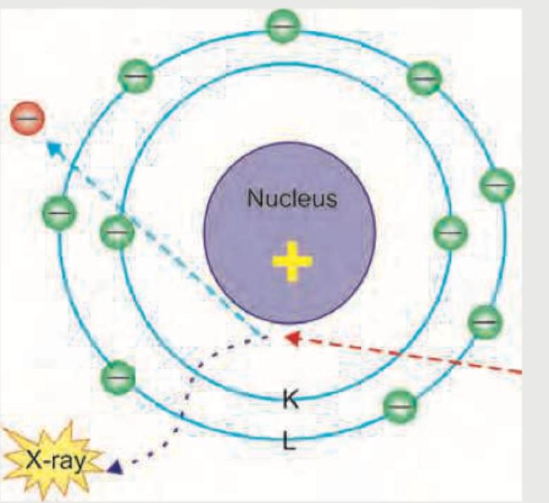
The following diagram shows production of X-rays. This is known as: (Recent NEET Pattern 2016-17)

A radiopaque density may be noticed in poisoning by which of the following agents?
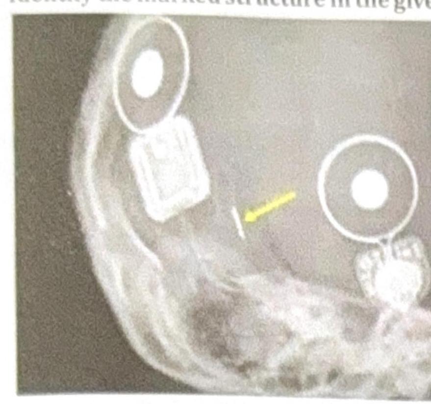
Identify the marked structure in the given image.

Which of the following typically results in the maximum radiation exposure?
Which of the following investigations work on the same principle?
A developer at high temperature will cause:
Which of the following reduces the development of unexposed Ag halide crystals, and also acts as an antifog agent?
At t = 0 there are 6 x 10^23 radioactive atoms of a substance, which decay with a disintegration constant (λ) equal to 0.01/sec. What would be the initial decay rate?
The longest half life is that of:
Explanation: ***CT scan*** - CT scan utilizes **ionizing radiation**, which carries potential risks including teratogenesis, fetal growth restriction, and childhood malignancy, particularly with high radiation doses (>100 mGy). - CT is **not absolutely contraindicated** but should be **avoided when alternative imaging is available** (e.g., ultrasound or non-contrast MRI). - When medically necessary (e.g., pulmonary embolism, acute appendicitis, severe trauma), CT can be performed with appropriate justification and dose reduction techniques. - Most diagnostic CT scans deliver fetal doses **below the threshold for deterministic effects** (<50 mGy), but the **ALARA principle** (As Low As Reasonably Achievable) applies. - Among the given options, CT carries the **highest radiation risk** and is the investigation most strongly discouraged unless essential. *MRI* - Non-contrast MRI uses **magnetic fields and radio waves** without ionizing radiation, making it **safe for diagnostic purposes** during pregnancy, particularly after the first trimester. - **Gadolinium contrast is contraindicated**, especially in the first trimester, as it crosses the placenta, remains in amniotic fluid, and has been associated with adverse fetal outcomes in some studies. - Non-contrast MRI is increasingly used for neurological, musculoskeletal, and abdominal imaging in pregnancy. *Ultrasound* - Ultrasound is the **safest and preferred** imaging modality in pregnancy, using high-frequency **sound waves** without ionizing radiation. - Essential for routine prenatal care, monitoring fetal growth, anatomical survey, and assessing placental location and amniotic fluid. - No known harmful effects to the fetus when used appropriately. *Doppler* - Doppler is a **safe and specialized type of ultrasound** that measures **blood flow velocity and vascular resistance** (e.g., umbilical artery, middle cerebral artery, uterine artery). - Crucial for evaluating fetal well-being in high-risk pregnancies, particularly in cases of **intrauterine growth restriction (IUGR)**, pre-eclampsia, or suspected fetal anemia. - No contraindication; thermal and mechanical indices should be monitored per safety guidelines.
Explanation: ***Bremsstrahlung effect*** - The diagram shows an **incoming electron** being decelerated as it passes near the nucleus of an atom, causing it to lose energy and emit an **X-ray photon**. - This process, where an electron is slowed down ("braked") by the electric field of the nucleus, resulting in the emission of radiation (X-rays), is precisely what is known as **Bremsstrahlung**, or "braking radiation." *Bragg effect* - The **Bragg effect** describes the phenomenon where X-rays are diffracted by the atoms in a **crystal lattice** at specific angles. - It is used in **X-ray crystallography** to determine the atomic and molecular structure of a crystal, and not the primary production of X-rays itself. *Compton effect* - The **Compton effect** involves the **scattering of a photon** (like an X-ray) by a charged particle, typically an electron, resulting in a decrease in the photon's energy and an increase in its wavelength. - This describes the **interaction of existing X-rays with matter**, not the mechanism of X-ray generation as depicted in the diagram. *Thomson scattering* - **Thomson scattering** is the elastic scattering of **electromagnetic radiation** by a free charged particle, usually an electron, producing radiation of the **same wavelength** (no energy loss). - It is a classical explanation for the scattering of light and does not account for the production of X-rays or the change in electron energy.
Explanation: ***Chloral hydrate*** - Due to its halogenated structure, **chloral hydrate** can be radio-opaque on X-rays, making it one of the "CHIPES" substances. - This property allows for radiological detection of its presence in the **gastrointestinal tract** following ingestion, particularly in large overdoses. *Chloroquine* - **Chloroquine** is not significantly radio-opaque and is generally not detectable on plain radiographs following overdose. - Clinical diagnosis of chloroquine poisoning relies on symptoms such as **hypotension**, **cardiac arrhythmias**, and **hypokalemia**, not radiological findings. *Phenazopyridine* - **Phenazopyridine** is a urinary analgesic that does not possess properties that render it radiographically detectable. - Its metabolism and excretion do not produce **radio-opaque metabolites** or complexes. *Ethylene glycol* - **Ethylene glycol** itself is not radio-opaque on plain X-rays, and its presence is typically diagnosed through laboratory tests like anion gap metabolic acidosis. - While it can lead to the formation of **calcium oxalate crystals** in the kidneys, these are typically microscopic and not visible as general radiopacities in the GI tract.
Explanation: ***Coil*** - The marked structure appears to be a **cochlear implant's internal coil**, which is common in X-ray imaging of these devices. - The **cochlear implant internal coil** is crucial for transmitting processed sound signals via electromagnetic induction to the electrode array within the cochlea. *Electrode* - An **electrode array** is typically a thin, flexible wire with multiple contacts inserted into the cochlea, which is not what the arrow is pointing to directly. - While electrodes are part of a cochlear implant, the marked structure's shape and position are more consistent with the **internal coil** that connects to the electrode array. *Magnet* - A **magnet** is present in a cochlear implant system, typically in both the external processor and internal receiver, to hold these two components together through the skin. - Magnets usually appear as dense, circular structures in X-rays, often seen more anteriorly or superiorly to the coil for external component alignment. *Processor* - The **processor** for a cochlear implant is an external device worn behind the ear, not an implanted component visible on an X-ray. It processes sound and sends it to the internal coil. - The structures seen in the X-ray are **implanted components** of the cochlear implant, not the external sound processor.
Explanation: ***PET CT*** - **PET CT (Positron Emission Tomography-Computed Tomography)** combines the radiation from both a PET scan (using radiotracers like FDG) and a CT scan, resulting in the highest typical radiation exposure among the listed options. - The integration of functional (PET) and anatomical (CT) imaging, while providing comprehensive diagnostic information, significantly increases the total absorbed dose (~20-30 mSv). *Chest X-ray* - A **chest X-ray** involves a very low dose of radiation (~0.1 mSv), making it one of the imaging modalities with the least radiation exposure. - Due to its low dose and widespread use, the benefits of chest X-rays in diagnosing pulmonary and cardiac conditions far outweigh the minimal radiation risk. *IV pyelography* - **Intravenous pyelography (IVP)**, or intravenous urography, uses X-rays and contrast dye to visualize the urinary tract, delivering a moderate radiation dose (~3-5 mSv). - While higher than a standard X-ray, its dose is significantly lower than that of complex combined imaging like PET-CT. *Barium Enema* - A **barium enema** involves multiple X-ray images of the large intestine after administering barium contrast, leading to a moderate to high radiation dose (~8-15 mSv). - The series of exposures required to adequately visualize the entire colon contributes to a higher cumulative dose compared to single-shot X-rays.
Explanation: ***CT and X-ray*** - Both **Computed Tomography (CT)** and **X-ray** imaging utilize **ionizing radiation** to generate images of the body's internal structures. - They work by passing X-ray beams through the patient, with different tissues absorbing the radiation to varying degrees, which is then detected to create an image. *MRI and PET Scan* - **Magnetic Resonance Imaging (MRI)** uses **strong magnetic fields and radio waves** to create detailed images of soft tissues, based on water content. - **Positron Emission Tomography (PET) scans** use **radioactive tracers** to visualize metabolic activity and blood flow, detecting gamma rays emitted from the patient. *CT and MRI* - **CT scans** use **ionizing radiation** (X-rays) to produce cross-sectional images. - **MRI scans** use **magnetic fields and radio waves** and do not involve ionizing radiation. *USG and HIDA Scan* - **Ultrasound (USG)** uses **high-frequency sound waves** to create real-time images of organs and structures. - **Hepatobiliary Iminodiacetic Acid (HIDA) scans** are a type of nuclear medicine study that uses a **radioactive tracer** to evaluate liver and gallbladder function.
Explanation: ***Very dark image*** - A developer solution at a **high temperature** accelerates the chemical reactions involved in reducing exposed silver halide crystals. - This over-development leads to an excessive amount of metallic silver being generated, resulting in an **overly dense** and thus very dark image. *Clear white spots on the film* - **White spots** on film typically indicate areas where the silver halide crystals were either not exposed to radiation or were not developed, often due to a **fixer spot** or **air bubble**. - High developer temperature causes over-development, not under-development or lack of development in specific areas. *Very light image* - A **very light image** suggests under-development, which can occur due to **low developer temperature**, insufficient developing time, or an exhausted developing solution. - Conversely, high developer temperature causes over-development, leading to a dark image. *Yellow stains* - **Yellow stains** on film are usually a sign of **insufficient rinsing** after fixing, allowing residual thiosulfate compounds to react with silver, or using an **exhausted fixer solution**. - While processing errors can occur, yellow stains are not a direct consequence of high developer temperature.
Explanation: ***Sodium bromide*** - As a **restrainer**, sodium bromide **reduces the development of unexposed silver halide crystals** by competing with the developing agents for adsorption onto the crystal surface. - It also acts as an **antifogging agent** by increasing the threshold potential required for the reduction of silver ions, thereby preventing the reduction of unexposed crystals that might otherwise be reduced due to minor defects or chemical fog. *Potassium hydroxide* - **Potassium hydroxide** is a **strong alkali** used as an **activator** in photographic developers to maintain a high pH. - It does not reduce the development of unexposed crystals or act as an antifogging agent; rather, its role is to **accelerate the development process**. *Sodium sulfite* - **Sodium sulfite** is primarily used as a **preservative** in photographic developers to prevent the oxidation of developing agents by atmospheric oxygen. - While it can help maintain the stability of the developer, it does not directly act as a restrainer or antifogging agent to control fog due to unexposed crystals. *Phenidone* - **Phenidone** is a **developing agent**, known for its high activity and super-additive effect when combined with hydroquinone. - Its function is to **reduce exposed silver halide crystals** to metallic silver, not to reduce the development of unexposed crystals or act as an antifogging agent.
Explanation: ***6 x 10^21*** - The **initial decay rate** (A) is calculated using the formula **A = λN**, where **λ** is the disintegration constant and **N** is the initial number of radioactive atoms. - Given **λ = 0.01/sec** and **N = 6 x 10^23** atoms, the decay rate is **0.01 x 6 x 10^23 = 6 x 10^21 decays/sec**. *6 x 10^19* - This value is significantly lower than the calculated initial decay rate, suggesting an error in calculation or an incorrect application of the decay rate formula. - It does not account for the product of the disintegration constant and the total number of atoms. *6 x 10^23* - This value represents the **initial number of radioactive atoms** (N), not the initial decay rate. - The decay rate is a measure of how many atoms decay per unit of time, which requires multiplying N by the disintegration constant λ. *6 x 10^22* - This value is an order of magnitude higher than the correct decay rate. - An error in multiplying by 0.01 (which is 10^-2) would lead to this incorrect result.
Explanation: ***Uranium*** - **Uranium-238**, a common isotope, has an incredibly long half-life of approximately **4.468 billion years**, which is comparable to the age of the Earth. - This extremely long half-life is due to its slow **alpha decay**, making it a very stable radioactive element. *Radon* - **Radon-222** has a relatively short half-life of about **3.8 days**. - Its short half-life makes it a significant indoor air pollutant as it rapidly decays into other radioactive isotopes. *Cesium* - **Cesium-137**, a product of nuclear fission, has a half-life of approximately **30 years**. - While longer than radon, its half-life is much shorter compared to uranium, meaning it decays significantly faster. *Radium* - **Radium-226**, a decay product of uranium, has a half-life of approximately **1,600 years**. - Although much longer than radon and cesium, it is still orders of magnitude shorter than the half-life of uranium-238.
Electromagnetic Radiation
Practice Questions
X-ray Production
Practice Questions
Interaction of Radiation with Matter
Practice Questions
Radiation Measurement Units
Practice Questions
Radiation Detectors
Practice Questions
Radiobiology Fundamentals
Practice Questions
Radiation Protection Principles
Practice Questions
Personnel Monitoring
Practice Questions
Shielding Design and Calculations
Practice Questions
Radiation Dose Optimization
Practice Questions
Regulatory Requirements
Practice Questions
Radiation Accidents Management
Practice Questions
Get full access to all questions, explanations, and performance tracking.
Start For Free