What is the ICRP recommended genetic dose of radiation exposure for the general population?
Which imaging modality involves the least radiation exposure?
What is the SI unit of dose equivalent of radiation?
On taking an X-ray, where is the grid placed?
Which of the following isotopes has the longest half-life?
Which material is commonly used for X-ray exposure prevention screens?
Which of the following is NOT mutagenic?
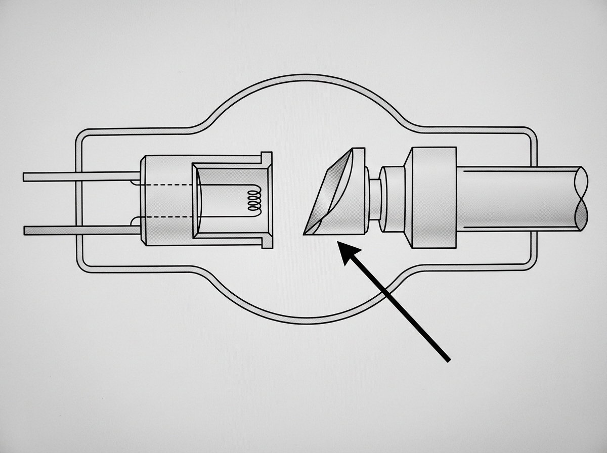
Identify the component indicated by the arrow.

An airline flight of 5 hours in the middle latitudes at an altitude of 12 km may result in an exposure of _____ µSv.
What type of particle does Phosphorus-32 emit?
Explanation: **Explanation:** The **International Commission on Radiological Protection (ICRP)** establishes guidelines to minimize the stochastic effects of radiation, particularly genetic mutations that can be passed to future generations. **Why Option A is Correct:** The recommended genetic dose limit for the general population is **5 rems (50 mSv) over a period of 30 years**. This specific timeframe (30 years) is chosen because it represents the average **human reproductive span** (generation time). The goal is to ensure that the cumulative radiation dose to the gonads of the population does not significantly increase the natural rate of genetic mutations. **Analysis of Incorrect Options:** * **Option B & D (30 rems):** This value is far too high for the general public. For context, the annual limit for occupational workers is 2 rems (20 mSv) per year, but applying a 30 rem limit to the entire population would lead to an unacceptable increase in the genetic burden. * **Option C (5 years):** While 5 years is the timeframe used to calculate the *occupational* dose limit (100 mSv over 5 years, not exceeding 50 mSv in any single year), it is not the standard interval for calculating population genetic risk. **High-Yield Clinical Pearls for NEET-PG:** * **Occupational Dose Limit:** 20 mSv per year (averaged over 5 years). * **Public Dose Limit:** 1 mSv per year. * **Pregnant Worker:** Once pregnancy is declared, the dose to the surface of the abdomen should not exceed **2 mSv** for the remainder of the pregnancy. * **Deterministic vs. Stochastic:** Genetic effects and carcinogenesis are **stochastic** (no threshold, probability increases with dose), whereas cataracts or skin erythema are **deterministic** (threshold-based).
Explanation: **Explanation:** The core concept behind this question is the distinction between **ionizing** and **non-ionizing** radiation. **MRI (Magnetic Resonance Imaging)** is the correct answer because it utilizes strong magnetic fields and radiofrequency (RF) pulses to generate images. These are forms of non-ionizing radiation, which do not have enough energy to remove electrons from atoms or damage DNA directly. Therefore, MRI involves **zero** ionizing radiation exposure. **Analysis of Incorrect Options:** * **CT Scan:** This modality uses multiple X-ray beams to create cross-sectional images. It carries the highest radiation burden among the options (e.g., a single chest CT is equivalent to ~70-100 chest X-rays). * **Arthrography:** This involves the use of fluoroscopy (continuous X-rays) to visualize joints after injecting contrast. While the dose is lower than CT, it still utilizes ionizing radiation. * **OPG (Orthopantomogram):** This is a panoramic dental X-ray. Although the effective dose is relatively low (approx. 0.014–0.024 mSv), it still involves exposure to ionizing X-rays. **High-Yield Clinical Pearls for NEET-PG:** * **Non-ionizing modalities:** MRI and Ultrasound (USG). These are the safest options for pregnant women and pediatric patients. * **Radiosensitivity:** Lymphocytes are the most radiosensitive cells in the body; nerve cells are the most radioresistant. * **ALARA Principle:** "As Low As Reasonably Achievable" is the fundamental rule of radiation protection. * **Deterministic vs. Stochastic effects:** Radiation-induced cancer is a **stochastic effect** (no threshold dose), while cataracts are a **deterministic effect** (threshold-dependent).
Explanation: **Explanation:** The **Sievert (Sv)** is the SI unit for **Dose Equivalent**. Dose equivalent is a calculated value used to represent the biological effect of ionizing radiation on human tissue. It is derived by multiplying the absorbed dose by a "quality factor" or "weighting factor" (Wᵣ), which accounts for the fact that different types of radiation (e.g., alpha particles vs. X-rays) cause different levels of biological damage even at the same absorbed dose. **Analysis of Options:** * **Sievert (Sv):** The SI unit for Dose Equivalent. (1 Sv = 100 rem). * **Rem (Roentgen Equivalent Man):** The **traditional/old unit** for Dose Equivalent. While it measures the same concept as the Sievert, it is not the SI unit. * **Gray (Gy):** The **SI unit for Absorbed Dose** (energy deposited per unit mass). 1 Gy = 1 Joule/kg. * **Rad (Radiation Absorbed Dose):** The **traditional/old unit** for Absorbed Dose. (1 Gy = 100 rad). **High-Yield Clinical Pearls for NEET-PG:** * **Exposure:** Measured in **Coulomb/kg** (SI) or **Roentgen** (Traditional). It measures the ionization of air. * **Effective Dose:** Also measured in **Sieverts**. It accounts for the varying radiosensitivity of different organs (using tissue weighting factors). * **Radioactivity:** Measured in **Becquerel (Bq)** (SI) or **Curie (Ci)** (Traditional). * **Rule of 100:** To convert SI to Traditional units, remember that **1 Gy = 100 rad** and **1 Sv = 100 rem**. * The annual public dose limit is **1 mSv**, while the occupational limit for radiation workers is **20 mSv per year** (averaged over 5 years).
Explanation: ### Explanation The primary purpose of a grid in radiography is to **reduce scatter radiation** (Compton scatter) from reaching the film, thereby improving image contrast and detail. **1. Why Option B is Correct:** When X-rays pass through a patient, they interact with tissues and scatter in various directions. If this scattered radiation reaches the film, it creates "fog," which degrades image quality. To prevent this, the grid—composed of alternating strips of lead (radiopaque) and plastic/aluminum (radiolucent)—is placed **between the patient and the film**. It allows the primary, useful beam to pass through while absorbing the angled, scattered rays. **2. Why Other Options are Incorrect:** * **Option A:** Placing the grid in front of the patient would filter the primary beam before it even interacts with the body, increasing patient dose without reducing scatter. * **Options C & D:** Placing the grid "lateral" to the patient or film is anatomically and physically irrelevant, as the grid must be perpendicular to the X-ray beam path to function. Placing it "behind the film" is useless because the radiation has already interacted with the detector. **3. Clinical Pearls for NEET-PG:** * **Grid Ratio:** Defined as the height of the lead strips to the distance between them ($H/D$). A higher grid ratio is more effective at removing scatter but requires a higher radiation dose (**Bucky Factor**). * **Bucky-Potter Diaphragm:** A moving grid used to blur out the grid lines on the final radiograph. * **Indications:** Grids are generally used when the body part thickness exceeds **10 cm** or when high kVp techniques are used. * **Grid Cut-off:** An undesirable loss of primary beam intensity caused by improper alignment of the X-ray tube and the grid.
Explanation: **Explanation:** The half-life of a radioactive isotope is the time required for its radioactivity to decrease to half of its initial value. In the context of radiation physics, isotopes with extremely long half-lives are typically naturally occurring primordial elements. **Why Uranium is Correct:** **Uranium-238**, the most common isotope of Uranium, has a half-life of approximately **4.5 billion years**. Even Uranium-235 has a half-life of about 700 million years. Because it is the parent element of several decay series (including the Radium and Radon series), it naturally possesses the longest stability among the options provided. **Analysis of Incorrect Options:** * **Radium (Ra-226):** A decay product of Uranium, it has a half-life of approximately **1,600 years**. While long in a clinical sense, it is significantly shorter than Uranium. * **Cesium (Cs-137):** A common byproduct of nuclear fission used in radiotherapy (brachytherapy), it has a half-life of approximately **30 years**. * **Radon (Rn-222):** A radioactive gas produced by the decay of Radium, it has a very short half-life of only **3.8 days**. **NEET-PG High-Yield Pearls:** * **Technetium-99m (Tc-99m):** The most commonly used isotope in Nuclear Medicine (SPECT); half-life is **6 hours**. * **Iodine-131:** Used for thyroid imaging and ablation; half-life is **8 days**. * **Cobalt-60:** Historically used in teletherapy; half-life is **5.27 years**. * **Iridium-192:** Most common isotope used in modern **Brachytherapy**; half-life is **74 days**. * **Rule of thumb:** After 10 half-lives, the radioactivity of a sample is considered negligible (less than 0.1% of original activity).
Explanation: **Explanation:** **Lead (Pb)** is the material of choice for radiation protection screens and personal protective equipment (PPE) due to its **high atomic number (Z=82)** and high density. According to the principles of radiation physics, the probability of **Photoelectric Absorption** increases significantly with the atomic number of the absorbing material ($Z^3$). Lead effectively attenuates X-ray photons by absorbing their energy, preventing them from reaching healthcare workers. Its high density also allows for a "High Stopping Power" in a relatively thin layer, making it practical for aprons, thyroid shields, and mobile screens. **Analysis of Incorrect Options:** * **Tungsten (B):** While Tungsten has a high atomic number (Z=74) and high melting point, it is primarily used as the **Target material in the X-ray tube anode** to produce X-rays, rather than for shielding screens. * **Manganese (C) & Titanium (D):** These metals have much lower atomic numbers (Z=25 and Z=22, respectively). They lack the density and electron cloud density required to provide adequate attenuation against high-energy diagnostic X-rays. **High-Yield Clinical Pearls for NEET-PG:** * **Standard Lead Equivalence:** Lead aprons typically provide **0.25 mm to 0.5 mm** of lead equivalence. A 0.5 mm apron can attenuate approximately 90-99% of scatter radiation. * **ALARA Principle:** Radiation protection follows the "As Low As Reasonably Achievable" principle, utilizing **Time, Distance, and Shielding.** * **Barium & Antimony:** These are often used in "Lead-free" or lightweight aprons as they are efficient at absorbing radiation at specific k-edge energies. * **Gonadal Shielding:** The most sensitive organs to radiation are the gonads, bone marrow, and the lens of the eye.
Explanation: ### Explanation The core concept behind this question is the distinction between **ionizing** and **non-ionizing** radiation. **Why Ultrasound is the Correct Answer:** Ultrasound is a form of **mechanical energy** consisting of high-frequency sound waves (above 20,000 Hz). Unlike electromagnetic radiation, ultrasound does not have enough energy to displace electrons from atoms or break chemical bonds in DNA. Therefore, it is **non-ionizing** and lacks mutagenic potential. In clinical practice, its safety profile makes it the modality of choice for fetal imaging. **Why the Other Options are Incorrect:** * **X-rays (Option A):** These are high-energy electromagnetic waves. They are **ionizing**, meaning they can cause direct DNA strand breaks or indirect damage via free radical production, leading to mutations or cell death. * **UV rays (Option B):** Although non-ionizing in the traditional sense of displacing inner-shell electrons, UV radiation (specifically UV-B) is absorbed by DNA, leading to the formation of **pyrimidine dimers**. This is a classic mutagenic mechanism. * **Beta rays (Option D):** These consist of high-energy electrons or positrons emitted during radioactive decay. As particulate **ionizing radiation**, they possess significant energy to cause genomic instability and mutations. **High-Yield Clinical Pearls for NEET-PG:** * **Safe in Pregnancy:** Ultrasound and MRI (non-ionizing) are considered safe. However, MRI is generally avoided in the first trimester unless essential. * **Radiosensitivity:** According to the **Law of Bergonie and Tribondeau**, cells that are rapidly dividing, undifferentiated, and have a long mitotic future (e.g., lymphocytes, germ cells, bone marrow) are the most sensitive to radiation. * **Teratogenesis:** The period of maximum sensitivity for radiation-induced teratogenesis (organogenesis) is **2 to 8 weeks** post-conception.
Explanation: ***Anode*** - The **anode** is the **positive electrode** in an X-ray tube where **X-rays are produced** when high-speed electrons from the cathode strike the **tungsten target**. - It consists of a **tungsten target** (due to its high atomic number and melting point) and can be **rotating** or **stationary** to dissipate heat effectively. *Cathode* - The **cathode** is the **negative electrode** that contains the **tungsten filament** which produces electrons when heated. - It is located **opposite** to the anode and **emits electrons** rather than receiving them like the anode does. *Focal spot* - The **focal spot** is the **small area on the anode target** where electrons actually strike and X-rays are produced. - It is a **specific region** within the anode, not the entire component that would be indicated by an arrow pointing to the whole structure. *X-ray tube* - The **X-ray tube** refers to the **entire vacuum tube assembly** containing both anode and cathode components. - It encompasses the **complete unit** including the glass envelope, not just the single component indicated by the arrow.
Explanation: **Explanation:** The correct answer is **25 µSv**. This question tests the understanding of background radiation exposure from cosmic rays, a high-yield topic in radiation physics. **1. Why 25 µSv is correct:** At high altitudes (like 12 km/39,000 ft), the Earth's atmosphere is thinner, providing less shielding against **cosmic radiation** (protons, alpha particles, and neutrons from space). The average dose rate at mid-latitudes at this altitude is approximately **5 µSv per hour**. * **Calculation:** 5 hours × 5 µSv/hour = **25 µSv**. For perspective, this is roughly equivalent to the radiation dose received from a single standard **Chest X-ray (PA view)**, which is approximately 20–100 µSv (0.02–0.1 mSv). **2. Why other options are incorrect:** * **50 µSv:** This would represent a 10-hour flight or a flight at much higher latitudes (near the poles), where the Earth’s magnetic field provides less protection. * **100 µSv:** This is the approximate dose of a screening mammogram or 4–5 Chest X-rays. * **250 µSv:** This is a significantly higher dose, closer to the annual exposure from natural terrestrial sources in some regions, and far exceeds a single short-duration flight. **Clinical Pearls for NEET-PG:** * **Annual Limit:** The general public's annual dose limit is **1 mSv**, while for radiation workers, it is **20 mSv/year** (averaged over 5 years). * **Natural Background Radiation:** The average annual exposure is ~**3 mSv**. The largest component is **Radon gas**. * **Altitude Effect:** Radiation exposure doubles for every 1,500–2,000 meters of increased elevation. * **ALARA Principle:** As Low As Reasonably Achievable (Time, Distance, Shielding).
Explanation: **Explanation:** **Phosphorus-32 ($^{32}$P)** is a pure **beta-emitter**. It undergoes radioactive decay by emitting a high-energy beta particle (electron) and a neutrino, transforming into stable Sulfur-32. Because it does not emit gamma rays, it is primarily used for therapeutic purposes rather than diagnostic imaging. * **Why Beta particles are correct:** $^{32}$P emits beta particles with a maximum energy of 1.71 MeV and an average tissue penetration of about 3–8 mm. This makes it ideal for treating superficial lesions or localized conditions where deep tissue penetration is undesirable. * **Why other options are incorrect:** * **Alpha particles:** These are heavy particles (Helium nuclei) used in agents like Radium-223. $^{32}$P is too light for alpha decay. * **Neutrons:** These are typically used in external beam neutron therapy or produced within nuclear reactors; they are not the primary emission of medical radiopharmaceuticals like $^{32}$P. * **X-rays:** X-rays originate from electron shell transitions, whereas $^{32}$P decay is a nuclear process. While Bremsstrahlung (secondary X-rays) can occur when beta particles hit lead shielding, the primary emission is beta. **High-Yield Clinical Pearls for NEET-PG:** * **Clinical Uses:** Historically used for **Polycythemia Vera**, essential thrombocythemia, and intracavitary treatment of malignant effusions. * **Route:** Can be administered intravenously or orally. * **Physical Half-life:** **14.3 days**. * **Shielding:** Since it is a beta-emitter, **plastic or Perspex** shielding is preferred over lead to minimize Bremsstrahlung radiation.
Electromagnetic Radiation
Practice Questions
X-ray Production
Practice Questions
Interaction of Radiation with Matter
Practice Questions
Radiation Measurement Units
Practice Questions
Radiation Detectors
Practice Questions
Radiobiology Fundamentals
Practice Questions
Radiation Protection Principles
Practice Questions
Personnel Monitoring
Practice Questions
Shielding Design and Calculations
Practice Questions
Radiation Dose Optimization
Practice Questions
Regulatory Requirements
Practice Questions
Radiation Accidents Management
Practice Questions
Get full access to all questions, explanations, and performance tracking.
Start For Free