
Radiation Physics and Protection — MCQs

On this page
Which of the following statements about MRI is incorrect?
An obese patient has heavy, thick bones. What technical factor should be increased on the X-ray machine to achieve a diagnostic image?
Which of the following radiological modalities is not considered safe in pregnancy?
Which of the following has the highest Hounsfield Unit?
All of the following have a naturally occurring decay product in a gaseous form, EXCEPT:
Amount of radioactivity absorbed by the body is measured by?
What protective equipment is used to prevent radiation exposure in an operation theatre?
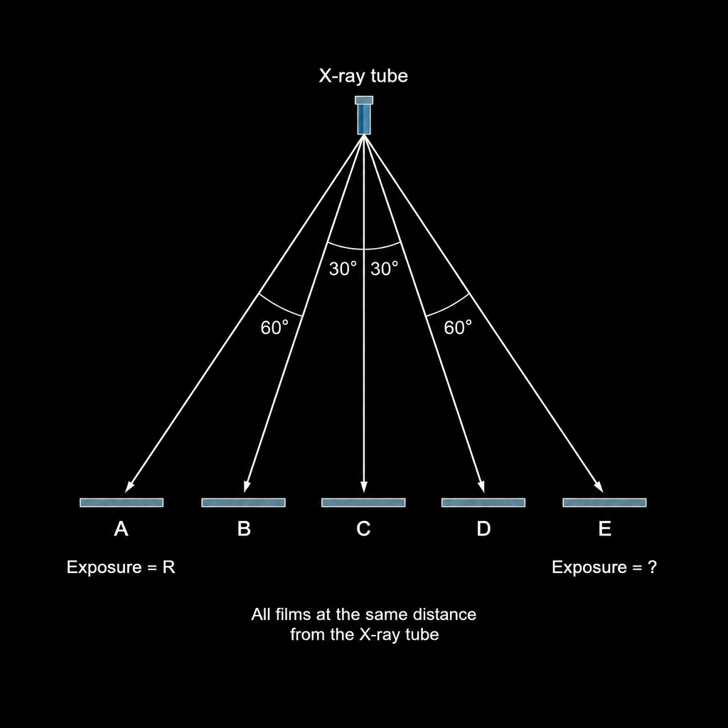
If the exposure of film A is R, then the exposure of film E is:
What is the conventional unit of Exposure?
A 50-year-old male patient complains of reduced mouth opening. The patient has a history of a road traffic accident one week prior. A panoramic X-ray was taken using an intensifying screen containing gadolinium and lanthanum. On interaction with X-rays, what does the content of this intensifying screen emit?
Practice by Chapter
Electromagnetic Radiation
Practice Questions
X-ray Production
Practice Questions
Interaction of Radiation with Matter
Practice Questions
Radiation Measurement Units
Practice Questions
Radiation Detectors
Practice Questions
Radiobiology Fundamentals
Practice Questions
Radiation Protection Principles
Practice Questions
Personnel Monitoring
Practice Questions
Shielding Design and Calculations
Practice Questions
Radiation Dose Optimization
Practice Questions
Regulatory Requirements
Practice Questions
Radiation Accidents Management
Practice Questions
Want unlimited practice?
Get full access to all questions, explanations, and performance tracking.
Scan to download app