
Radiation Physics and Protection — MCQs

On this page
What is the acceptable safe dose of radiation during pregnancy?
What is the recommended thickness of lead in a thyroid collar for radiation protection?
High resolution is obtained with which of the following methods?
On taking an X-ray, where is the grid placed?
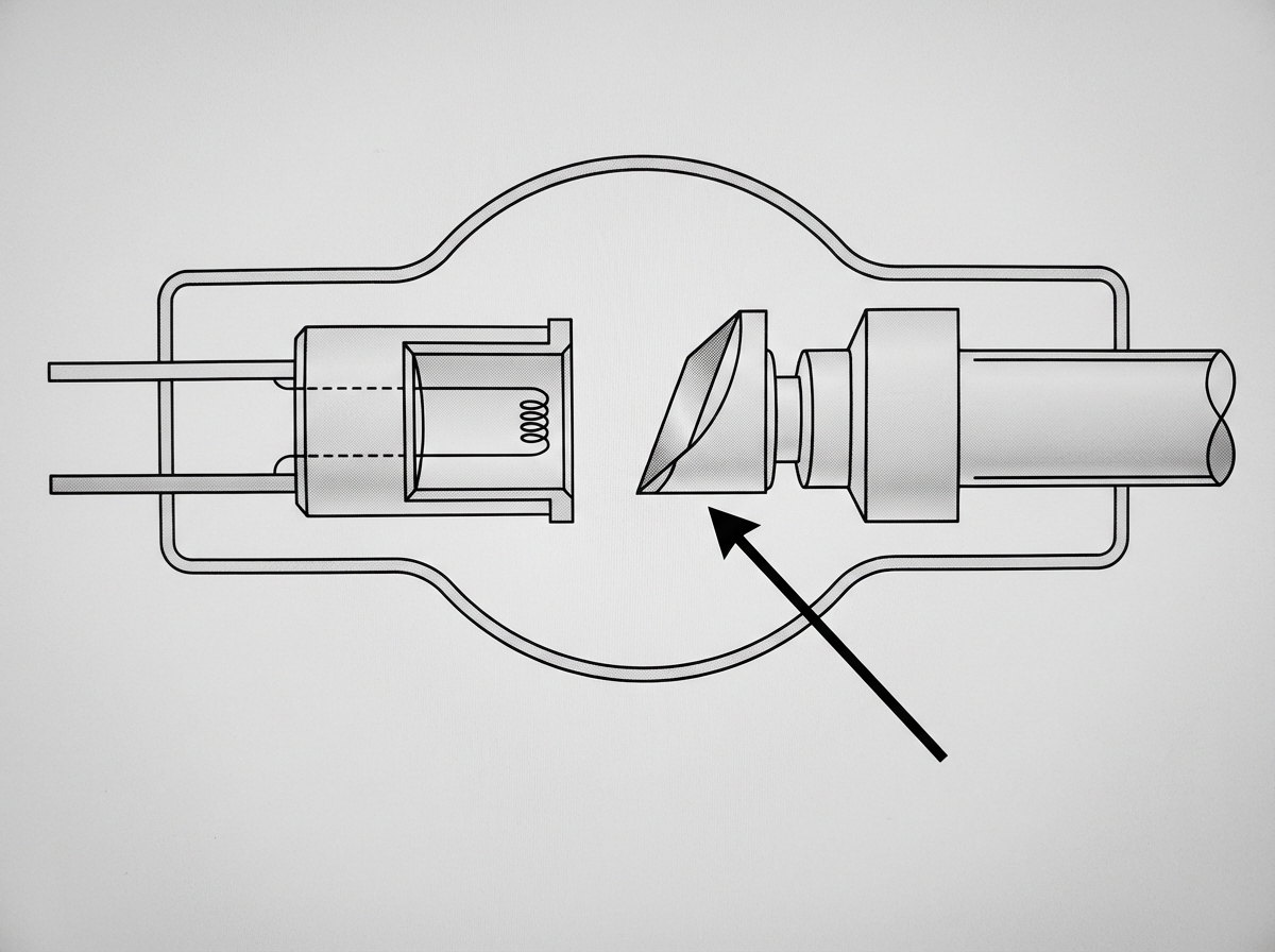
Identify the component indicated by the arrow.
Nuclear Magnetic Resonance (NMR) is based on which principle?
Regarding the interaction of X-rays with matter, which phenomenon occurs maximally?
Radiation exposure is the least in which of the following procedures?
One gray is equivalent to how many rads?
Hounsfield units are used in which imaging modality?
Practice by Chapter
Electromagnetic Radiation
Practice Questions
X-ray Production
Practice Questions
Interaction of Radiation with Matter
Practice Questions
Radiation Measurement Units
Practice Questions
Radiation Detectors
Practice Questions
Radiobiology Fundamentals
Practice Questions
Radiation Protection Principles
Practice Questions
Personnel Monitoring
Practice Questions
Shielding Design and Calculations
Practice Questions
Radiation Dose Optimization
Practice Questions
Regulatory Requirements
Practice Questions
Radiation Accidents Management
Practice Questions
Want unlimited practice?
Get full access to all questions, explanations, and performance tracking.
Scan to download app