
Pediatric Radiology — MCQs

On this page
In croup, the characteristic radiographic finding of subglottic narrowing on a frontal chest X-ray creates a tapering appearance of the upper trachea. This diagnostic sign is commonly referred to as which of the following?

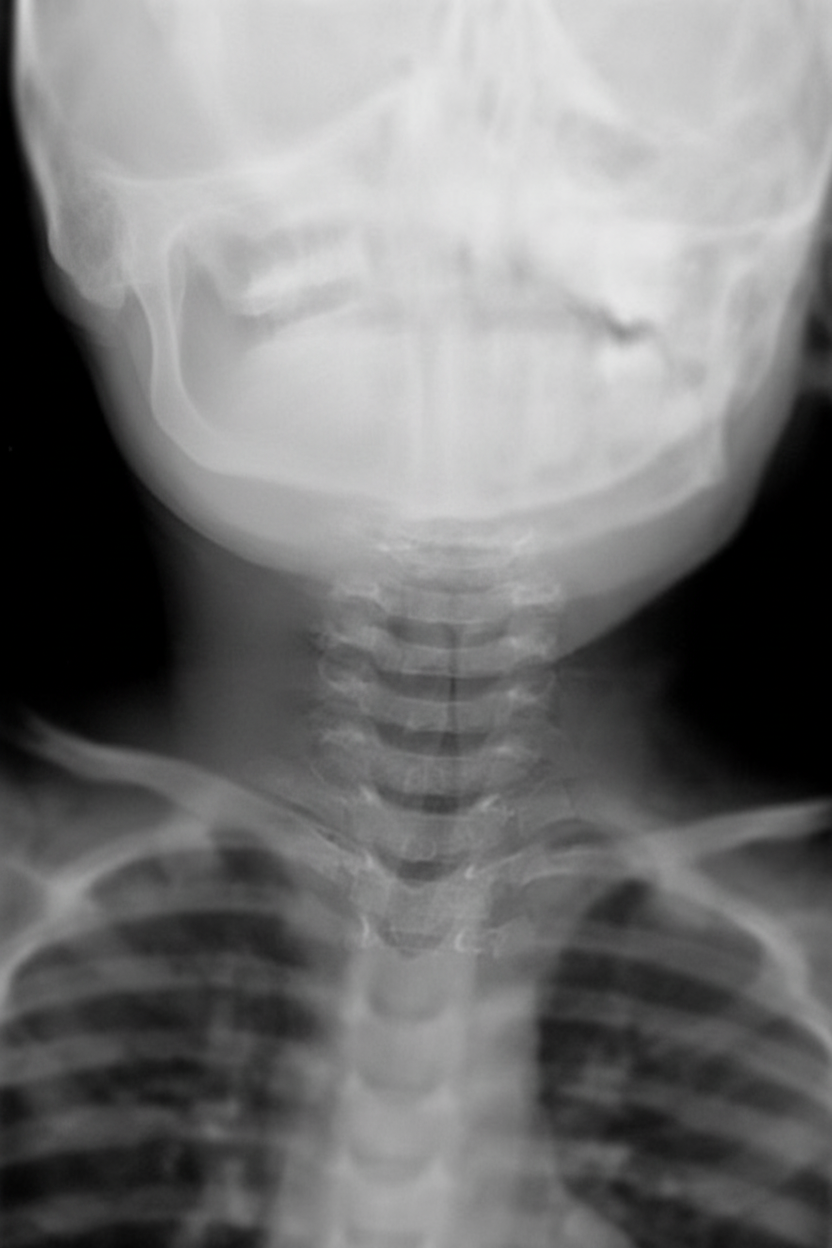
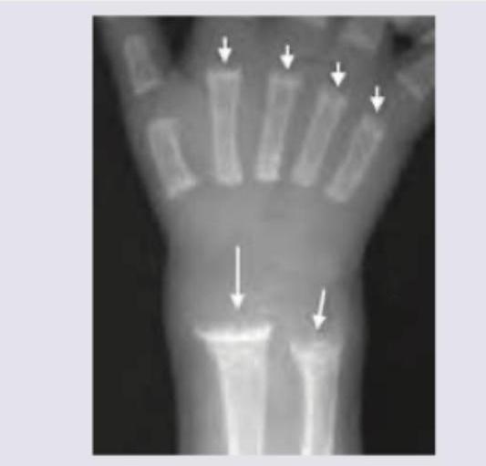
The neck X-ray of a patient shows:
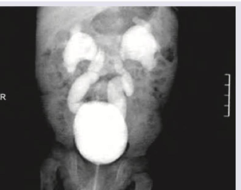
The following image shows presence of:
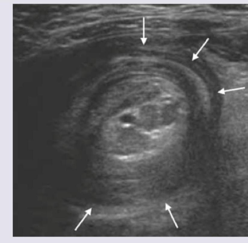
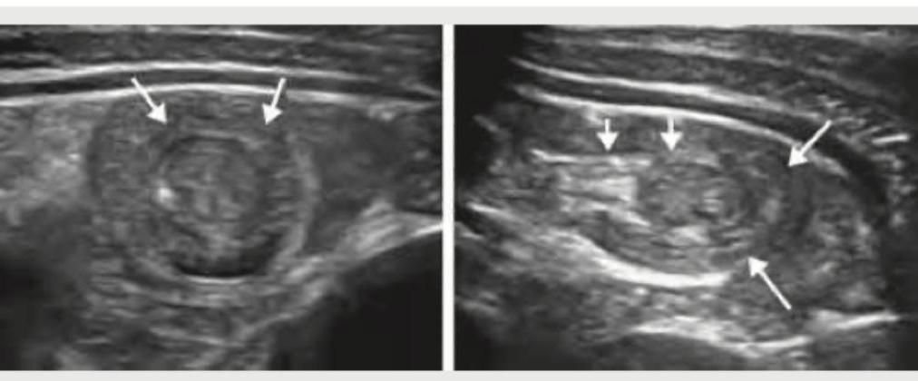
The ultrasound finding of a 7 -month-old child with abdominal pain and mass in the upper abdomen is shown below. What is the diagnosis? (NEET Pattern 2018)
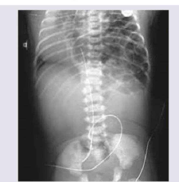
The figure shows:
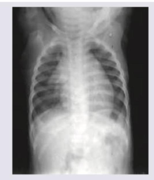
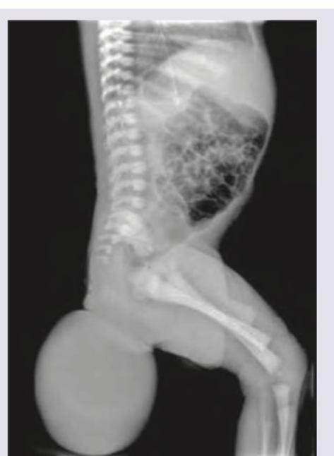
Infantogram shows presence of:
All statements about the disease shown are wrong except:
Identify the condition on the basis of infantogram shown in the image:
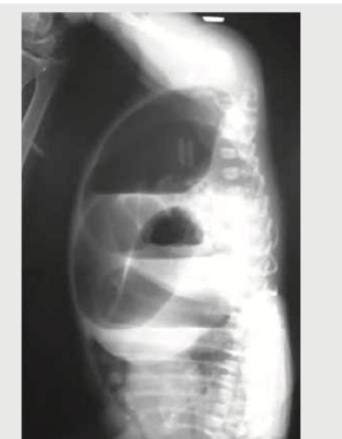
What does the following image show?
USG abdomen of a 9-month-old child as shown below denotes:
Practice by Chapter
Normal Pediatric Developmental Anatomy
Practice Questions
Neonatal Imaging
Practice Questions
Pediatric Chest Imaging
Practice Questions
Pediatric Abdominal Imaging
Practice Questions
Pediatric Musculoskeletal Imaging
Practice Questions
Pediatric Neuroradiology
Practice Questions
Congenital Heart Disease Imaging
Practice Questions
Pediatric Oncology Imaging
Practice Questions
Child Abuse Imaging
Practice Questions
Pediatric Interventional Radiology
Practice Questions
Radiation Protection in Pediatrics
Practice Questions
Sedation in Pediatric Imaging
Practice Questions
Want unlimited practice?
Get full access to all questions, explanations, and performance tracking.
Scan to download app