
Pediatric Radiology — MCQs

On this page
In a newborn, what is the typical time frame for gas to reach the colonic end?
A child presents with multiple vertebral anomalies and a posterior mediastinal mass. What is the likely diagnosis?
Antenatal detection of bone fractures on ultrasound suggests which of the following conditions?
What is the characteristic radiographic sign of acute laryngotracheobronchitis?
Antenatal ultrasound demonstrating a double bubble appearance is typically indicative of which of the following conditions?
Time sector scanning of neonates is preferred because of which most practical reason?
What is the characteristic radiological sign observed in Croup?
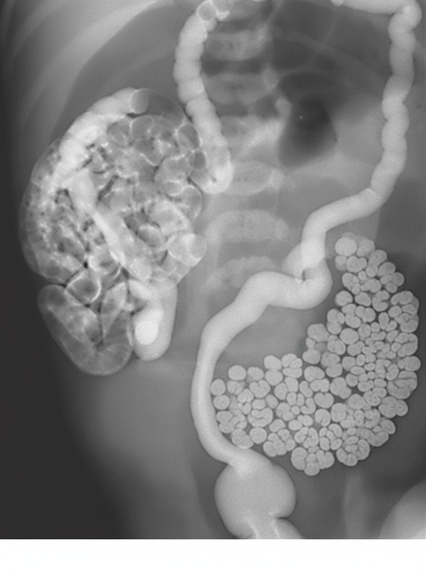
A contrast enema is performed on a one-day-old infant presenting with bilious emesis and abdominal distension. Based on the findings, which of the following is the MOST likely diagnosis?
What is the most common cause of a mass in the posterior mediastinum in children?
Reflux of urine into the pelvis and calyces with mild dilatation and minimal blunting of fornices indicates which grade of Vesicoureteral Reflux (VUR)?
Practice by Chapter
Normal Pediatric Developmental Anatomy
Practice Questions
Neonatal Imaging
Practice Questions
Pediatric Chest Imaging
Practice Questions
Pediatric Abdominal Imaging
Practice Questions
Pediatric Musculoskeletal Imaging
Practice Questions
Pediatric Neuroradiology
Practice Questions
Congenital Heart Disease Imaging
Practice Questions
Pediatric Oncology Imaging
Practice Questions
Child Abuse Imaging
Practice Questions
Pediatric Interventional Radiology
Practice Questions
Radiation Protection in Pediatrics
Practice Questions
Sedation in Pediatric Imaging
Practice Questions
Want unlimited practice?
Get full access to all questions, explanations, and performance tracking.
Scan to download app