
Pediatric Radiology — MCQs

On this page
Radiological hallmark of primary tuberculosis in childhood is?
True about hypertrophic pyloric stenosis on ultrasound
The 'figure of eight' appearance on chest X-ray is commonly seen in which congenital heart defect?
What is indicated by the 'double bubble sign' on prenatal ultrasound?
What is the first-line imaging modality for the evaluation of pyloric stenosis in infants?
Which of the following is the most appropriate initial imaging study for a child with suspected pyloric stenosis?
Which imaging modality is most commonly used to diagnose ectopic kidneys in children?
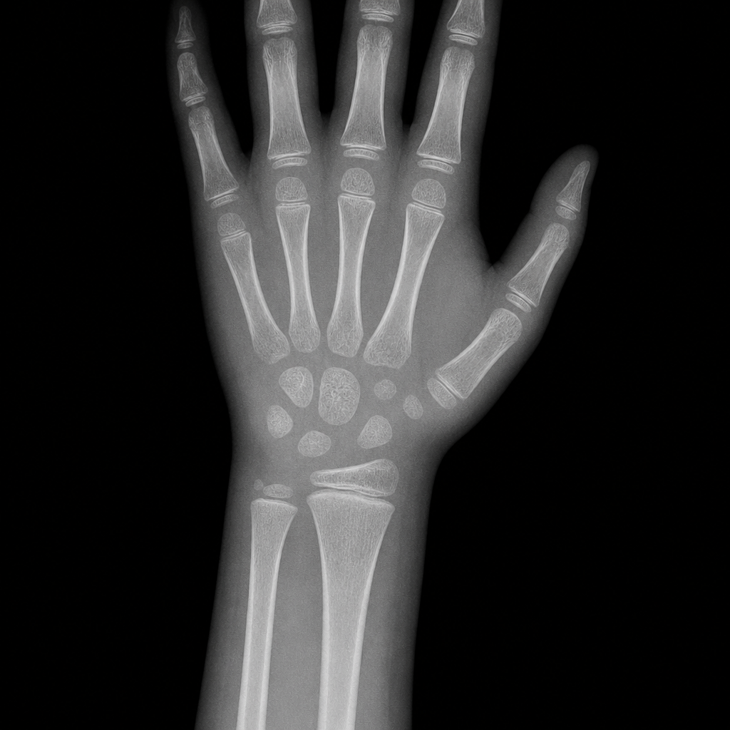
In the following X-ray of the wrist (image provided), what is the exact age of the child?
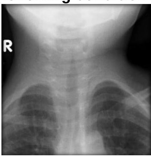
Steeple sign is seen in which of the following conditions?
Which of the following is NOT a typical ultrasonographic finding in autosomal recessive polycystic kidney disease (ARPKD)?
Practice by Chapter
Normal Pediatric Developmental Anatomy
Practice Questions
Neonatal Imaging
Practice Questions
Pediatric Chest Imaging
Practice Questions
Pediatric Abdominal Imaging
Practice Questions
Pediatric Musculoskeletal Imaging
Practice Questions
Pediatric Neuroradiology
Practice Questions
Congenital Heart Disease Imaging
Practice Questions
Pediatric Oncology Imaging
Practice Questions
Child Abuse Imaging
Practice Questions
Pediatric Interventional Radiology
Practice Questions
Radiation Protection in Pediatrics
Practice Questions
Sedation in Pediatric Imaging
Practice Questions
Want unlimited practice?
Get full access to all questions, explanations, and performance tracking.
Scan to download app