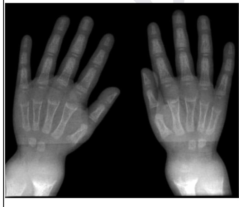
A child presents with poor growth and swelling at joints. A radiograph of his wrist is given below. Lab investigations reveal serum ALP levels of >1500. What is the possible diagnosis?

What is the investigation of choice for diagnosing a stress fracture?
A patient is brought to the emergency following a head-on collision road traffic accident. His BP is 90/60 mmHg. Tachycardia is present. Most likely diagnosis is
A man presents to the emergency department with a head injury following a vehicular accident. What is the investigation of choice?
Which of the following is a characteristic feature of Battered Baby Syndrome (Non-Accidental Injury)?
An intrauterine scan at the 13th week of pregnancy showed a fetus with multiple long bone fractures. What is commonly associated with this finding?
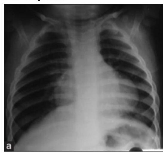
A young child presented with mild intermittent upper abdominal pain. X-ray is given below. What is the diagnosis?

Shape of extradural hematoma on NCCT is?
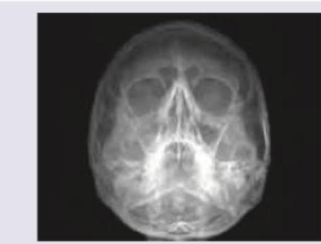
The following X-ray is used to evaluate \qquad sinus?

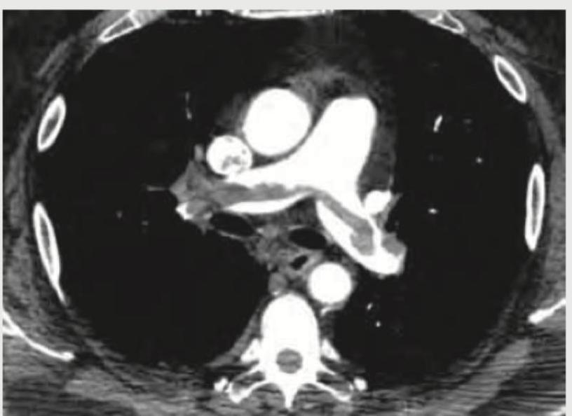
A 35-year-old male with history of 4 weeks of immobilization for fracture of femur develops sudden onset breathlessness and blood in sputum. CT angiography shows? (Recent NEET Pattem 2018-19)

Explanation: ***Rickets*** - The combination of **poor growth**, **joint swelling**, and **elevated alkaline phosphatase (ALP)** in a child strongly indicates rickets, a condition of defective bone mineralization in growing bones. - The radiograph of the wrist would likely show typical findings like **widened growth plates**, **fraying** and **cupping of metaphyses**, and **decreased bone density**, which are characteristic of rickets. *Osteoporosis* - This condition is characterized by **reduced bone mass** and **fragile bones**, typically seen in older adults or due to secondary causes, and is not primarily linked to joint swelling in children. - While ALP levels can be normal or slightly elevated in osteoporosis, a level of >1500 is highly suggestive of active bone formation or breakdown, not typically seen in osteoporosis. *Osteomalacia* - This is defective bone mineralization in adults after growth plates have fused, leading to **bone softening** and **pain**, typically not presenting with joint swelling as a primary symptom. - While it also involves high ALP and bone demineralization, the clinical context of a *child* with growth issues points more specifically to rickets. *Osteogenesis imperfecta* - This is a group of **genetic disorders** characterized by **brittle bones** that fracture easily, often accompanied by **blue sclerae** and **hearing loss**, which are not mentioned in the presentation. - While bone fragility is present, it does not typically cause the described joint swelling or the significantly elevated ALP levels seen in this case.
Explanation: ***MRI*** - **Magnetic Resonance Imaging (MRI)** is the most sensitive and specific imaging modality for diagnosing **stress fractures**, especially in their early stages. - It can detect **bone marrow edema** and **periosteal reactions** indicative of stress injury before cortical changes are visible on plain radiographs. *X-ray* - **X-rays** are often the initial investigation, but they have low sensitivity for **stress fractures** in the early stages as bone changes may not be apparent for several weeks. - A positive X-ray for stress fracture typically shows a **sclerotic line** or **periosteal reaction**, but this indicates a more advanced injury. *CT scan* - **CT scans** provide excellent detail of **cortical bone** and can detect subtle fractures not seen on X-rays. - While more sensitive than X-rays, CT has **higher radiation exposure** and is generally less sensitive than MRI for early detection of **bone marrow edema** associated with stress injuries. *Bone scan* - **Bone scans** (scintigraphy) are highly sensitive for detecting increased **osteoblastic activity** associated with stress fractures. - However, they are **less specific** as various conditions can cause increased uptake, and they do not provide detailed anatomical information, making MRI superior for definitive diagnosis and staging.
Explanation: ***Intra-abdominal bleeding*** - Following a **head-on collision**, hypotension (BP 90/60 mmHg) and tachycardia are classic signs of **hypovolemic shock**, most commonly due to significant internal bleeding. - The **abdomen** is a common site for massive blood loss after blunt trauma, as it can contain large volumes of blood without obvious external signs. *SDH (Subdural Hematoma)* - While a subdural hematoma can occur after head trauma, significant **intracranial bleeding** typically causes signs of increased intracranial pressure (e.g., headache, altered mental status, neurological deficits), and often leads to **hypertension with bradycardia** (Cushing's reflex), not hypotension and tachycardia. - The primary hemodynamic response to an isolated SDH would not be profound hypotension and tachycardia unless there was a co-existing systemic injury. *EDH (Epidural Hematoma)* - An epidural hematoma is also an intracranial injury that causes signs of **increased intracranial pressure**, such as headache, vomiting, and a potential "lucid interval." - Like SDH, it would not typically cause **hypotension and tachycardia** as the primary hemodynamic response, as it does not lead to significant blood loss from the circulatory system. *Intracranial hemorrhage* - This is a general term for bleeding within the skull, encompassing conditions like SDH and EDH. - While it is a severe injury, isolated intracranial hemorrhage generally does not cause **hypotension and tachycardia** because the cranial vault has limited space, and therefore, blood loss is not sufficient to produce systemic shock. Instead, it often leads to signs of **increased intracranial pressure** including **hypertension and bradycardia**.
Explanation: ***NCCT*** - **Non-contrast Computed Tomography (NCCT)** of the head is the **investigation of choice** for acute head trauma due to its rapid acquisition, wide availability, and excellent sensitivity for detecting acute hemorrhage, fractures, and mass effects. - It rapidly identifies life-threatening conditions such as **epidural, subdural, and intracerebral hemorrhages**, which require immediate intervention. *MRI* - **MRI** is superior for detecting subtle brain tissue injuries, diffuse axonal injury, and non-hemorrhagic lesions but is generally **not the first-line investigation** in acute trauma due to longer scan times, limited availability in the emergency setting, and inability to detect acute hemorrhage as clearly as CT. - Its use is typically reserved for follow-up studies or when CT findings are inconclusive or specific soft tissue detail is required. *CECT* - **Contrast-enhanced CT (CECT)** of the head is reserved for specific indications like evaluating vascular lesions (e.g., aneurysms, arteriovenous malformations) or tumors, which are generally **not the primary concern** in the initial assessment of acute head trauma. - Administering contrast agents can delay imaging, may pose risks to patients with renal impairment or allergies, and does not significantly improve the detection of acute traumatic hemorrhage compared to NCCT. *X-ray* - **X-rays** of the skull are useful for detecting **skull fractures**, but they provide **limited information** regarding intracranial injuries or soft tissue damage, which are critical in head trauma. - They have largely been superseded by CT scans, which offer a more comprehensive view of both bony structures and intracranial contents.
Explanation: ***Bruises of varying ages*** - The presence of bruises at **different stages of healing** is a hallmark indicator of **non-accidental trauma** or Battered Baby Syndrome, as it suggests repeated injuries occurring over time rather than a single incident. - **Forensic significance**: Fresh bruises (red/purple) alongside older bruises (yellow/green/brown) indicate multiple episodes of trauma, which is inconsistent with the caregiver's explanation of a single accidental event. - Other classic features include fractures (especially metaphyseal/corner fractures, rib fractures), subdural hematomas, retinal hemorrhages, and injuries in protected body areas. *Stab injury* - While a stab injury represents severe trauma requiring forensic investigation, it is **not characteristic** of the typical presentation pattern of Battered Baby Syndrome. - Stab wounds indicate a specific violent act rather than the pattern of **repeated blunt force trauma** that defines the syndrome. - Battered Baby Syndrome classically involves injuries from shaking, hitting, or blunt trauma rather than penetrating injuries. *Firearm injury* - A firearm injury is a distinct acute traumatic event that does not represent the **chronic, repetitive abuse pattern** seen in Battered Baby Syndrome. - Such injuries are typically isolated incidents rather than part of ongoing physical abuse with varied injury ages. - The syndrome is characterized by multiple injuries at different healing stages from repeated episodes, not single penetrating trauma. *None of the options* - This option is incorrect because "bruises of varying ages" is a **well-established forensic indicator** for diagnosing Battered Baby Syndrome in medical literature and practice. - The presence of injuries at multiple stages of healing is one of the most important diagnostic features that raises suspicion for non-accidental injury in pediatric forensic medicine.
Explanation: ***Osteogenesis imperfecta*** - **Multiple long bone fractures** detected early in pregnancy are a classic presentation of **osteogenesis imperfecta (OI)**, a genetic disorder characterized by **bone fragility**. - OI is primarily caused by mutations in genes encoding **type I collagen**, leading to defective bone formation. *Achondroplasia* - This condition is a form of **dwarfism** characterized by disproportionately short limbs and a normal-sized trunk, resulting from a mutation in the **FGFR3 gene**. - While it affects bone growth, it typically does not cause **multiple fractures** prenatally. *Marfan syndrome* - This is a connective tissue disorder affecting the skeletal, ocular, and cardiovascular systems, characterized by **tall stature**, **long limbs and fingers**, and **aortic root dilation**. - It results from a mutation in the **fibrillin-1 gene** and is not primarily associated with prenatal long bone fractures. *Cretinism* - This is a historical term for **congenital hypothyroidism**, which results from severely deficient thyroid hormone production in a newborn. - It leads to developmental delays, growth retardation, and intellectual disability, but not to **multiple bone fractures**.
Explanation: ***Morgagni hernia*** - The X-ray shows a **gas-filled lesion** in the **right cardiophrenic angle**, which is characteristic of a Morgagni hernia, where abdominal contents (often colon or omentum) herniate through the foramen of Morgagni. - The mild intermittent **upper abdominal pain** in a child is consistent with the infrequent or non-specific symptoms these hernias can present, as they are often discovered incidentally. *Bochdalek hernia* - **Bochdalek hernias** typically occur posteriorly and laterally, predominately on the **left side**, and are usually identified in the **neonatal period** with severe respiratory distress. - The radiographic appearance would be of abdominal contents (bowel loops, liver, spleen) largely filling the ipsilateral hemithorax, causing significant mediastinal shift, which is not seen here. *Gastric volvulus* - **Gastric volvulus** involves abnormal rotation of the stomach, often presenting with acute symptoms like **epigastric pain, vomiting, and inability to pass a nasogastric tube (Borchardt's triad)**. - Radiographically, it would show a **distended stomach** with an abnormal position, often high in the chest, but without the distinct localized air-filled mass in the cardiophrenic angle. *Eventration of diaphragm* - **Diaphragmatic eventration** is an abnormal elevation of part or all of an intact hemidiaphragm, usually due to muscular hypoplasia. - The X-ray would show a **uniformly elevated hemidiaphragm** with normal continuity, and there would be no discrete air-filled structures above the diaphragm to suggest herniated bowel.
Explanation: ***Lens shaped*** - An **extradural hematoma** (EDH) appears **biconvex** or **lens-shaped** because it is limited by the cranial sutures, where the dura mater is tightly attached to the inner table of the skull. - This characteristic shape helps differentiate it from other intracranial hemorrhages on **non-contrast CT (NCCT)** scans. *Bean shaped* - While descriptive, "bean-shaped" is not the standard or most accurate descriptor for an EDH, which is typically more organized and sharply delineated due to its confinement. - This term might be loosely applied to other lesions but lacks the precision needed for a confident diagnosis of EDH. *Crescent shaped* - A **crescent shape** is characteristic of a **subdural hematoma (SDH)**, which spreads more freely over the brain surface as it is located between the dura and arachnoid mater, not limited by sutures. - This shape indicates bleeding across suture lines, a key differentiator from EDH. *Medially concave* - This describes the typical appearance of a **subdural hematoma** (SDH), where the collection of blood conforms to the curvature of the brain surface and can extend across suture lines. - An EDH is typically **convex** towards the brain parenchyma because of the dura's adherence to the skull at the sutures.
Explanation: ***Maxillary*** - The image provided is a **Waters' view** (occipitomental view) X-ray of the paranasal sinuses, which is primarily used to visualize the **maxillary sinuses**. - In a Waters' view, the **petrous ridges** (dense bone at the base of the skull) are projected below the maxillary sinuses, allowing for a clear view of these sinuses. *Frontal* - While the **frontal sinuses** are visible in a Waters' view, they are generally better visualized in a **Caldwell view** (occipitofrontal view) or lateral view. - In this projection, their visualization can be obscured by other bony structures, and they are not the primary focus. *Ethmoidal* - The **ethmoidal sinuses** are typically comprised of multiple small air cells located between the orbits and are best seen on a **Caldwell view** or specialized oblique views. - In a Waters' view, their evaluation is limited due to superimposition of other facial bones. *Sphenoidal* - The **sphenoidal sinuses** are located deep within the skull, inferior to the sella turcica, and are quite difficult to visualize on standard plain radiographs like the Waters' view. - They are best assessed using a **lateral view of the skull** or advanced imaging like **CT scans**.
Explanation: ***Acute cor-pulmonale*** - **4 weeks of immobilization** is a major risk factor for **deep vein thrombosis (DVT)** leading to **pulmonary embolism (PE)** - **CT pulmonary angiography** is the gold standard investigation for PE, showing filling defects in pulmonary arteries - Massive or submassive PE causes acute **right ventricular strain** = **acute cor-pulmonale** - Clinical presentation of **sudden breathlessness** and **hemoptysis** is classic for pulmonary thromboembolism - The timing (4 weeks post-immobilization) fits thromboembolism, not fat embolism *Fat embolism* - Occurs **acutely within 24-72 hours** after long bone fracture (especially femur/tibia) - The **4-week delay** makes fat embolism extremely unlikely - Presents with **respiratory distress, petechial rash, and neurological symptoms** (Gurd's criteria) - CT findings show diffuse ground-glass opacities, not typical filling defects seen on CT angiography *Pulmonary oedema* - Caused by **left heart failure** or **ARDS**, showing bilateral interstitial and alveolar fluid - Would show diffuse bilateral infiltrates on imaging, not filling defects in pulmonary vessels - **Hemoptysis** is uncommon in cardiogenic pulmonary edema - No clear cardiac history or precipitant in this patient *Aortic dissection* - Involves a tear in the aortic intima with blood dissecting through the aortic wall - Presents with **sudden severe chest/back pain**, not primarily with hemoptysis - CT angiography would show **aortic flap and false lumen**, not pulmonary vascular abnormalities - Unrelated to femur fracture or prolonged immobilization
Get full access to all questions, explanations, and performance tracking.
Start For Free