Investigation of choice for locating Parathyroid gland:
Which of the following conditions is the most common complication of radioiodine treatment for Graves' disease?
A 26-year-old male presents to the outpatient department with a discrete thyroid swelling. On neck ultrasound, an isolated cystic swelling of the gland is seen. What is the risk of malignancy associated with this finding?
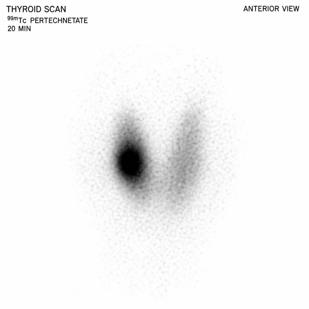
What is a gamma camera used for?
Most sensitive investigation for preoperative localization of abnormal parathyroid glands is
Which one of the following conditions is diagnosed by Tc99m Pertechnetate Scintigraphy?
A thyroid FNA shows 'bubble gum' colloid. Which nuclear feature would best support papillary thyroid carcinoma?
A 45-year-old female presents with a 2 cm thyroid nodule. Which TIRADS category has >95% risk of malignancy?
Which of the following ultrasound features of a thyroid nodule is not suggestive of malignancy?
Which artificial radioisotopes are used in nuclear medicine?
Explanation: ***Sestamibi scan*** - The **Sestamibi scan** (Technetium-99m Sestamibi scintigraphy) is the investigation of choice for localizing **hyperfunctioning parathyroid glands**, especially in cases of primary hyperparathyroidism. - This nuclear medicine scan uses a radiotracer that is preferentially taken up and retained by **abnormal (adenomatous or hyperplastic) parathyroid tissue**, allowing for its differentiation from normal thyroid tissue. *USG* - **Ultrasound (USG)** can visualize enlarged parathyroid glands, but its accuracy is highly dependent on the operator and the gland's location. - While useful for initial screening or guiding biopsies, it is less sensitive than Sestamibi for identifying **ectopic or smaller adenomas**. *CAT Scan* - **Computed Tomography (CT) scans** can identify enlarged parathyroid glands and rule out other neck masses, but it is not specific for parathyroid tissue. - CT involves **radiation exposure** and may not reliably distinguish hyperplastic parathyroid tissue from lymph nodes or thyroid nodules without contrast. *Angiography* - **Angiography** is an invasive procedure primarily used to visualize blood vessels and is generally not the primary investigation for locating parathyroid glands. - It might be rarely used in very complex cases to localize **ectopic glands with specific vascular supply**, but it carries higher risks and is less sensitive than nuclear imaging.
Explanation: ***Hypothyroidism*** - **Radioiodine (RAI) therapy** destroys overactive thyroid cells, making it highly effective for Graves' disease but often leading to a permanent state of **hypothyroidism** post-treatment. - The goal of RAI is to eliminate the source of excess hormone production, and while effective, it frequently necessitates lifelong **thyroid hormone replacement**. *Thyroid storm* - **Thyroid storm** is a rare, life-threatening complication, usually seen in untreated or undertreated hyperthyroidism or during acute stress, not typically a direct outcome of effective RAI. - While a transient increase in thyroid hormones can occur shortly after RAI, a full-blown thyroid storm is infrequent with proper preparation and management. *Thyroid cancer* - There is no significant evidence to suggest an increased risk of **thyroid cancer** in adults following therapeutic doses of radioiodine for Graves' disease [1]. - The radiation dose is targeted primarily at the thyroid gland, and studies have shown no clear link to increased malignancy [1]. *Subacute thyroiditis* - **Subacute thyroiditis** (also known as de Quervain's thyroiditis) is typically a post-viral inflammatory condition of the thyroid, characterized by pain and tenderness in the thyroid gland [2]. - It does not directly result from radioiodine treatment; however, some patients may experience a transient inflammatory response (radiation thyroiditis) after RAI, which is usually mild and self-limiting, not true subacute thyroiditis.
Explanation: ***3%*** - **Purely cystic thyroid nodules** (as described in this case with "isolated cystic swelling") have a **very low risk of malignancy**, typically **2-3%** or less. - According to **ATA guidelines** and **TIRADS classification**, purely cystic nodules are considered **low suspicion** lesions. - The cystic nature suggests a **benign process** such as a degenerated adenoma, colloid cyst, or simple cyst. - **Fine needle aspiration (FNA)** may still be considered if the nodule is >2 cm or has any suspicious solid components, but is often not required for purely cystic lesions. *48%* - This percentage is **significantly higher** than the actual malignancy risk for a purely cystic thyroid swelling. - Such a **high risk** would typically be associated with **solid nodules** exhibiting highly suspicious ultrasound features such as: - Microcalcifications - Irregular or spiculated margins - Taller-than-wide shape - Marked hypoechogenicity - Extrathyroidal extension *24%* - This percentage represents a **moderate to high risk** of malignancy, which is **not characteristic** of an isolated purely cystic thyroid swelling. - A risk in this range might be seen with: - **Mixed solid-cystic nodules** with predominantly solid components - Solid nodules with **intermediate suspicious features** on ultrasound *12%* - While lower than 24% or 48%, 12% is still **considerably higher** than the generally accepted malignancy risk for purely cystic thyroid nodules. - This risk level could be plausible for: - **Predominantly cystic nodules** with some eccentric solid components - Solid nodules with **mildly suspicious** features on ultrasound
Explanation: ***Organ imaging*** - A **gamma camera**, also known as a Anger camera, is a device used in **nuclear medicine** to image the distribution of gamma-emitting radioisotopes within the body. - This allows for the visualization and assessment of organ function and structure, such as in **bone scans** or **myocardial perfusion studies**. *Measuring radioactivity* - While a gamma camera detects gamma rays, its primary function is **spatial imaging** of radiotracer distribution, not simply quantifying general radioactivity levels. - Devices like **Geiger counters** or **scintillation counters** are more commonly used for general measurement of radioactivity. *RIA (Radioimmunoassay)* - **Radioimmunoassay (RIA)** is an in vitro technique used for measuring the concentration of specific substances (like hormones or drugs) in a sample, often using gamma-emitting tracers. - RIA primarily uses **beta counters** or **gamma counters** sensitive to small samples, not large-field-of-view gamma cameras. *Monitoring surface contamination* - Monitoring surface contamination typically involves handheld detectors like **Geiger-Müller counters** or **portable survey meters**. - These devices are designed for detecting radiation on surfaces, whereas a gamma camera is optimized for **internal imaging** within a patient.
Explanation: ***(99mTc) labelled Sestamibi isotope scan*** - This scan uses a **radioactive tracer** that is preferentially taken up and retained by hyperfunctioning parathyroid tissue, making it highly sensitive for identifying **abnormal parathyroid glands**, especially parathyroid adenomas. - It is particularly useful for detecting **ectopic parathyroid glands** and in cases of persistent or recurrent hyperparathyroidism. *Neck ultrasound* - While useful for localizing parathyroid glands, its sensitivity can be limited by **operator dependence**, gland size, and location (e.g., retrosternal). - It is generally good for initial screening but not as sensitive as Sestamibi for identifying all abnormal glands, especially those located in challenging areas. *CT scan* - CT scans can visualize larger parathyroid adenomas, but their sensitivity is lower than Sestamibi scans for smaller lesions or those with **atypical locations**. - It is often used as a **second-line imaging modality** when Sestamibi is inconclusive or to complement findings. *MRI* - MRI can provide detailed anatomical information and identify parathyroid glands, but its sensitivity for detecting abnormal parathyroid tissue is generally **comparable to or slightly less** than CT and inferior to Sestamibi scanning. - It may be considered in cases of unclear findings from other modalities or when radiation exposure is a concern.
Explanation: ***Meckel's diverticulum*** - **Meckel's diverticulum** often contains **ectopic gastric mucosa**, which has parietal cells that secrete acid. - **Tc99m Pertechnetate** is taken up by these gastric parietal cells, allowing visualization of the diverticulum on scintigraphy. *Pharyngeal diverticulum* - A **pharyngeal diverticulum**, such as **Zenker's diverticulum**, is a pouch-like herniation of the pharyngeal mucosa; it does not contain ectopic gastric mucosa. - Diagnosis is typically made through **barium swallow studies** or **endoscopy**, not scintigraphy. *Duodenal diverticulum* - A **duodenal diverticulum** is a common pouch-like protrusion in the duodenum and usually lacks ectopic gastric mucosa. - It is often asymptomatic and diagnosed incidentally on **upper endoscopy** or **cross-sectional imaging** (CT, MRI). *Colonic diverticulum* - **Colonic diverticula** are small, bulging pouches common in the large intestine that do not contain gastric tissue. - They are typically diagnosed with **colonoscopy** or **CT colonography**, particularly in cases of diverticulitis.
Explanation: ***Ground glass nuclei*** - **Ground glass nuclei**, also known as **Orphan Annie eye nuclei** [1][2], are the most **characteristic and recognized nuclear feature** of **papillary thyroid carcinoma (PTC)** on FNA cytology. [1] - This appearance results from **fine, evenly dispersed chromatin** that gives the nucleus a clear, empty, or translucent appearance with a prominent nuclear membrane. [1] - Among the given options, this is the **single best feature** that would support a PTC diagnosis when 'bubble gum' colloid is present. *Nuclear grooves* - **Nuclear grooves** are a common and highly supportive feature of PTC, particularly when **prominent and numerous**. - However, as a **single finding**, they are less definitive than ground glass nuclei, as grooves can occasionally be seen in benign conditions (though usually less prominent). - In combination with other features, nuclear grooves are highly specific for PTC. *Prominent nucleoli* - **Prominent nucleoli** are more frequently associated with **follicular neoplasms**, **medullary thyroid carcinoma**, or anaplastic thyroid carcinoma. - Classical PTC typically has **inconspicuous nucleoli**, so prominent nucleoli would suggest an alternative diagnosis or a tall cell variant of PTC. *Salt and pepper chromatin* - **Salt and pepper chromatin** (finely stippled chromatin) is a classic cytological feature of **medullary thyroid carcinoma** (MTC). - This chromatin pattern reflects neuroendocrine differentiation and is distinct from the nuclear characteristics of PTC. **References:** [1] Kumar V, Abbas AK, et al.. Robbins and Cotran Pathologic Basis of Disease. 9th ed. The Endocrine System, pp. 1098-1100. [2] Cross SS. Underwood's Pathology: A Clinical Approach. 6th ed. Common Clinical Problems From Liver And Biliary System Disease, pp. 429-430.
Explanation: ***TIRADS 5*** - A **TIRADS 5** classification indicates a **highly suspicious** nodule with features strongly suggestive of **malignancy**. - This category corresponds to a **>95% risk of malignancy**, necessitating further investigation such as fine-needle aspiration (FNA). *TIRADS 4* - **TIRADS 4** nodules are classified as **moderately suspicious** for malignancy, with a risk ranging from **5% to 50%**. - While requiring follow-up and often FNA, the risk is significantly lower than for TIRADS 5. *TIRADS 2* - **TIRADS 2** nodules are considered **benign**, with a **0% risk of malignancy** (or extremely low). - These nodules typically have features like **spongiform appearance** or purely cystic composition. *TIRADS 3* - **TIRADS 3** nodules are classified as **mildly suspicious**, with a malignancy risk between **0% and 5%**. - They often have some indeterminate features but are predominantly considered to be low risk.
Explanation: ***Hyperechogenicity*** - A **hyperechoic** thyroid nodule appears brighter than the surrounding parenchyma on ultrasound, typically indicating a benign lesion, such as a **colloid nodule**. - This feature suggests a higher reflection of sound waves, characteristic of tissues rich in **fluid or colloid material**. *Hypoechogenicity* - **Hypoechoic** nodules appear darker than the surrounding thyroid tissue, which is a strong indicator of malignancy due to their often dense cellular structure. - This feature is associated with a higher risk of thyroid cancer and often prompts further investigation with **fine-needle aspiration (FNA)**. *Microcalcification* - The presence of **microcalcifications** (tiny, bright spots) within a thyroid nodule is one of the most specific ultrasound signs of **papillary thyroid carcinoma**. - These calcifications, often punctate, represent psammoma bodies, which are a histopathological hallmark of this common thyroid cancer. *Nonhomogeneous* - A **nonhomogeneous** (heterogeneous) echotexture within a thyroid nodule, characterized by irregular internal architecture, can be suggestive of malignancy. - This often indicates disorganized cellular growth, fibrosis, or cystic degeneration with solid components, which are features seen in various thyroid cancers.
Explanation: ### Explanation **Correct Answer: C. Plutonium** In nuclear medicine, radioisotopes are categorized as either **natural** (found in nature) or **artificial** (man-made via nuclear reactors or cyclotrons). **Plutonium (specifically Pu-238)** is an artificial radioisotope produced in nuclear reactors. While not used as a diagnostic tracer or therapeutic agent for internal administration, it has a significant historical and niche clinical application as a power source for **Radioisotope Thermoelectric Generators (RTGs)** in long-lived **cardiac pacemakers**. Its high energy density and long half-life made it ideal for devices requiring decades of operation without battery replacement. **Analysis of Incorrect Options:** * **A. Radium:** This is a **naturally occurring** radioactive metal found in uranium ores. While Radium-223 is used in treating bone metastases (Xofigo), the element itself is classified as natural. * **B. Uranium:** This is a **naturally occurring** heavy metal. It is the raw material used to produce artificial isotopes but is not used directly in clinical nuclear medicine. * **C. Iridium:** While Iridium-192 is used in Brachytherapy, it is generally classified as a transition metal used in "sealed sources" for radiotherapy rather than being the classic example of an "artificial radioisotope" in the context of general nuclear medicine tracers (like Technetium-99m). However, in the context of this specific question, Plutonium is the most distinct "artificial/man-made" element. **High-Yield Clinical Pearls for NEET-PG:** * **Technetium-99m (Tc-99m):** The most commonly used artificial radioisotope in diagnostic nuclear medicine (produced in a Mo-99/Tc-99m generator). * **Cyclotron-produced isotopes:** Include F-18 (used in PET scans), I-123, and Thallium-201. * **Reactor-produced isotopes:** Include I-131, Mo-99, and Xenon-133. * **Therapeutic Alpha Emitter:** Radium-223 is the first alpha-emitting radiopharmaceutical approved to improve survival in castration-resistant prostate cancer with bone metastases.
Get full access to all questions, explanations, and performance tracking.
Start For Free