Cardiotoxicity caused by radiotherapy & chemotherapy is best detected by
MUGA scan is not useful in:
Hot spot in heart is seen in which scan
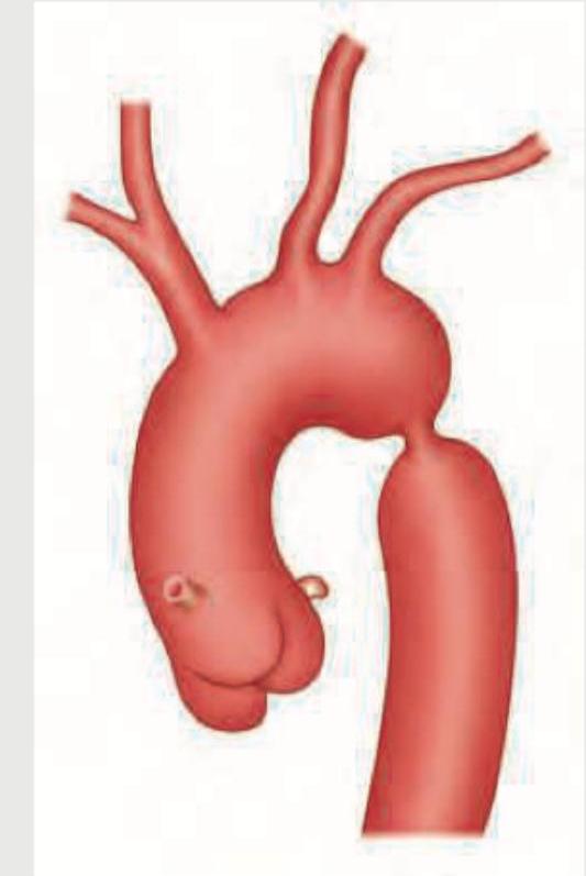
In the condition shown below, rib notching is present in which of the following ribs? (AIIMS Nov 2015)

Which artificial radioisotopes are used in nuclear medicine?
Increased radio-isotope uptake is seen in which of the following conditions?
Which of the following isotopes is radioactive?
Precautions advised after outpatient Radioiodine (I-131) therapy are all, EXCEPT:
Which of the following is a functional investigation?
Which of the following is the best method for measuring renal Glomerular Filtration Rate (GFR)?
Explanation: ***ECHO*** - **Echocardiography (ECHO)** is the primary and most widely used non-invasive method for detecting cardiotoxicity due to its ability to assess **left ventricular ejection fraction (LVEF)**, a key indicator of cardiac function, and structural changes. - It is crucial for **baseline assessment**, monitoring during treatment, and follow-up, identifying both systolic and diastolic dysfunction effectively. *Endomyocardial Biopsy* - While **endomyocardial biopsy** is considered the gold standard for definitive diagnosis of some cardiomyopathies (e.g., myocarditis), it is **invasive** and carries risks such as perforation, tamponade, and arrhythmias. - It is usually reserved for cases where other non-invasive tests are inconclusive and there's a strong clinical suspicion of severe cardiac disease, or for research, not routine monitoring of cardiotoxicity. *ECG* - An **ECG** can detect arrhythmias and ischemic changes but is **not sensitive or specific** enough to reliably detect early or subtle changes in cardiac function characteristic of cardiotoxicity. - It may show changes secondary to heart failure, but it does not directly measure ejection fraction or assess overall cardiac mechanical function. *Radionuclide Scan* - **Radionuclide scans**, specifically **MUGA (Multigated Acquisition)** scans, can accurately measure **LVEF** and are an alternative to ECHO, particularly when ECHO images are suboptimal [1]. - However, they involve **radiation exposure**, making them less ideal for frequent monitoring compared to echocardiography, especially in cancer patients who are already exposed to radiation.
Explanation: ***Regional wall perfusion*** - A MUGA scan assesses **ventricular function** through blood pool imaging, evaluating wall motion and ejection fraction. - It does not directly visualize or quantify myocardial perfusion, which is the flow of blood through the coronary arteries to the heart muscle. *Stroke volume* - A MUGA scan accurately measures **end-diastolic volume** and **end-systolic volume**, from which stroke volume (EDV – ESV) can be calculated. - This parameter directly reflects the amount of blood pumped out by the ventricle with each beat. *Left ventricular ejection fraction* - The MUGA scan is considered a gold standard for calculating **left ventricular ejection fraction** (LVEF), a key indicator of cardiac pump function. - It uses a count-based method from gated blood pool images to determine the percentage of blood ejected from the left ventricle. *Regional wall motion* - MUGA scans are highly effective in assessing **regional wall motion abnormalities**, identifying areas of **hypokinesis**, **akinesis**, or **dyskinesis**. - This is crucial for diagnosing and monitoring conditions like myocardial ischemia or infarction, and is a primary utility of the scan.
Explanation: ***Tc pyrophosphate scan*** - A **technetium-99m pyrophosphate (Tc-PYP) scan** demonstrates a "hot spot" in the heart in cases of **acute myocardial infarction** due to the tracer binding to calcium deposits in necrotic cardiomyocytes. - This hot spot indicates recent myocardial damage and is particularly useful in diagnosing **amyloidosis** (specifically transthyretin cardiac amyloidosis) where the tracer binds to amyloid fibrils. *Thallium* - **Thallium-201** is used in myocardial perfusion imaging to assess areas of reduced blood flow or infarction, creating a "cold spot" (decreased uptake). - It acts as a potassium analog and is taken up by viable myocardial cells, thus areas of ischemia or necrosis appear as defects rather than hot spots. *Gallium* - **Gallium-67** scans are primarily used to detect infection and inflammation, as well as certain tumors. - While it can accumulate in areas of inflammation in the heart (e.g., myocarditis), it does not create a characteristic "hot spot" associated with acute myocardial infarction. *Albumin labelled* - **Technetium-99m labeled albumin** (e.g., Technetium-99m macroaggregated albumin, MAA) is typically used for lung perfusion scans to diagnose pulmonary embolism or for gastrointestinal bleeding studies. - It is not used for direct assessment of myocardial damage or to create a "hot spot" in the heart for ischemic events.
Explanation: **3rd to 9th ribs** - The image provided depicts **coarctation of the aorta**, characterized by a narrowing of the aorta, typically distal to the origin of the left subclavian artery. - In coarctation of the aorta, collateral circulation develops through the **intercostal arteries** to bypass the constriction, leading to their enlargement and subsequent erosion of the inferior margins of the **3rd to 9th ribs**, a finding known as "rib notching." *1st to 9th ribs* - While rib notching affects upper ribs, it typically **spares the 1st and 2nd ribs** because the superior intercostal arteries (which supply these ribs) originate directly from the subclavian artery, often proximal to the coarctation, so they do not participate in collateral circulation as significantly. - The pattern of notching is usually more concentrated in the mid-thoracic region. *11th and 12th ribs* - Rib notching from coarctation of the aorta is rarely observed in the **floating ribs** (11th and 12th ribs). - These ribs have a different anatomical relationship with the pleura and typically do not bear the brunt of increased collateral flow from the intercostal arteries in the same way as the higher ribs. *All ribs* - Rib notching is a localized phenomenon reflecting increased blood flow through specific intercostal arteries involved in collateral circulation due to aortic coarctation. - Therefore, it does **not affect all ribs**, and its absence in certain ribs (like the 1st, 2nd, 11th, and 12th) helps differentiate this condition radiologically.
Explanation: ### Explanation **Correct Answer: C. Plutonium** In nuclear medicine, radioisotopes are categorized as either **natural** (found in nature) or **artificial** (man-made via nuclear reactors or cyclotrons). **Plutonium (specifically Pu-238)** is an artificial radioisotope produced in nuclear reactors. While not used as a diagnostic tracer or therapeutic agent for internal administration, it has a significant historical and niche clinical application as a power source for **Radioisotope Thermoelectric Generators (RTGs)** in long-lived **cardiac pacemakers**. Its high energy density and long half-life made it ideal for devices requiring decades of operation without battery replacement. **Analysis of Incorrect Options:** * **A. Radium:** This is a **naturally occurring** radioactive metal found in uranium ores. While Radium-223 is used in treating bone metastases (Xofigo), the element itself is classified as natural. * **B. Uranium:** This is a **naturally occurring** heavy metal. It is the raw material used to produce artificial isotopes but is not used directly in clinical nuclear medicine. * **C. Iridium:** While Iridium-192 is used in Brachytherapy, it is generally classified as a transition metal used in "sealed sources" for radiotherapy rather than being the classic example of an "artificial radioisotope" in the context of general nuclear medicine tracers (like Technetium-99m). However, in the context of this specific question, Plutonium is the most distinct "artificial/man-made" element. **High-Yield Clinical Pearls for NEET-PG:** * **Technetium-99m (Tc-99m):** The most commonly used artificial radioisotope in diagnostic nuclear medicine (produced in a Mo-99/Tc-99m generator). * **Cyclotron-produced isotopes:** Include F-18 (used in PET scans), I-123, and Thallium-201. * **Reactor-produced isotopes:** Include I-131, Mo-99, and Xenon-133. * **Therapeutic Alpha Emitter:** Radium-223 is the first alpha-emitting radiopharmaceutical approved to improve survival in castration-resistant prostate cancer with bone metastases.
Explanation: **Explanation:** In nuclear medicine, bone scintigraphy (Bone Scan) using **99mTc-MDP** (Methylene Diphosphonate) is the gold standard for assessing bone turnover. The uptake of the radiopharmaceutical depends on two primary factors: **blood flow** and **osteoblastic activity** (bone formation). **Why Pseudoarthrosis is the Correct Answer:** Pseudoarthrosis (a "false joint" resulting from non-union of a fracture) is characterized by persistent mechanical stress and abnormal motion at the fracture site. This leads to continuous, localized **reactive osteoblastic activity** and increased vascularity as the body attempts to heal the bone. On a bone scan, this manifests as a focal area of **increased radio-isotope uptake** (a "hot spot"). **Analysis of Incorrect Options:** * **Osteoclastoma (Giant Cell Tumor):** While GCT can show uptake, it typically presents with a "cold" center (photopenia) due to extensive bone destruction and hemorrhage, surrounded by a rim of increased uptake. * **Enchondroma:** These are benign cartilaginous tumors. They are typically **"cold"** or show very minimal uptake unless they are complicated by a pathological fracture or undergo malignant transformation. * **Ewing’s Sarcoma:** While Ewing’s sarcoma generally shows increased uptake due to its aggressive nature, in the context of this specific question (often derived from standard textbooks like *Bailey & Love* or *Maheshwari*), **Pseudoarthrosis** is the classic teaching example for identifying active bone remodeling in non-malignant conditions. **NEET-PG High-Yield Pearls:** * **Hot Spots (Increased Uptake):** Osteoblastic metastases (Prostate CA), Osteoid Osteoma (Double density sign), Paget’s Disease, and Fractures. * **Cold Spots (Decreased Uptake):** Multiple Myeloma (often missed on bone scans), Renal Cell Carcinoma metastases, and early Avascular Necrosis (AVN). * **Three-Phase Bone Scan:** Used to differentiate Cellulitis (increased uptake in first two phases) from Osteomyelitis (increased uptake in all three phases).
Explanation: **Explanation:** The correct answer is **Cobalt-60**. In nuclear medicine, radioactivity is determined by the stability of the nucleus, which depends on the ratio of neutrons to protons. **1. Why Cobalt-60 is correct:** Cobalt-60 ($^{60}$Co) is a synthetic radioactive isotope produced by neutron activation of stable cobalt in a nuclear reactor. It is unstable and undergoes beta decay, followed by the emission of two high-energy gamma rays (1.17 MeV and 1.33 MeV). Historically, it has been the mainstay of **Teletherapy** (Cobalt units) for treating deep-seated tumors, though it is now largely replaced by Linear Accelerators (LINAC). **2. Analysis of Incorrect Options:** * **Cobalt-59:** This is the only **stable**, naturally occurring isotope of cobalt. It is not radioactive. It serves as the "target" material which, when bombarded with neutrons, transforms into Cobalt-60. * **Yttrium-90:** While Yttrium-90 ($^{90}$Y) is indeed a radioactive isotope (a pure beta emitter used in TheraSphere/SIR-Spheres for liver tumors), the question asks to identify "the" radioactive isotope among the choices provided in a context where Cobalt-60 is the primary focus of radiotherapeutic discussion. *Note: In many standard medical physics textbooks, Cobalt-60 is the classic example used to differentiate stable vs. unstable isotopes.* **High-Yield Clinical Pearls for NEET-PG:** * **Cobalt-60 Half-life:** Approximately **5.27 years**. * **Decay Product:** It decays into stable **Nickel-60**. * **Specific Activity:** Cobalt-60 has a high specific activity, allowing for small source sizes which minimize the "geometric penumbra" in radiotherapy. * **Gamma Energy:** Average energy is **1.25 MeV** (mean of 1.17 and 1.33). * **Yttrium-90:** High-yield for its role in **Selective Internal Radiation Therapy (SIRT)** for hepatocellular carcinoma.
Explanation: **Explanation:** Radioiodine (I-131) therapy is commonly used for hyperthyroidism and thyroid carcinoma. Post-therapy precautions are designed to minimize radiation exposure to others (ALARA principle) and prevent environmental contamination. **Why Option C is the Correct Answer:** Patients are actually **encouraged** to use household chlorine bleach to clean toilets and sinks after use. I-131 is excreted primarily through urine and saliva. Chlorine bleach effectively decontaminates the surfaces by chemically reacting with the radioiodine, reducing the risk of indirect exposure to family members. Therefore, "avoiding" it is incorrect advice. **Analysis of Other Options:** * **Option A (Treatment Certificate):** Patients must carry a treatment certificate for up to **90 days**. Modern security sensors (e.g., at airports or international borders) are highly sensitive and can detect residual gamma radiation from the patient’s body for several weeks. * **Option B (Social Distancing):** Patients are advised to maintain a distance of **2 meters (6 feet)** from others, especially children and pregnant women, for a specified period (usually 3–7 days depending on the dose) to minimize external gamma radiation exposure. * **Option C (Contraception):** Female patients must avoid pregnancy for at least **6 months**, and males should use contraception for **3–4 months** (to allow for one full cycle of spermatogenesis) to prevent potential genetic damage to the fetus. **Clinical Pearls for NEET-PG:** * **Mechanism:** I-131 emits both **Beta particles** (therapeutic effect/tissue destruction) and **Gamma rays** (diagnostic/safety concern). * **Half-life:** The physical half-life of I-131 is **8.02 days**. * **Contraindication:** Radioiodine is strictly **contraindicated in pregnancy** (crosses the placenta and destroys the fetal thyroid) and **breastfeeding**. * **Hydration:** Patients are advised to increase fluid intake to facilitate the renal clearance of unbound I-131.
Explanation: ### Explanation **Correct Answer: D. PET scan** **Why PET scan is the correct answer:** Positron Emission Tomography (PET) is a **functional (molecular) imaging** modality. Unlike conventional imaging, which visualizes anatomical structures, PET scans detect metabolic and biochemical activity within tissues. It utilizes radiopharmaceuticals, most commonly **18F-Fluorodeoxyglucose (18F-FDG)**, which is a glucose analog. Since malignant cells and inflamed tissues have higher metabolic rates, they take up more FDG, appearing as "hot spots" on the scan. This allows for the detection of disease processes (like malignancy or ischemia) even before structural changes become visible on CT or MRI. **Why other options are incorrect:** * **A, B, and C (CT, MRI, USG):** These are primarily **structural (anatomical) imaging** modalities. They provide detailed information regarding the size, shape, position, and morphology of organs and lesions. While advanced sequences like Functional MRI (fMRI) or Dynamic Contrast-Enhanced CT exist, the standard modalities listed are categorized as anatomical investigations. **High-Yield Clinical Pearls for NEET-PG:** * **Hybrid Imaging:** Modern PET is almost always combined with CT (**PET-CT**) to provide both functional and precise anatomical localization. * **Gold Standard:** PET scan is the gold standard for **staging, restaging, and monitoring treatment response** in various cancers. * **Myocardial Viability:** PET using FDG is the gold standard for assessing myocardial viability in patients with ischemic heart disease. * **Brain Imaging:** It is used to differentiate between radiation necrosis and tumor recurrence, and in the evaluation of dementia (e.g., Alzheimer’s). * **Cyclotron:** The radioisotopes used in PET (like 18F, 11C, 15O) are produced in a cyclotron and have very short half-lives.
Explanation: **Explanation:** The measurement of Glomerular Filtration Rate (GFR) requires a substance that is exclusively filtered by the glomerulus, without being secreted or reabsorbed by the renal tubules. **Why Option C is correct:** **Technetium-99m DTPA (Diethylenetriaminepentaacetic acid)** is the radiopharmaceutical of choice for GFR estimation in nuclear medicine. It is handled by the kidneys almost entirely through **glomerular filtration** (approx. 95-98%). By measuring the rate at which the tracer is cleared from the blood or accumulates in the kidneys using a gamma camera (Gates’ method), an accurate GFR can be calculated. **Why other options are incorrect:** * **Option A (Tc-99m DMSA):** This is a **static renal imaging** agent. It binds to the proximal convoluted tubules and remains in the renal cortex. It is the gold standard for detecting **renal scarring** and evaluating renal morphology, not GFR. * **Option B (Tc-99m Pyrophosphate):** This is primarily a **bone scanning agent** (and used for detecting myocardial amyloidosis). It is not used for functional renal imaging. * **Option D (Creatinine Clearance):** While used clinically, it often **overestimates GFR** because creatinine is not only filtered but also secreted by the tubules. In the context of "best method" among the choices provided (specifically within Radiology/Nuclear Medicine), DTPA provides a more precise, real-time functional assessment. **High-Yield Clinical Pearls for NEET-PG:** * **Tc-99m MAG3:** The agent of choice for **Renal Plasma Flow (ERPF)** and evaluating obstructive uropathy (Diuretic renography). * **Gold Standard for GFR:** **Inulin clearance** (though rarely used clinically due to its invasive nature). * **Captopril Scan:** Used with DTPA or MAG3 to diagnose **Renovascular Hypertension** (Renal Artery Stenosis).
Get full access to all questions, explanations, and performance tracking.
Start For Free