What is the investigation of choice for diagnosing a stress fracture?
What is the best imaging modality for detecting early osteomyelitis?
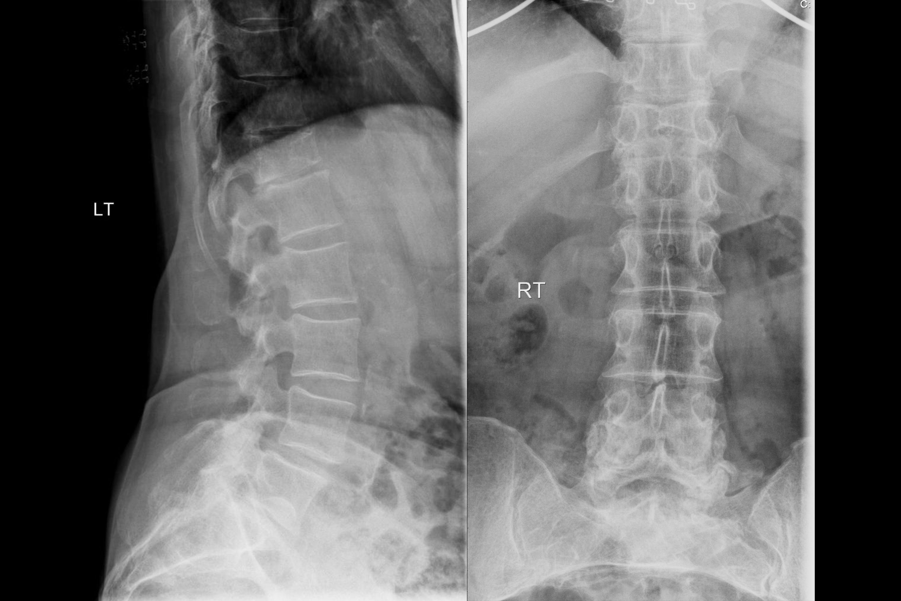
A 75-year-old female has chronic backache. X-ray of the spine is shown. What is the most likely diagnosis?

What is the investigation of choice in a patient with traumatic paraplegia?
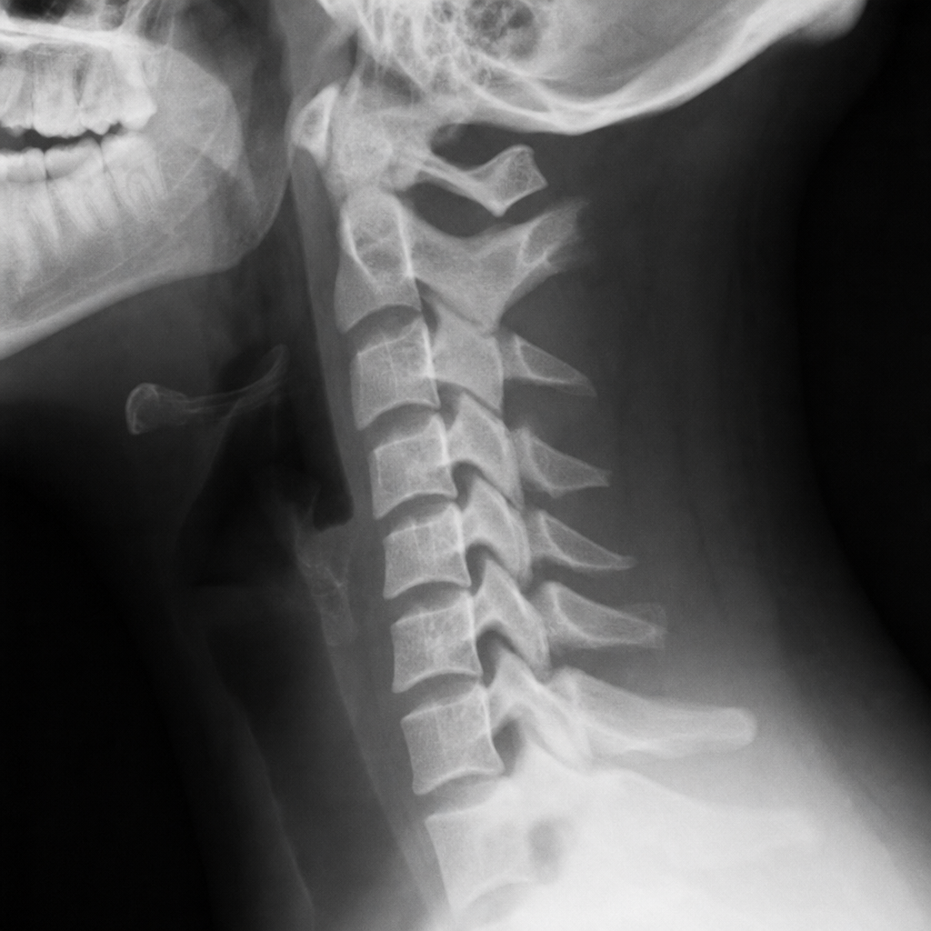
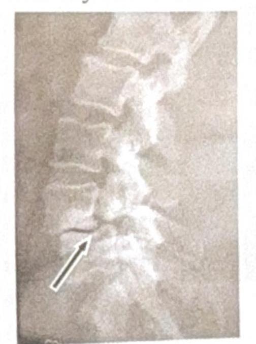
Identify the condition shown in the image:

After chronic use of steroids severe pain in right hip with immobility is due to
Spine MRI shows 'pencil-sharpened' vertebral bodies and 'H-shaped' vertebrae on T1-weighted images. Most likely diagnosis?
A patient with a history of chronic ear infection now presents with manifestations, including headache and vomiting. A CT brain image is shown. What is the most probable diagnosis?

Time of Flight technique is employed in —
What is the primary imaging modality used for diagnosing urethral trauma?
Explanation: ***MRI*** - **Magnetic Resonance Imaging (MRI)** is the most sensitive and specific imaging modality for diagnosing **stress fractures**, especially in their early stages. - It can detect **bone marrow edema** and **periosteal reactions** indicative of stress injury before cortical changes are visible on plain radiographs. *X-ray* - **X-rays** are often the initial investigation, but they have low sensitivity for **stress fractures** in the early stages as bone changes may not be apparent for several weeks. - A positive X-ray for stress fracture typically shows a **sclerotic line** or **periosteal reaction**, but this indicates a more advanced injury. *CT scan* - **CT scans** provide excellent detail of **cortical bone** and can detect subtle fractures not seen on X-rays. - While more sensitive than X-rays, CT has **higher radiation exposure** and is generally less sensitive than MRI for early detection of **bone marrow edema** associated with stress injuries. *Bone scan* - **Bone scans** (scintigraphy) are highly sensitive for detecting increased **osteoblastic activity** associated with stress fractures. - However, they are **less specific** as various conditions can cause increased uptake, and they do not provide detailed anatomical information, making MRI superior for definitive diagnosis and staging.
Explanation: ***MRI*** - **Magnetic Resonance Imaging (MRI)** is considered the **gold standard** for detecting early osteomyelitis due to its excellent soft tissue contrast and ability to visualize **bone marrow edema**, which is an early sign of infection. - It can identify changes within **3-5 days** of infection onset, much earlier than other modalities. *CT scan* - While useful for showing **bone destruction**, cortical integrity, and sequestra, **CT scans** are less sensitive than MRI for detecting early marrow edema. - Its ability to diagnose osteomyelitis is usually delayed until significant **bony changes** have occurred, typically around 1-2 weeks. *X-ray* - **Plain radiographs** are often the initial imaging study but are **insensitive** for early osteomyelitis, showing changes only after 10-14 days or more. - Early findings on X-rays can be subtle, such as **periosteal elevation** or **soft tissue swelling**, but frank bone destruction is a late finding. *Bone scintigraphy* - **Bone scintigraphy** (e.g., technetium-99m) is sensitive for detecting increased bone turnover associated with infection but lacks **specificity**, as it can be positive in other conditions like trauma or tumors. - While it can detect changes earlier than X-rays, typically within 2-3 days, it cannot clearly differentiate infection from other processes, and its spatial resolution is poor compared to MRI.
Explanation: ***Osteoporosis*** - The X-ray shows diffuse **osteopenia** (reduced bone density) and **vertebral compression fractures**, particularly visible in the lateral view, which are characteristic findings in elderly patients with osteoporosis and chronic backache. - The vertebral bodies appear **demineralized** and some exhibit a loss of height, suggesting collapse due to weakened bone structure. *Spondylodiscitis* - This condition involves **inflammation of the vertebral body and adjacent intervertebral disc**, typically showing **erosions** of the vertebral endplates and **narrowing of the disc space** on X-ray, which are not clearly evident here as the primary issue. - While it can cause back pain, the dominant finding on this X-ray is widespread bone density loss and fractures, rather than localized infection-related changes. *Pott's spine* - Pott's spine (**tuberculous spondylitis**) is a form of osteomyelitis that causes **destruction of vertebral bodies** and adjacent discs, often leading to a **gibbus deformity** (sharp posterior angulation of the spine). - The X-ray does not show extensive vertebral destruction, paraspinal abscess formation, or typical kyphotic deformity associated with Pott's spine. *Spondylolisthesis* - Spondylolisthesis is characterized by the **forward slippage of one vertebral body over another**, often due to a defect in the pars interarticularis. - While there may be some degenerative changes, there is no clear evidence of significant anterior translation of a vertebral body on the lateral X-ray that would indicate spondylolisthesis.
Explanation: ***MRI scan*** - An **MRI scan** provides superior imaging of **soft tissues**, including the spinal cord, nerves, and ligaments, which are crucial for assessing damage in **traumatic paraplegia**. - It is essential for detecting **spinal cord compression**, hemorrhage, edema, and ligamentous injuries that may not be visible on other imaging modalities. *Plain X-ray* - A **plain X-ray** primarily visualizes bony structures and can detect major **fractures or dislocations** but offers limited information about the spinal cord or soft tissue damage. - It may miss subtle bony injuries and provides no information on **spinal cord integrity**, which is critical in paraplegia. *Myelography* - **Myelography** involves injecting contrast dye into the spinal canal and then performing X-rays or CT scans, which is an **invasive procedure** with potential risks. - While it can demonstrate **spinal cord compression** indirectly, it has largely been replaced by MRI due to its invasiveness and MRI's direct visualization capabilities. *CT scan* - A **CT scan** is excellent for evaluating **bony injuries**, such as vertebral fractures and alignment, with good detail. - However, it is less effective than MRI for directly visualizing the **spinal cord itself** and assessing soft tissue damage, which is paramount in paraplegia.
Explanation: ***Spondylolysis*** * The image shows a **break in the pars interarticularis** of a vertebra, indicated by the arrow, which is characteristic of spondylolysis. * This condition is a **stress fracture** or defect in the pars interarticularis, a bony segment connecting the superior and inferior articular facets. *Renal osteodystrophy* * Renal osteodystrophy refers to a spectrum of **bone abnormalities** that occur in chronic kidney disease, not a specific vertebral fracture pattern. * It typically involves features such as **osteomalacia**, **osteitis fibrosa cystica**, or **osteoporosis**, which are not directly depicted as a fracture in this image. *Spondylolisthesis* * Spondylolisthesis is the **anterior slippage** of one vertebral body over another, which can be caused by bilateral spondylolysis but is not directly shown as a slip in this specific image. * The image distinctly highlights the **fracture line** itself, rather than the displacement of the vertebral body. *Tuberculosis (TB)* * Spinal tuberculosis (Pott's disease) typically presents with **destruction of vertebral bodies**, disc space narrowing, and often a **paravertebral abscess**. * The image does not show these features; instead, it demonstrates a clear **bony defect** in the pars interarticularis.
Explanation: ***Avascular necrosis*** - Chronic **steroid use** is a major risk factor for avascular necrosis (AVN), particularly affecting the **femoral head** of the hip. - Reduced blood supply leads to bone death, resulting in severe pain and impaired mobility. *Perthes disease* - This is a condition of idiopathic **avascular necrosis of the femoral head** occurring in **children**, primarily between ages 4-10. - It is not associated with steroid use and typically presents in a different age group. *Hip dislocation* - Hip dislocation presents with **acute, severe pain** and an inability to bear weight or move the hip, often due to significant trauma. - While it causes immobility, it is an **acute traumatic event** rather than a chronic consequence of steroid use. *Osteoarthritis* - Osteoarthritis is a degenerative joint disease characterized by **cartilage breakdown** and joint pain that typically **worsens with activity** and improves with rest. - While chronic hip pain can be due to osteoarthritis, its direct link to steroid use for severe pain and immobility as described is less prominent than AVN.
Explanation: ***Sickle cell disease*** - **Sickle cell disease** can lead to vertebral body changes due to **bone infarction** and **hyperplasia of hematopoietic marrow**, causing central depression and characteristic 'H-shaped' or 'pencil-sharpened' vertebrae. - The abnormal hemoglobin in sickle cell anemia causes red blood cells to stiffen and form a crescent or "sickle" shape, leading to a host of debilitating symptoms and early death. *Thalassemia* - **Thalassemia** can cause widespread marrow expansion leading to generalized osteopenia and widened medullary spaces, but typically does not result in the focal 'H-shaped' vertebral changes seen with infarction. - While it also causes anemia and bone changes, the specific vertebral findings described are not characteristic of thalassemia. *Osteopetrosis* - **Osteopetrosis** is characterized by **increased bone density** and brittle bones, often described as a "stone bone" appearance. - This condition leads to thickened, sclerotic bones and does not produce the 'H-shaped' or 'pencil-sharpened' vertebral deformities. *Paget's disease* - **Paget's disease** is characterized by disorganized bone remodeling, leading to bone enlargement and deformity with a characteristic **"cotton wool" appearance** on imaging. - While it affects vertebrae, it typically results in cortical thickening and coarsened trabeculae, not the specific 'H-shaped' deformity.
Explanation: ***Temporal lobe Abscess*** - The CT scan shows a **ring-enhancing lesion** with significant surrounding edema, which is characteristic of a **brain abscess**. - Given the history of a **chronic ear infection**, the temporal lobe is a common site for bacterial spread from the mastoid air cells or middle ear. *Meningitis* - Meningitis involves inflammation of the **meninges** and typically presents with diffuse changes on imaging, such as sulcal effacement or leptomeningeal enhancement, rather than a focal, encapsulated lesion. - While it can cause headache and vomiting, the CT image does not show findings typical of meningitis. *Extradural Abscess* - An extradural (or epidural) abscess is located **between the dura mater and the skull bone**. - It would typically appear as a collection outside the brain parenchyma, potentially causing mass effect but distinct from an intraparenchymal lesion seen in the image. *Cerebral Abscess* - The image does show a **cerebral abscess**, but this option is less specific than "Temporal lobe abscess." - The question asks for the **most probable diagnosis**, and combining the imaging findings with the patient's history of ear infection points to a specific location within the cerebrum.
Explanation: ***MR imaging*** - The **Time of Flight (TOF)** technique is a type of **magnetic resonance angiography (MRA)** that exploits the phenomenon of **flow-related enhancement** of fresh, unsaturated blood entering an imaging slice. - It is used to visualize blood flow without the need for an external contrast agent, making it particularly useful for assessing vessels in the brain and neck. *Spiral CT* - **Spiral CT** (helical CT) involves continuous data acquisition as the patient moves through the gantry, creating a spiral path of X-ray projection data. - While it has revolutionised CT angiography, it does not employ the Time of Flight principle, which is specific to MR imaging. *Digital radiography* - **Digital radiography** uses X-rays to create images, which are captured by digital sensors rather than photographic film. - This technique primarily focuses on structural imaging and does not involve the physical principles (like spin physics of protons in a magnetic field) necessary for Time of Flight applications. *CT angiography* - **CT angiography** uses **iodinated contrast material** injected intravenously to visualize blood vessels with high resolution using X-rays. - Unlike Time of Flight MRA, it relies on the contrast enhancement of flowing blood with an exogenous agent, not on the intrinsic properties of blood flow within a magnetic field.
Explanation: ***Ascending urethrogram*** - An **ascending urethrogram** (also known as a retrograde urethrogram) is the **gold standard** for diagnosing urethral trauma. - It involves injecting contrast material directly into the urethra to visualize its integrity and identify any extravasation, strictures, or ruptures. *Descending urethrogram* - A descending urethrogram (or voiding cystourethrogram) is primarily used to evaluate the **bladder and urethra during urination**, often for vesicoureteral reflux or bladder neck dysfunction. - It is not the primary diagnostic tool for acute urethral trauma, as it requires the patient to void, which might be painful or difficult with an injured urethra. *USG* - **Ultrasound** (USG) can be used to assess the presence of peri-urethral hematomas or fluid collections but is generally **not sufficient to definitively diagnose urethral integrity** or the exact location and extent of a tear. - Its utility in urethral trauma is limited compared to direct contrast imaging of the urethra. *CT scan* - A **CT scan** of the pelvis can identify associated injuries, such as **pelvic fractures** or hematomas, that often accompany urethral trauma. - However, it is **less sensitive for direct visualization of the urethral lumen** and diagnosing the extent of a urethral injury compared to an ascending urethrogram.
Get full access to all questions, explanations, and performance tracking.
Start For Free