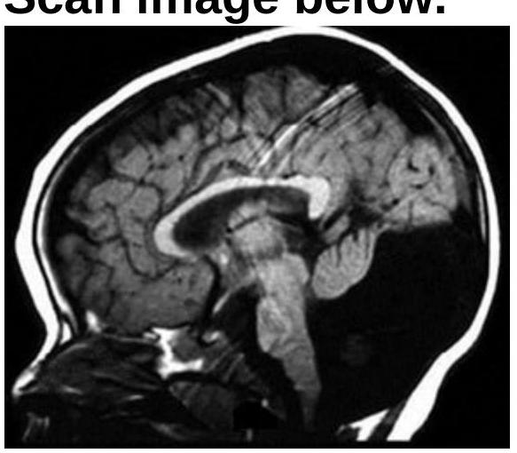
Identify the condition shown in the CT scan image.

Which of the following abnormalities is most commonly detected as a vascular malformation on skull MRI?
Tigroid pattern on MRI is seen in -

On imaging, diffuse axonal injury is characterized by -
What is the CT scan finding in a carotid cavernous sinus fistula?
Which imaging modality is most effective in differentiating between epidermoid cyst and arachnoid cyst?
What are the X-ray findings associated with chronic otitis media?
All the following are true of craniopharyngioma except:
Which condition is characterized by a specific radiological appearance resembling a sunburst pattern?
Investigation of choice for intramedullary SOL is -
Explanation: ***Dandy-Walker malformation*** - The image shows an enlarged posterior fossa with **cystic dilation of the fourth ventricle** and **absence/hypoplasia of the cerebellar vermis**, which are classic features of Dandy-Walker malformation. - The elevated tentorium and upward displacement of the transverse sinuses are also characteristic, contributing to the distinct appearance. *Cerebellar vermis hypoplasia* - While cerebellar vermis hypoplasia is a component of Dandy-Walker malformation, it is not the sole, defining feature. - Dandy-Walker also includes cystic dilation of the fourth ventricle and an enlarged posterior fossa, which are evident in the image and go beyond isolated vermis hypoplasia. *Mega cisterna magna* - A **mega cisterna magna** is a benign enlargement of the cisterna magna, which is the space between the cerebellum and the medulla oblongata. - Unlike in Dandy-Walker malformation, a mega cisterna magna usually does not involve displacement of the tentorium or hypoplasia of the cerebellar vermis, and the fourth ventricle is typically normal in shape and size. *None of the options* - The image clearly displays the diagnostic hallmarks of Dandy-Walker malformation, making this option incorrect.
Explanation: ***Vein of Galen abnormalities*** - **Vein of Galen malformations (VOGM)** are the **most common symptomatic cerebral vascular malformations in neonates and infants**, accounting for approximately **30% of pediatric cerebral vascular malformations**. - They often present with **high-output cardiac failure**, hydrocephalus, or seizures in early life. - On **MRI**, they appear as a large, abnormal midline venous pouch in the quadrigeminal cistern with characteristic **flow voids** on T2-weighted images. - VOGM represents an **arteriovenous shunt** to a persistent embryonic vein (median prosencephalic vein of Markowski). *Dandy Walker malformation* - This is a **congenital brain malformation** involving the cerebellum and fourth ventricle, characterized by hypoplasia of the cerebellar vermis, cystic dilatation of the fourth ventricle, and an enlarged posterior fossa. - While readily seen on MRI, it is a **structural developmental anomaly**, not a vascular malformation. *Pneumocephalus presence* - **Pneumocephalus** refers to the presence of **air within the intracranial cavity**, usually resulting from trauma, neurosurgery, or skull base fractures. - It is an **acquired condition**, not a congenital vascular malformation, and appears as dark signal (air) on all MRI sequences. *Crouzon syndrome features* - **Crouzon syndrome** is a genetic disorder (FGFR2 mutation) characterized by **craniosynostosis** with premature fusion of coronal and sagittal sutures, leading to brachycephaly and midface hypoplasia. - It is a **craniofacial skeletal disorder**, not a vascular malformation.
Explanation: ***Metachromatic leukodystrophy*** - The **tigroid pattern** on MRI, characterized by **perivascular sparing** within demyelinated white matter, is highly characteristic of metachromatic leukodystrophy. - This pattern results from the accumulation of **sulfatides** in oligodendrocytes and macrophages, leading to central demyelination with spared U-fibers and white matter adjacent to vessels. *Wilson's disease* - Wilson's disease involves **copper accumulation** and typically presents with abnormalities in the **basal ganglia**, thalami, and brainstem. - While it causes neurodegeneration, it does not produce a characteristic tigroid demyelination pattern. *Parkinsonism* - Parkinsonism refers to a group of neurological disorders characterized by motor symptoms like **bradykinesia, rigidity, tremor, and postural instability**. - MRI findings in Parkinsonism often show **nigral degeneration** but do not typically involve a tigroid pattern of leukodystrophy. *GB syndrome* - **Guillain-Barré syndrome (GBS)** is an acute autoimmune peripheral neuropathy affecting the **peripheral nerves and nerve roots**. - It does not involve central nervous system demyelination or display a tigroid pattern on brain MRI.
Explanation: ***Multiple small petechial hemorrhages*** - Diffuse axonal injury (DAI) is characterized on imaging by numerous **small petechial hemorrhages** at the **gray-white matter junction**, **corpus callosum**, and **brainstem**. - These microhemorrhages are the **hallmark imaging finding** and are best visualized on MRI (GRE/SWI sequences), though they can be seen on CT as small hyperdensities. - This is the **most specific** and characteristic finding that defines DAI. *Patchy ill-defined low density lesion mixed with small hyperdensities of petechial hemorrhage* - While this description can be seen in DAI (combining edema with microhemorrhages), it is **less specific** and could represent other entities. - This mixed pattern is more commonly associated with **contusions** where there is more prominent parenchymal injury with larger areas of edema and hemorrhage. - DAI classically shows predominantly **small punctate hemorrhages** rather than larger patchy low-density lesions. *Crescentic extra-axial hematoma* - A crescentic extra-axial collection describes a **subdural hematoma**, which is an entirely separate entity from DAI. - Subdural hematomas are located between the dura and arachnoid membranes, whereas DAI involves direct **axonal shearing injury** within brain parenchyma. *White matter lucencies* - This is a **non-specific finding** that can occur in many conditions including ischemia, demyelination, and chronic small vessel disease. - While DAI can cause white matter edema leading to lucencies, this does not capture the **characteristic petechial hemorrhages** that define the condition on imaging.
Explanation: ***Enlarged superior ophthalmic vein*** * A carotid cavernous sinus fistula (CCSF) causes **high-pressure arterial blood** to shunt into the **venous system** of the cavernous sinus. * This increased pressure leads to retrograde flow and congestion in the draining veins, most notably the **superior ophthalmic vein**, causing its dilation. *Enlarged inferior ophthalmic vein* * While the inferior ophthalmic vein can also be involved in some cases of CCSF, the **superior ophthalmic vein** is typically more prominent and consistently affected due to its primary drainage pathway. * The inferior ophthalmic vein is less frequently the primary or most notable imaging finding compared to the superior ophthalmic vein. *Enlarged superior ophthalmic artery* * A CCSF involves an abnormal connection between the **carotid artery** (an artery) and the **cavernous sinus** (a venous structure), leading to venous, not arterial, dilation. * Arteries in the orbital region, like the superior ophthalmic artery, would not typically enlarge as a direct result of increased venous pressure in the cavernous sinus. *Enlarged inferior ophthalmic artery* * Similar to the superior ophthalmic artery, the inferior ophthalmic artery is an **arterial structure** and would not enlarge due to a high-flow arteriovenous shunt within the cavernous sinus. * The pathological changes in CCSF are primarily observed in the **venous drainage pathways**.
Explanation: ***Magnetic Resonance Imaging (MRI)*** - **MRI** is superior for differentiating epidermoid and arachnoid cysts due to its excellent **soft tissue contrast** and ability to detect fluid signal characteristics. - **Diffusion-weighted imaging (DWI)**, a specific MRI sequence, is crucial; **epidermoid cysts** show high signal on DWI (diffusion restriction) due to their keratin content, while **arachnoid cysts** follow CSF signal on all sequences and do not restrict diffusion. *Ultrasound (USG)* - **Ultrasound** has limited utility for intracranial lesions in adults due to the bony skull, which prevents sound wave penetration. - It may be used in infants through the fontanelles but lacks the resolution and specific sequences needed to characterize these cysts. *Computed Tomography (CT) scan* - While a **CT scan** can identify these cysts as hypodense lesions, it lacks the detailed **tissue characterization** and signal patterns provided by MRI. - Both cysts would appear as **hypodense lesions**, making differentiation difficult based on density alone, especially after contrast administration. *Positron Emission Tomography (PET) scan* - A **PET scan** primarily assesses metabolic activity and would not effectively differentiate between benign **non-metabolic cysts** like epidermoid and arachnoid cysts. - It is more commonly used for detecting **malignancies** or assessing brain activity patterns, not for structural differentiation of fluid-filled lesions.
Explanation: ***Sclerosis with cavity in mastoid*** - Chronic otitis media leads to **long-standing inflammation** and **destruction** of the mastoid air cells, resulting in dense, **sclerotic bone** with cavity formation due to bone erosion. - This is the **characteristic X-ray finding** in chronic otitis media, indicating osseous remodeling and bone destruction from persistent infection. - The sclerosis represents reactive bone formation, while cavities form from **coalescence** of destroyed air cells. *Honeycombing of mastoid* - Honeycombing describes a **normal, well-pneumatized mastoid** with numerous small, distinct air cells visible on X-ray. - This appearance indicates a healthy mastoid bone with good aeration and is **inconsistent** with chronic inflammation. - Chronic otitis media causes bone remodeling and sclerosis, **not** preserved pneumatization. *Clear-cut distinct bony partition between cells* - This describes **normal mastoid anatomy** where air cells are well-defined and separated by thin, intact bony septa. - In chronic otitis media, these septa are typically **eroded or thickened** by inflammation, leading to loss of distinctness. - The inflammatory process causes destruction and sclerosis, **not** preservation of normal architecture. *Increased pneumatization of mastoid cells* - Increased pneumatization indicates **excessive air cell development**, which is opposite to the changes seen in chronic infection. - Chronic otitis media causes **destruction and sclerosis** of air cells, not increased pneumatization. - This would be seen in normal developmental variants, not chronic inflammatory disease.
Explanation: ***Present in sella or infra-sellar location*** - Craniopharyngiomas are typically located in the **suprasellar region**, above the **sella turcica**, where they can compress the optic chiasm. - While they can extend into the sella, their primary location is rarely exclusively intrasellar or infrasellar. *Derived from Rathke's pouch* - This statement is true; craniopharyngiomas arise from remnants of **Rathke's pouch**, the embryonic precursor of the anterior pituitary gland. - This origin explains their characteristic location near the pituitary stalk and third ventricle. *Contains epithelial cells* - This statement is true as **craniopharyngiomas** are benign **epithelial tumors**, specifically adamantinomatous or papillary types. - They are composed of stratified squamous epithelium, often with calcifications and cystic components. *Causes visual disturbances* - This statement is true because the **suprasellar location** of a craniopharyngioma often leads to compression of the **optic chiasm**, resulting in characteristic visual field deficits like bitemporal hemianopsia. - Visual disturbances are a common presenting symptom due to their proximity to the visual pathways.
Explanation: ***Osteosarcoma*** - **Osteosarcoma** is known for its classic radiological findings, including the **sunburst (rising sun)** or **spiculated periosteal reaction**, where new bone forms perpendicular to the cortex. - Another characteristic finding is **Codman's triangle**, which is a triangular elevation of the periosteum visible on X-ray. *Chondrosarcoma* - **Chondrosarcomas** are typically characterized by a **"rings and arcs"** pattern of calcification within the cartilaginous matrix on imaging studies. - They tend to appear as lobular masses with endosteal scalloping and soft tissue components rather than the sunburst pattern. *Ewing's sarcoma* - **Ewing's sarcoma** classically presents with an **"onion skin" (lamellated)** periosteal reaction due to layers of parallel new bone formation. - It often appears as an ill-defined lytic lesion with cortical destruction, differing from the sunburst appearance. *Fibrosarcoma* - **Fibrosarcomas** are typically **lytic lesions** with aggressive cortical destruction and soft tissue involvement. - They generally do not produce characteristic periosteal reactions like the sunburst or onion skin appearance, often presenting as non-specific destructive lesions.
Explanation: ***MRI*** - **Magnetic Resonance Imaging (MRI)** is the investigation of choice for intramedullary lesions due to its superior **soft tissue contrast** and ability to visualize the **spinal cord** parenchyma. - It provides detailed information on lesion size, location, and internal characteristics, which is crucial for diagnosis and surgical planning. *USG* - **Ultrasound (USG)** has limited utility for intramedullary lesions as a primary diagnostic tool because **bone impedes sound waves**, making it difficult to visualize structures within the spinal canal. - It might be used for neonatal spinal screening or intraoperative guidance, but not for definitive diagnosis of intramedullary lesions in adults. *CT* - **Computed Tomography (CT)** excels at visualizing **bone structures** and calcifications, but it provides less detailed information about soft tissue elements like the spinal cord compared to MRI. - While it can identify bony changes associated with intramedullary lesions, it is not the preferred initial imaging modality for characterizing the lesion itself. *X-ray* - **X-rays** provide basic imaging of bone but offer essentially **no visualization of soft tissues** such as the spinal cord or intramedullary lesions. - They are primarily used to identify gross bony abnormalities like fractures or severe degenerative changes, not for subtle intraspinal pathologies.
Neuroanatomy for Radiologists
Practice Questions
Cerebrovascular Diseases
Practice Questions
Intracranial Tumors
Practice Questions
CNS Infections
Practice Questions
Demyelinating and Degenerative Diseases
Practice Questions
Head Trauma Imaging
Practice Questions
Spine Imaging: Trauma and Degenerative Disease
Practice Questions
Spine Tumors and Infections
Practice Questions
Pediatric Neuroradiology
Practice Questions
Congenital CNS Anomalies
Practice Questions
Functional Neuroimaging
Practice Questions
Neurointerventional Procedures
Practice Questions
Get full access to all questions, explanations, and performance tracking.
Start For Free