Which imaging modality is most sensitive for detecting early ischemic stroke?
Which of the following lesions is typically hypodense on a non-contrast CT scan?
Which imaging modality is considered the best for evaluating spinal cord compression and soft tissue abnormalities?
A 6-year-old child presents with a severe headache and vomiting. An MRI shows a cystic lesion in the cerebellum with an enhancing mural nodule. What is the likely diagnosis?
In evaluating a patient with suspected multiple sclerosis, which of the following findings would typically be seen on MRI?
A 45-year-old male patient presents with a sudden severe headache, nuchal rigidity, and loss of consciousness. A CT scan shows hyperdensity in the subarachnoid spaces. What is the next best investigation?
Which imaging modality is most appropriate for evaluating a suspected acoustic neuroma?
A 35-year-old female presents with a history of seizures and headaches. An MRI shows a well-defined cystic lesion with an enhancing mural nodule in the temporal lobe. What is the most likely diagnosis?
What is the primary use of T2-weighted imaging in MRI?
How does cerebrospinal fluid (CSF) appear on T1 and T2 weighted MRI images?
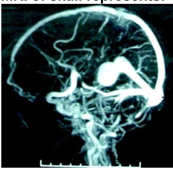
Explanation: ***MRI with DWI*** - **Diffusion-weighted imaging (DWI)** within an MRI scan is highly sensitive in detecting **cytotoxic edema** within minutes of **ischemic stroke** onset. This makes it crucial for early diagnosis and treatment decisions. - DWI can identify areas of restricted water diffusion, which is a hallmark of acute cellular injury due to **ischemia**, even before changes are visible on conventional T1 or T2-weighted MRI sequences. *CT* - While frequently used in acute stroke settings, **non-contrast CT** is primarily used to **rule out hemorrhagic stroke** and may only show subtle or no signs of acute ischemia in the first few hours. - Early ischemic changes on CT, often referred to as the **"ischemic penumbra"**, may appear hours after stroke onset, making it less sensitive for very early detection compared to DWI. *Ultrasound* - **Transcranial Doppler (TCD) ultrasound** can evaluate blood flow velocities in intracranial arteries and detect stenoses or occlusions but is not a primary imaging modality for directly visualizing brain parenchymal ischemia. - Cervical ultrasound (e.g., **carotid duplex**) assesses extracranial vessels but cannot directly detect **ischemic changes** within the brain tissue itself. *PET scan* - **PET (Positron Emission Tomography)** can assess brain metabolism and blood flow but is typically not the preferred or most sensitive modality for **early detection of acute ischemic stroke** due to its complexity, cost, and limited availability in emergency settings. - PET is more commonly used in research or for assessing chronic conditions and **metabolic abnormalities**, rather than acute stroke diagnosis.
Explanation: ***Chronic subdural hematoma*** - A **chronic subdural hematoma** (> 3 weeks old) is typically **hypodense** (dark) on a non-contrast CT scan because the blood products have aged, broken down, and become diluted with CSF-like fluid. - Its appearance is usually **crescent-shaped**, conforming to the brain surface, and located between the dura and arachnoid mater. - This hypodense appearance distinguishes it from acute hemorrhages. *Epidural hematoma* - An **acute epidural hematoma** is typically **hyperdense** (bright) on a non-contrast CT scan due to freshly clotted blood with high protein content. - It has a characteristic **lenticular (lens-shaped)** appearance located between the skull and dura mater. - Epidural hematomas present acutely and are evacuated surgically, rarely progressing to chronic hypodense stages. *Subdural hematoma* - An **acute subdural hematoma** is typically **hyperdense** on CT with a **crescent-shaped** appearance. - As time progresses: acute (0-3 days) = hyperdense, subacute (3-21 days) = isodense, chronic (> 3 weeks) = hypodense. - Without the qualifier "chronic," this refers to acute subdural, which is hyperdense. *Subarachnoid hemorrhage* - A **subarachnoid hemorrhage** appears **hyperdense** within the subarachnoid spaces, filling sulci, cisterns, and fissures on non-contrast CT. - Blood in the subarachnoid space is typically acute and hyperdense at presentation. - SAH is a medical emergency requiring immediate intervention.
Explanation: ***MRI (Correct Answer)*** - **Magnetic Resonance Imaging (MRI)** is superior for visualizing **soft tissues** such as the spinal cord, nerves, ligaments, and intervertebral discs. - It provides excellent contrast resolution, making it the **gold standard** for detecting and characterizing **spinal cord compression**, nerve root impingement, and other soft tissue abnormalities. - MRI is non-invasive and does not use ionizing radiation, making it safer for repeated imaging when needed. *CT scan (Incorrect)* - **Computed Tomography (CT) scans** excel at imaging **bone structures** and showing **bony impingement** but are less effective for direct visualization of the spinal cord itself. - While it can indirectly show compression through bone changes, its ability to depict actual cord and nerve tissue is limited compared to MRI. *X-ray (Incorrect)* - **X-rays** primarily show **bone structures** and are useful for detecting fractures, dislocations, or degenerative changes in the vertebrae. - They provide very limited information about **soft tissues** like the spinal cord or intervertebral discs and cannot directly visualize spinal cord compression. *Bone scan (Incorrect)* - A **bone scan** is a nuclear medicine test that detects **increased metabolic activity** in bones, often used to identify infections, tumors, or fractures that may not be visible on standard X-rays. - It does not directly visualize the **spinal cord** or soft tissue abnormalities and is not used for assessing spinal cord compression.
Explanation: ***Pilocytic astrocytoma*** - This is the most common **cerebellar tumor in children** and classically presents as a **cystic lesion with an enhancing mural nodule** on imaging. - The symptoms of **headache and vomiting** are indicative of increased intracranial pressure, often caused by the tumor obstructing CSF flow in the posterior fossa. *Medulloblastoma* - While also a common pediatric cerebellar tumor, medulloblastoma typically presents as a **solid, enhancing mass** in the midline of the cerebellum, not typically cystic with a mural nodule. - It is known for its tendency to **disseminate via CSF pathways** to other parts of the brain and spine. *Glioblastoma* - This is a highly malignant primary brain tumor that is **rare in children** and typically found in the **cerebral hemispheres** in adults, not the cerebellum. - Imaging usually shows an **irregularly enhancing mass with central necrosis** and surrounding edema. *Ependymoma* - Ependymomas can occur in the posterior fossa in children but typically arise from the **floor of the fourth ventricle** and often show heterogeneous enhancement, sometimes with calcifications. - They are less commonly characterized by the classic **cystic lesion with an enhancing mural nodule** seen in pilocytic astrocytomas.
Explanation: ***Multiple periventricular white matter lesions*** - **Multiple sclerosis (MS)** is characterized by **demyelination** in the central nervous system, which appears as bright lesions in the **periventricular white matter** on T2-weighted MRI. - The presence of multiple lesions disseminated in space and time is a key diagnostic criterion for MS, reflecting distinct episodes of demyelination. *Bilateral acoustic neuromas* - **Bilateral acoustic neuromas** are a hallmark finding of **Neurofibromatosis type 2 (NF2)**, a genetic disorder, not multiple sclerosis. - These tumors arise from the vestibulocochlear nerves and typically cause hearing loss and balance issues. *Hypointensities in the basal ganglia* - **Hypointensities in the basal ganglia** are often associated with conditions involving **iron deposition** or calcification, such as **Parkinson's disease** at later stages or metabolic disorders. - This finding is not characteristic of demyelinating lesions seen in multiple sclerosis. *Single enhancing brainstem lesion* - A **single enhancing lesion** in the brainstem can be seen in various conditions, including tumors, infections, or inflammatory processes like **acute disseminated encephalomyelitis (ADEM)**. - While MS can cause brainstem lesions, the finding of only a single enhancing lesion would not be typical for confirmed MS, which usually presents with multiple lesions.
Explanation: ***CT angiography*** - A CT scan showing **hyperdensity in the subarachnoid spaces** confirms a **subarachnoid hemorrhage (SAH)**. - **CT angiography** is the most appropriate next step to rapidly identify the source of bleeding, typically an **aneurysm**, crucial for guiding immediate management. *MRI* - While MRI can provide detailed images, it is generally **slower to perform** and less accessible in an emergency setting for initial diagnostic evaluation of acute SAH. - It is often reserved for cases where the CT is negative but clinical suspicion of SAH remains high, or for evaluating **SAH mimics**. *Lumbar puncture* - A lumbar puncture would confirm the presence of blood or xanthochromia in the **cerebrospinal fluid (CSF)**, but it is typically performed only if the initial CT scan is negative and clinical suspicion for SAH remains high. - Given the positive CT scan showing hyperdensity indicative of SAH, a lumbar puncture is **unnecessary and delays definitive diagnosis** of the bleeding source. *Digital subtraction angiography* - **Digital subtraction angiography (DSA)** is considered the **gold standard** for detecting intracranial aneurysms. - However, it is an **invasive procedure** and takes longer to perform than CT angiography, making it less suitable as the *immediate* next best investigation in an acute, unstable presentation where speed is critical.
Explanation: ***MRI*** - **Magnetic Resonance Imaging (MRI)** is the gold standard for diagnosing acoustic neuromas due to its superior soft tissue contrast and ability to visualize the **internal auditory canal** and **cerebellopontine angle**. - MRI with **gadolinium contrast** can detect even small acoustic neuromas, which appear as enhancing lesions. *CT scan* - While CT can show larger tumors indirectly by revealing **bone erosion** or **widening of the internal auditory canal**, it has poor soft tissue resolution for early detection of acoustic neuromas. - CT scans expose the patient to **ionizing radiation**, and its primary role is usually limited to evaluating bone structures or for patients who cannot undergo MRI. *X-ray* - **Plain X-rays** (e.g., skull films) are not effective for diagnosing acoustic neuromas as they only show bone and cannot visualize soft tissue tumors in this region. - They lack the necessary detail and sensitivity to detect such a tumor, especially in its early stages. *Ultrasound* - **Ultrasound** is not suitable for evaluating intracranial structures like acoustic neuromas because the **skull bone** obstructs the transmission of sound waves. - Its utility is primarily in visualizing superficial soft tissue structures or organs in the abdomen and neck.
Explanation: ***Low-grade astrocytoma*** - The **cyst with enhancing mural nodule** appearance in the temporal lobe of a young adult with seizures is highly characteristic of **pleomorphic xanthoastrocytoma (PXA)**, a subtype of low-grade astrocytoma (WHO Grade II). - PXA typically presents as a **superficial, cortically-based cystic mass** with a solid enhancing nodule, often with **dural involvement** ("dural tail" sign). - The patient's age (35-year-old female) and presenting symptoms of **seizures** and **headaches** are consistent with the slow-growing nature of these tumors, which often irritate cortical structures. - PXA has a **favorable prognosis** compared to other astrocytomas and commonly occurs in the **temporal lobe**. *Glioblastoma multiforme* - **Glioblastoma multiforme (GBM)** is a highly aggressive WHO Grade IV tumor that typically presents with **thick irregular ring enhancement**, **central necrosis**, and **extensive surrounding vasogenic edema**. - GBM shows rapid clinical progression and would not typically present as a well-defined cyst with mural nodule. - The imaging appearance described is more consistent with a low-grade tumor. *Meningioma* - **Meningiomas** are **extra-axial (dural-based) tumors** that demonstrate **uniform, homogeneous enhancement** with a characteristic **dural tail** sign. - They are typically **solid masses** (not cystic) that compress rather than infiltrate brain parenchyma. - The described cystic lesion with mural nodule is more consistent with an intra-axial tumor like PXA. *Oligodendroglioma* - **Oligodendrogliomas** characteristically show **calcifications** (present in 70-90% of cases) and are commonly located in the **frontal lobes**. - They typically appear as **heterogeneous, infiltrative masses** rather than well-defined cystic lesions with mural nodules. - While they can cause seizures, the imaging appearance described is not typical for oligodendroglioma.
Explanation: ***Excellent for detecting pathology*** - T2-weighted imaging highlights areas with **increased water content**, which is characteristic of many pathological processes like **inflammation, edema, and tumors**. - Pathological tissues often appear **bright** (hyperintense) on T2-weighted images due to their higher water content, making them distinguishable from normal tissues. *Useful for both pathology and anatomy* - While T2-weighted imaging is excellent for pathology, its primary strength lies in its **sensitivity to water**, which makes it less ideal for detailed anatomical bone or fatty tissue differentiation compared to T1-weighted images. - T1-weighted images are generally superior for visualizing **normal anatomy** due to good contrast between fat (bright) and water (dark). *Not useful for either* - This statement is incorrect as T2-weighted imaging is a fundamental sequence in MRI, providing crucial information about **fluid and pathology**. - It plays a vital role in the diagnosis and characterization of a wide range of diseases. *Good for visualizing normal anatomy* - T2-weighted imaging is less optimal for visualizing normal anatomy compared to T1-weighted imaging due to the **bright signal from free water** (e.g., CSF), which can obscure fine anatomical details. - While some anatomical structures are visible, its main utility is in **pathological detection** rather than fine anatomical delineation.
Explanation: ***Hypointense on T1 weighted image and hyperintense on T2 weighted image*** - **CSF** appears **dark (hypointense)** on **T1-weighted MRI** due to its long T1 relaxation time, which means it returns to its equilibrium magnetization slowly. - On **T2-weighted MRI**, CSF appears **bright (hyperintense)** because of its prolonged T2 relaxation time, indicating that it retains its transverse magnetization longer. *Hyperintense on T1 weighted image and hypointense on T2 weighted image* - This description typically applies to **fat** (hyperintense on T1) and may describe areas with short T2 relaxation times, which is not characteristic of CSF. - If CSF were hyperintense on T1, it would indicate a very short T1 relaxation time, which is contrary to its fluid properties. *Hyperintense on both T1 and T2 weighted images* - Substances that appear **bright on both T1 and T2** are generally those with high lipid content or certain types of hemorrhagic products (e.g., **subacute hemorrhage**). - This appearance is inconsistent with the behavior of cerebrospinal fluid. *Hypointense on both T1 and T2 weighted images* - Material that is **dark on both T1 and T2** typically includes **cortical bone**, **air**, or areas of **calcification**, which have very little free water or fast signal decay. - CSF, being primarily water, shows a distinct signal behavior on T1 and T2 images rather than appearing dark on both.
Neuroanatomy for Radiologists
Practice Questions
Cerebrovascular Diseases
Practice Questions
Intracranial Tumors
Practice Questions
CNS Infections
Practice Questions
Demyelinating and Degenerative Diseases
Practice Questions
Head Trauma Imaging
Practice Questions
Spine Imaging: Trauma and Degenerative Disease
Practice Questions
Spine Tumors and Infections
Practice Questions
Pediatric Neuroradiology
Practice Questions
Congenital CNS Anomalies
Practice Questions
Functional Neuroimaging
Practice Questions
Neurointerventional Procedures
Practice Questions
Get full access to all questions, explanations, and performance tracking.
Start For Free