
Neuroradiology — MCQs

On this page
Which of the following conditions typically presents with spinal cord edema on MRI?
What is the imaging modality of choice for determining the etiology of subarachnoid hemorrhage?
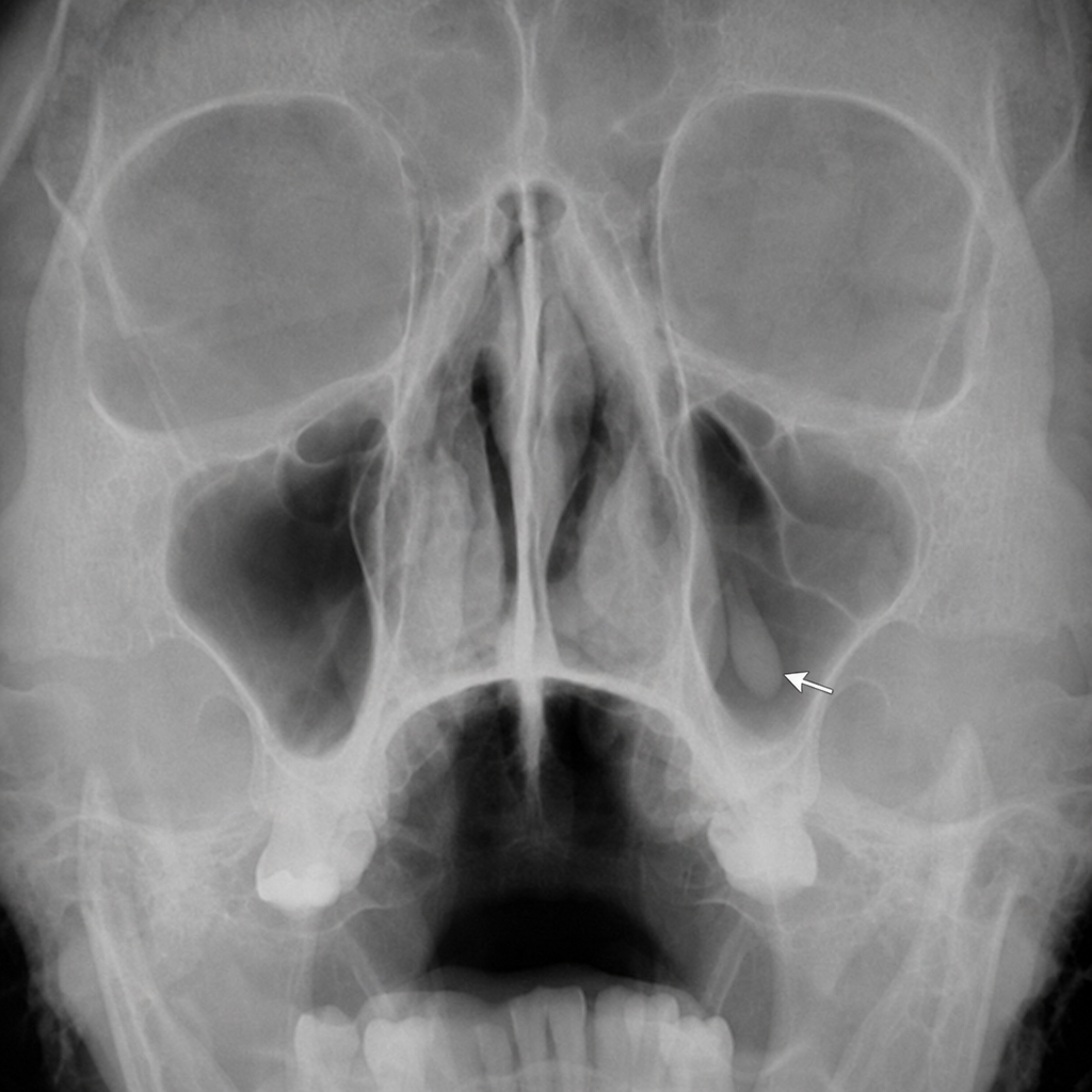
Tear-drop appearance on a PNS x-ray is seen in?
Absent swallow tail sign is seen in which of the following conditions?
Which of the following tumors shows a radiological sign called "pneumosinus dilatans"?
Which condition is associated with a 'wine glass' appearance on T2-weighted MRI of the brain?
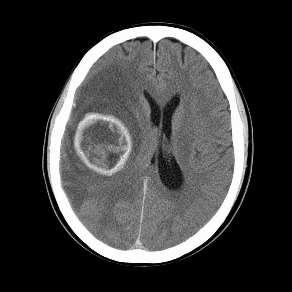
A 63-year-old man complains of worsening headache and right-sided weakness. Based on the CT head shown, the most likely diagnosis is:
Widened neural foramina are frequently seen in:
Practice by Chapter
Neuroanatomy for Radiologists
Practice Questions
Cerebrovascular Diseases
Practice Questions
Intracranial Tumors
Practice Questions
CNS Infections
Practice Questions
Demyelinating and Degenerative Diseases
Practice Questions
Head Trauma Imaging
Practice Questions
Spine Imaging: Trauma and Degenerative Disease
Practice Questions
Spine Tumors and Infections
Practice Questions
Pediatric Neuroradiology
Practice Questions
Congenital CNS Anomalies
Practice Questions
Functional Neuroimaging
Practice Questions
Neurointerventional Procedures
Practice Questions
Want unlimited practice?
Get full access to all questions, explanations, and performance tracking.
Scan to download app