Which of the following is NOT a typical radiological finding in meningioma?
Lytic "punched-out" or "beaten-metal" lesions are seen in a skull X-ray, the most likely cause is:
CT findings in Epidural Hematoma (EDH) are typically described as?
Upstream and downstream hydrocephalus is seen in which of the following conditions?
A 35-year-old female presented with a history of multiple transient ischemic attacks. What is the most likely diagnosis suggested by the given carotid angiogram?

What are the characteristic imaging findings in Alzheimer's disease?
What condition presents with a hyperdense biconvex appearance on a cranial CT scan?
Which of the following conditions is characterized by bracket calcification?
What are the characteristic features of tubercular meningitis on MR imaging?
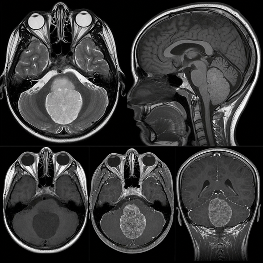
A 10-year-old male patient presents with a 20-day history of headache and vomiting. Examination reveals an ataxic gait. NCCT shows a fourth ventricular mass causing obstructive hydrocephalus. CE-MRI is performed to characterize the lesion. What is the most probable diagnosis?
Explanation: ### Explanation Meningiomas are the most common extra-axial, typically benign intracranial tumors arising from the arachnoid cap cells. The correct answer is **None of the above** because all three features listed (Calcification, Hyperostosis, and Vascularity) are classic, high-yield radiological hallmarks of meningiomas. **Analysis of Options:** * **Calcification (Option A):** Seen in approximately 20–25% of cases. On CT, meningiomas often appear hyperdense due to psammomatous calcification (sand-like microscopic calcifications). * **Hyperostosis (Option B):** This is a characteristic reactive thickening or bony outgrowth of the overlying calvarium. It is a crucial diagnostic clue that helps differentiate meningioma from other dural-based lesions. * **Vascularity (Option C):** Meningiomas are highly vascular tumors. On angiography, they typically show a "sunburst" or "spoked-wheel" appearance of vessels, often supplied by the external carotid artery (e.g., middle meningeal artery). They also show intense, homogenous enhancement on contrast-enhanced CT and MRI. **High-Yield Clinical Pearls for NEET-PG:** * **Dural Tail Sign:** A classic MRI finding representing reactive thickening of the adjacent dura (seen in ~60-70% of cases). * **CSF Cleft Sign:** A rim of CSF between the tumor and the brain parenchyma, confirming its **extra-axial** location. * **Location:** Most commonly found along the parasagittal/falcine region. * **Associations:** Multiple meningiomas are strongly associated with **Neurofibromatosis Type 2 (NF2)**. * **Histology:** Look for **Psammoma bodies** (concentric lamellated calcifications).
Explanation: **Explanation:** The correct answer is **Eosinophilic Granuloma (EG)**, the most common and localized form of Langerhans Cell Histiocytosis (LCH). **Why Eosinophilic Granuloma is correct:** In EG, the proliferation of Langerhans cells leads to focal bone destruction. On a skull X-ray, this typically manifests as **"punched-out" lytic lesions** that lack a sclerotic margin. A classic radiological feature is the **"beveled edge" appearance**, caused by unequal involvement of the inner and outer tables of the skull. When multiple such lesions coalesce, they create a map-like appearance known as a **"geographic skull."** **Analysis of Incorrect Options:** * **Multiple Myeloma:** While it also presents with "punched-out" lesions, they are typically smaller, uniform in size ("raindrop skull"), and occur in older adults. EG is more common in children and young adults. * **Metastasis:** Osteolytic metastases (e.g., from breast or lung cancer) usually have ill-defined, moth-eaten borders rather than the sharp, "punched-out" margins seen in EG. * **Osteosarcoma:** This is primarily a bone-forming (osteoblastic) tumor. It typically presents with a "sunburst" periosteal reaction or Codman’s triangle, rather than simple lytic lesions. **High-Yield Clinical Pearls for NEET-PG:** * **Beveled Edge Sign:** Pathognomonic for Eosinophilic Granuloma. * **Button Sequestrum:** A central piece of residual bone within a lytic lesion, also seen in EG (and osteomyelitis). * **Hand-Schüller-Christian Disease:** A triad of LCH consisting of exophthalmos, diabetes insipidus, and lytic bone lesions. * **Letterer-Siwe Disease:** The acute, multisystem, and often fatal form of LCH seen in infants.
Explanation: **Explanation:** **1. Why "Hyperdense lesion" is correct:** In the acute phase, an Epidural Hematoma (EDH) consists of fresh, clotted blood. On a Non-Contrast Computed Tomography (NCCT) scan, acute blood has high attenuation (typically 60–80 Hounsfield Units) due to the high concentration of hemoglobin. Therefore, it appears as a **hyperdense (bright white)** area. This is the primary radiological description of the density of the lesion. **2. Why the other options are incorrect:** * **Crescent-shaped collection:** This describes a **Subdural Hematoma (SDH)**. SDHs are not restricted by dural attachments and spread along the brain surface, following its contour. * **Concavo-convex collection:** This is a geometric misnomer in this context. An EDH is classically described as **Biconvex or Lentiform (lens-shaped)**. It is convex on both sides because the blood is under high pressure (usually arterial) and is limited by the cranial sutures where the dura is firmly attached to the skull. * **None of the above:** Incorrect, as hyperdensity is a hallmark feature of acute intracranial hemorrhage. **3. Clinical Pearls for NEET-PG:** * **Source of Bleed:** Most commonly the **Middle Meningeal Artery** (associated with temporal bone fractures at the Pterion). * **Clinical Presentation:** Classically associated with a **"Lucid Interval"** (initial loss of consciousness followed by a period of recovery before rapid deterioration). * **Anatomical Boundary:** EDH **does not cross cranial sutures** (because the dura is attached there) but **can cross the midline** (dural reflections like the falx). * **Swirl Sign:** The presence of hypodense areas within the hyperdense clot indicates active, hyperacute bleeding (unclotted blood).
Explanation: **Explanation:** The correct answer is **Choroid plexus papilloma (CPP)**. **Why it is correct:** Hydrocephalus is typically classified as obstructive (non-communicating) or non-obstructive (communicating). However, Choroid Plexus Papilloma is unique because it causes hydrocephalus through **three distinct mechanisms**, leading to both upstream and downstream dilatation: 1. **Overproduction of CSF:** CPP is a functional tumor that secretes CSF at a rate exceeding the absorption capacity of the arachnoid villi. This leads to generalized (downstream) ventricular enlargement. 2. **Direct Obstruction:** The physical mass of the tumor (commonly in the lateral ventricles in children or the 4th ventricle in adults) blocks CSF flow, causing "upstream" dilatation of the ventricles behind the lesion. 3. **Recurrent Hemorrhage:** These tumors are prone to minor bleeds, leading to arachnoiditis and impaired CSF resorption (communicating hydrocephalus). **Why the other options are incorrect:** * **Ependymoma:** Typically causes **obstructive (upstream)** hydrocephalus by blocking the 4th ventricle. It does not cause CSF overproduction. * **Pineal Germinoma:** Causes **obstructive (upstream)** hydrocephalus by compressing the Aqueduct of Sylvius (Parinaud syndrome is a common clinical association). * **Meningioma:** Usually causes focal mass effect or edema; it does not typically cause generalized hydrocephalus unless it obstructs a major venous sinus or a specific CSF pathway (e.g., Foramen of Monro). **High-Yield Clinical Pearls for NEET-PG:** * **Most common site for CPP:** Lateral ventricle (Atrium) in children; 4th ventricle in adults. * **Imaging Hallmark:** A cauliflower-like, intensely enhancing intraventricular mass on CT/MRI. * **Choroid Plexus Carcinoma:** Suggested by parenchymal invasion and vasogenic edema (rare in benign CPP). * **Key Concept:** CPP is the only brain tumor where hydrocephalus persists even if the CSF pathway is surgically bypassed, due to the ongoing overproduction of fluid.
Explanation: ***Fibromuscular dysplasia*** - Classic **"string of beads"** appearance on angiography in a **young female** with TIAs is pathognomonic for fibromuscular dysplasia. - This **non-atherosclerotic** vascular disease commonly affects the **renal** and **carotid arteries** in women aged 20-60 years. *Atherosclerosis* - Typically presents as **smooth, eccentric plaques** rather than the characteristic beaded appearance. - More common in **older patients** with traditional cardiovascular risk factors like smoking, diabetes, and hypertension. *Thrombosis* - Would appear as an **abrupt cutoff** or **filling defect** within the vessel lumen on angiography. - Usually associated with **acute onset** symptoms rather than recurrent transient episodes. *Carotid dissection* - Angiographic findings include **tapering stenosis** with a characteristic **flame-shaped** narrowing or **double lumen** sign. - Often presents with **sudden severe headache** or neck pain along with neurological symptoms.
Explanation: **Explanation:** Alzheimer’s Disease (AD) is the most common cause of dementia, characterized pathologically by amyloid plaques and neurofibrillary tangles. On neuroimaging (MRI), the hallmark finding is **disproportionate atrophy of the medial temporal lobes** (specifically the hippocampus, entorhinal cortex, and amygdala) and the **parietal lobes** (specifically the precuneus and posterior cingulate gyrus). 1. **Why Option A is correct:** The earliest structural changes occur in the hippocampus (temporal lobe), leading to memory deficits. As the disease progresses, atrophy spreads to the temporoparietal association cortex. This pattern results in the characteristic widening of the choroid fissure and temporal horns of the lateral ventricles. 2. **Why other options are wrong:** * **Occipital lobe (B & D):** The primary visual cortex in the occipital lobe is typically spared in AD. Occipital involvement is more characteristic of Posterior Cortical Atrophy (a variant) or Lewy Body Dementia. * **Frontal lobe (C):** While frontal atrophy can occur in late-stage AD, primary frontal and temporal pole atrophy is the hallmark of **Frontotemporal Dementia (FTD)**, not AD. **High-Yield Clinical Pearls for NEET-PG:** * **MTA Score (Scheltens Scale):** Used to grade Medial Temporal lobe Atrophy on coronal T1 MRI. * **FDG-PET Finding:** Characterized by **hypometabolism** in the temporoparietal regions and posterior cingulate gyrus. * **Amyloid PET:** Uses tracers like Pittsburgh Compound B (PiB) to visualize amyloid deposition. * **Key Sign:** "Ex-vacuo" ventriculomegaly due to significant cortical loss.
Explanation: **Explanation:** **Extradural Hemorrhage (EDH)** is the correct answer because it occurs in the potential space between the skull and the dura mater. Since the dura is firmly attached to the cranial sutures, the expanding hematoma cannot cross suture lines. This anatomical restriction forces the blood to expand inward toward the brain, resulting in a characteristic **biconvex (lentiform or lemon-shaped)**, hyperdense appearance on a non-contrast CT (NCCT). **Analysis of Incorrect Options:** * **Subdural Hemorrhage (SDH):** Occurs between the dura and arachnoid mater. Because this space is not limited by sutures, the blood spreads widely over the hemisphere, appearing **crescent-shaped (concavo-convex)**. * **Foreign Body:** While often hyperdense (e.g., metal or glass), foreign bodies have distinct shapes related to the object itself and do not typically conform to a biconvex anatomical plane. * **Intracerebral Hemorrhage (ICH):** This is bleeding within the brain parenchyma. It appears as a hyperdense area surrounded by edema but lacks the specific biconvex peripheral configuration. **High-Yield Clinical Pearls for NEET-PG:** * **Vessel Involved:** Most commonly the **Middle Meningeal Artery** (due to fracture at the Pterion). * **Clinical Presentation:** Classically associated with a **"Lucid Interval"** (a period of temporary improvement before rapid deterioration). * **Suture Lines:** EDH **does not** cross sutures but **can** cross the midline (dural attachments). Conversely, SDH **can** cross sutures but **cannot** cross the midline. * **Management:** Urgent surgical evacuation (burr hole or craniotomy) is required if the volume is significant, as it can lead to transtentorial herniation.
Explanation: **Explanation:** **Corpus Callosal Lipoma** is the correct answer. This is a rare congenital lesion resulting from the persistence of the primitive meningeal tissue. On a frontal skull radiograph or a coronal CT scan, it classically presents with **"bracket calcification."** This occurs because the lipoma is often associated with dysgenesis of the corpus callosum, and curvilinear calcifications form along its peripheral margins, resembling parentheses or brackets surrounding the fatty mass. **Analysis of Incorrect Options:** * **Sturge-Weber Syndrome:** Characterized by **"tram-track"** or "railroad track" calcifications. These are cortical and subcortical gyriform calcifications, typically in the occipital or parietal lobes, due to underlying leptomeningeal angiomatosis. * **Cytomegalovirus (CMV) Infection:** Characterized by **periventricular calcifications**. CMV targets the germinal matrix near the ventricles, unlike Toxoplasmosis, which typically causes scattered intracranial calcifications. * **Paragonimus Cyst:** Characterized by **"soap-bubble"** calcifications. This parasitic infection (lung fluke) can migrate to the brain, leading to clustered, ring-like calcified lesions. **High-Yield Pearls for NEET-PG:** * **Lipoma Imaging:** On CT, it shows fat density (negative Hounsfield units, -50 to -100). On MRI, it is hyperintense on T1 and T2, dropping signal on fat-suppression sequences. * **Associated Findings:** Over 50% of callosal lipomas are associated with **corpus callosal agenesis/dysgenesis**. * **Location:** The pericallosal region is the most common site for intracranial lipomas. * **Other Calcification Patterns:** * *Toxoplasmosis:* Scattered/Diffuse. * *Tuberous Sclerosis:* Subependymal nodules ("candle guttering"). * *Oligodendroglioma:* Chunky, intratumoral calcification.
Explanation: **Explanation:** Tubercular Meningitis (TBM) is the most common form of neurotuberculosis and presents with a classic triad of radiological findings on MRI. 1. **Exudates in Basal Cisterns (Option A):** This is the hallmark of TBM. Thick, gelatinous inflammatory exudates accumulate preferentially in the **basal cisterns** (suprasellar, perimesencephalic, and sylvian fissures). On post-contrast T1-weighted images, these appear as intense, shaggy enhancement. 2. **Leptomeningeal Enhancement (Option B):** While the basal involvement is characteristic, TBM also causes diffuse enhancement of the pia and arachnoid mater over the cerebral convexities and within the sulci. 3. **Hydrocephalus (Option C):** This is the most common complication of TBM. It is usually **communicating** type (due to exudates obstructing the basal cisterns and arachnoid villi) but can be non-communicating if the Aqueduct of Sylvius is blocked. Since all three features are characteristic of the disease process, **Option D** is the correct answer. **High-Yield Clinical Pearls for NEET-PG:** * **Vasculitis & Infarcts:** TBM often involves the "Medial Striate" and "Thalamoperforating" arteries, leading to infarcts in the **Basal Ganglia** and **Internal Capsule** (the "Tubercular Zone"). * **Tuberculomas:** These may coexist and show "ring enhancement" with a central area of necrosis. * **MR Spectroscopy:** TBM exudates and tuberculomas typically show a prominent **Lipid-Lactate peak** at 1.3 ppm, which is highly suggestive of mycobacterial infection. * **Complication:** Spinal arachnoiditis is a common sequela, often visualized as "clumping" of the cauda equina nerve roots.
Explanation: ### Explanation The clinical presentation of a posterior fossa mass in a child, specifically located within the **fourth ventricle**, points toward two primary differentials: Ependymoma and Medulloblastoma. **Why Ependymoma is the correct answer:** Ependymomas typically arise from the floor of the fourth ventricle. A key diagnostic feature on imaging is their "plastic" nature—they tend to squeeze through the foramina of Luschka and Magendie into the cerebellopontine angle or the cisterna magna (the **"toothpaste sign"**). On NCCT, they often show **coarse calcifications** (seen in ~50% of cases), which is a significant differentiator from other posterior fossa tumors. **Analysis of Incorrect Options:** * **Medulloblastoma:** While also a fourth ventricular mass, it typically arises from the **roof** (vermis) and is highly cellular, appearing hyperdense on NCCT. It usually expands the ventricle rather than tracking through the foramina. * **Pilocytic Astrocytoma:** This is the most common pediatric cerebellar tumor, but it usually presents as a **cystic lesion with a bright enhancing mural nodule** in the cerebellar hemisphere, not primarily within the fourth ventricle. * **Hemangioblastoma:** These are rare in children (mostly seen in adults or associated with Von Hippel-Lindau syndrome). They appear as highly vascular cystic lesions with an enhancing nodule, similar to astrocytomas but in an older age group. **NEET-PG Clinical Pearls:** * **Location:** Medulloblastoma = Roof of 4th ventricle; Ependymoma = Floor of 4th ventricle. * **Calcification:** Most common in Ependymoma among posterior fossa tumors. * **Imaging Sign:** "Plastic Ependymoma" or "Toothpaste Sign" is pathognomonic for its extension through foramina. * **Hydrocephalus:** Both Medulloblastoma and Ependymoma cause obstructive hydrocephalus due to their midline location.
Neuroanatomy for Radiologists
Practice Questions
Cerebrovascular Diseases
Practice Questions
Intracranial Tumors
Practice Questions
CNS Infections
Practice Questions
Demyelinating and Degenerative Diseases
Practice Questions
Head Trauma Imaging
Practice Questions
Spine Imaging: Trauma and Degenerative Disease
Practice Questions
Spine Tumors and Infections
Practice Questions
Pediatric Neuroradiology
Practice Questions
Congenital CNS Anomalies
Practice Questions
Functional Neuroimaging
Practice Questions
Neurointerventional Procedures
Practice Questions
Get full access to all questions, explanations, and performance tracking.
Start For Free