
Neuroradiology — MCQs

On this page
In which of the following diseases is the NAA peak seen?
On MRI, which of the following shows diffusion restriction?
Which of the following conditions is known to cause intracranial calcification visible on a skull X-ray?
Which primary brain parenchymal tumor is most commonly associated with calcifications seen on imaging studies?
Tram track appearance on a CT scan of the head is seen in which condition?
Primary investigation of choice in a patient with suspected subarachnoid hemorrhage should be:
Which of the following conditions is most commonly associated with the "doughnut" sign seen on a brain scan?
A patient with suspected subarachnoid haemorrhage presents with blood isolated in the fourth ventricle on a CT scan. Aneurysmal rupture is likely to have resulted from which of the following?
A middle-aged person is rushed to the emergency department with a history of loss of motor power in the left upper and lower limb since the last 30 minutes. The imaging modality of choice to plan appropriate treatment would be
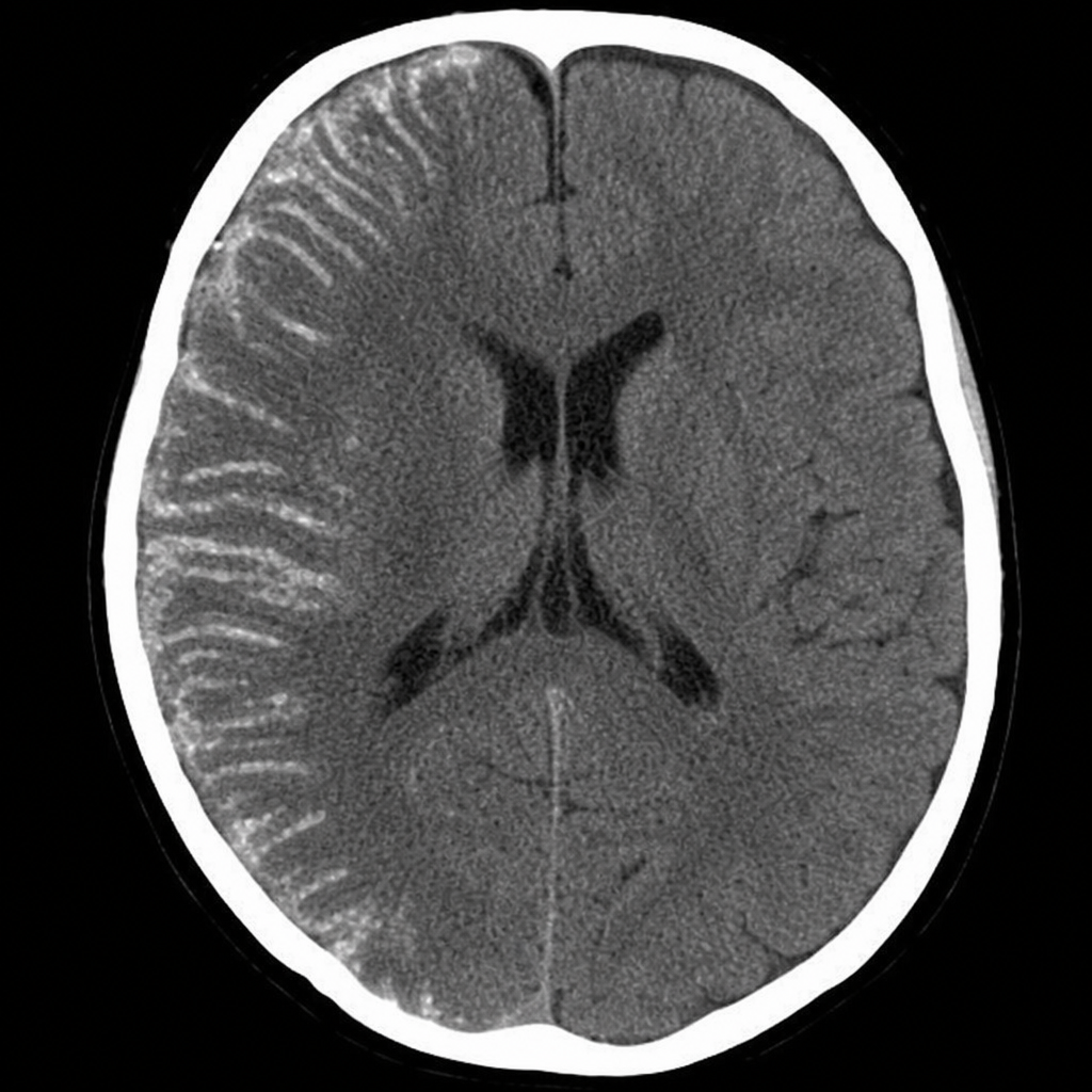
CT scan of a child with intellectual disability who has recurrent seizures and a facial hemangioma. What is the diagnosis?

Practice by Chapter
Neuroanatomy for Radiologists
Practice Questions
Cerebrovascular Diseases
Practice Questions
Intracranial Tumors
Practice Questions
CNS Infections
Practice Questions
Demyelinating and Degenerative Diseases
Practice Questions
Head Trauma Imaging
Practice Questions
Spine Imaging: Trauma and Degenerative Disease
Practice Questions
Spine Tumors and Infections
Practice Questions
Pediatric Neuroradiology
Practice Questions
Congenital CNS Anomalies
Practice Questions
Functional Neuroimaging
Practice Questions
Neurointerventional Procedures
Practice Questions
Want unlimited practice?
Get full access to all questions, explanations, and performance tracking.
Scan to download app