
Neuroradiology — MCQs

On this page
Which imaging modality is most appropriate for evaluating a suspected acoustic neuroma?
A 35-year-old female presents with a history of seizures and headaches. An MRI shows a well-defined cystic lesion with an enhancing mural nodule in the temporal lobe. What is the most likely diagnosis?
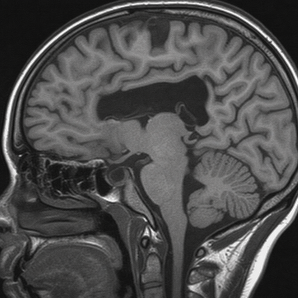
Identify the condition based on the provided image.
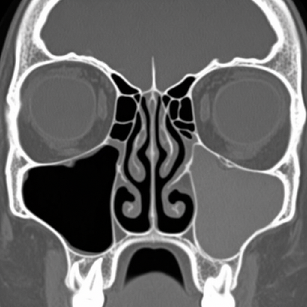
What is the most likely finding in the CT image of the left maxillary sinus in a patient with a history of allergic rhinitis?
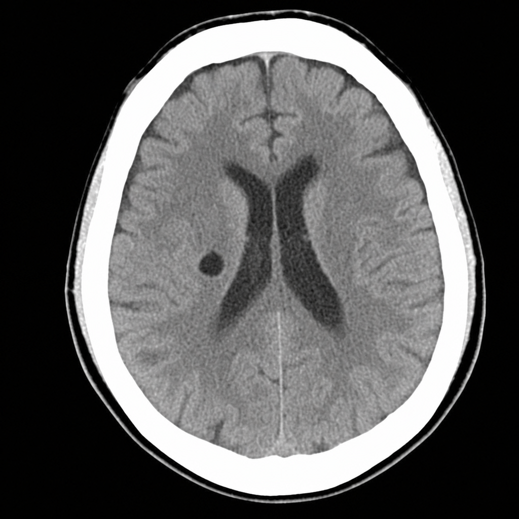
Identify the condition in the image below?
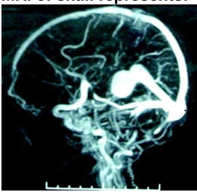
What condition is shown in the MR angiogram of the skull?
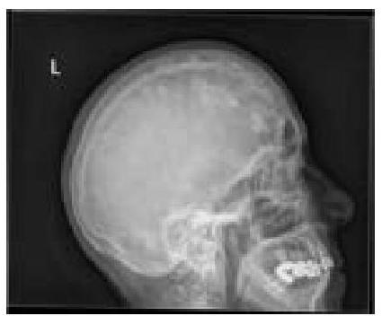
What type of lesions in the skull calvarium can be identified on this X-ray?
How does cerebrospinal fluid (CSF) appear on T1 and T2 weighted MRI images?
Stenver's view is used for -
Which of the following provides excellent details about the chemodectomas?
Practice by Chapter
Neuroanatomy for Radiologists
Practice Questions
Cerebrovascular Diseases
Practice Questions
Intracranial Tumors
Practice Questions
CNS Infections
Practice Questions
Demyelinating and Degenerative Diseases
Practice Questions
Head Trauma Imaging
Practice Questions
Spine Imaging: Trauma and Degenerative Disease
Practice Questions
Spine Tumors and Infections
Practice Questions
Pediatric Neuroradiology
Practice Questions
Congenital CNS Anomalies
Practice Questions
Functional Neuroimaging
Practice Questions
Neurointerventional Procedures
Practice Questions
Want unlimited practice?
Get full access to all questions, explanations, and performance tracking.
Scan to download app