
Neuroradiology — MCQs

On this page
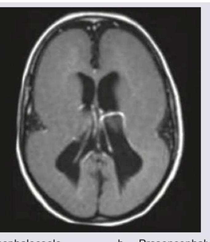
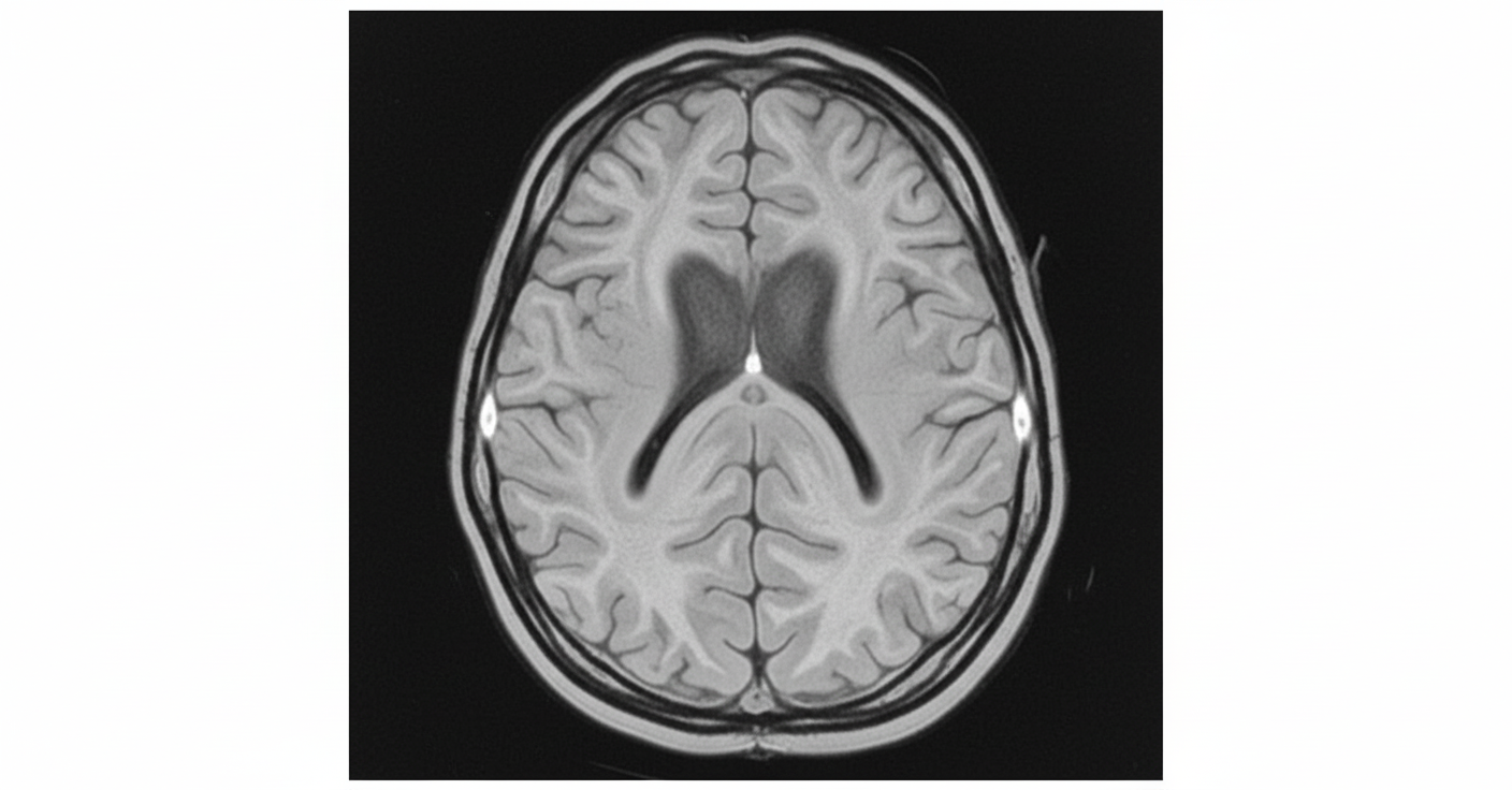
Identify the following abnormality?
What is the most likely diagnosis in an imaging study showing a large posterior fossa?
The MRI image shows a smooth brain surface with absence of normal gyri and sulci. What is the diagnosis?
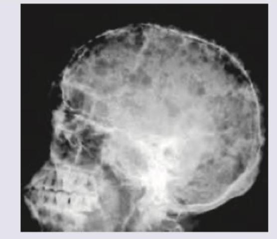
X-ray skull shows: (Recent NEET Pattern 2016-17)
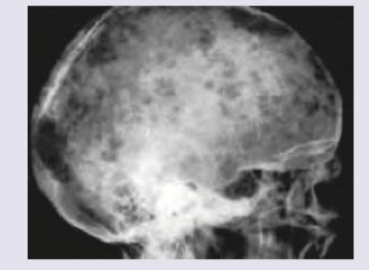
What is shown in this skull X-ray?
What is the probable diagnosis for shown below the image?

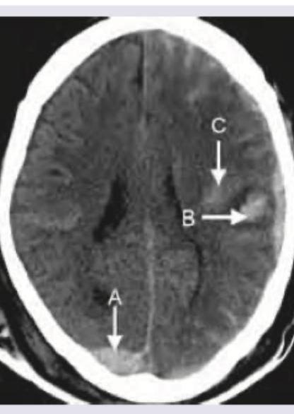
Which is correct about the intracranial bleeding shown below?
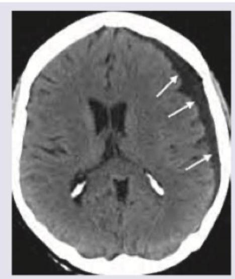
A 55-year-old diabetic patient develops sudden onset hemiparesis and facial asymmetry. NCCT scan shows:
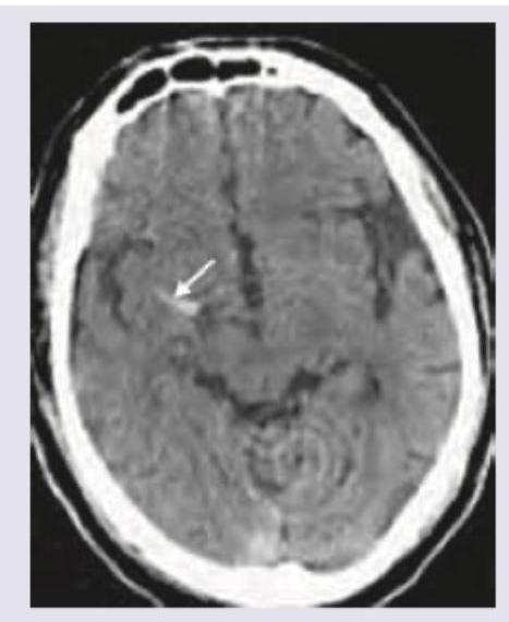
What is the correct diagnosis based on the image shown below?
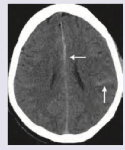
Which of the following is correct about the image shown below?
Practice by Chapter
Neuroanatomy for Radiologists
Practice Questions
Cerebrovascular Diseases
Practice Questions
Intracranial Tumors
Practice Questions
CNS Infections
Practice Questions
Demyelinating and Degenerative Diseases
Practice Questions
Head Trauma Imaging
Practice Questions
Spine Imaging: Trauma and Degenerative Disease
Practice Questions
Spine Tumors and Infections
Practice Questions
Pediatric Neuroradiology
Practice Questions
Congenital CNS Anomalies
Practice Questions
Functional Neuroimaging
Practice Questions
Neurointerventional Procedures
Practice Questions
Want unlimited practice?
Get full access to all questions, explanations, and performance tracking.
Scan to download app