
Neuroradiology — MCQs

On this page
A 30-year-old female patient involved in a road traffic accident had nausea and vomiting. She was brought unconscious to the emergency room. The NCCT brain reveals the following findings. What is the most likely diagnosis? 
A child presents with seizures. Contrast-enhanced CT reveals a cystic lesion with a dot sign. What is the most likely diagnosis?
Eye of the tiger appearance on MRI is associated with:
A 30-year-old male patient presents to OPD with complaints of recurrent headache and nausea, MRI of brain shown below. What is the diagnosis?
Which of the following is the investigation of choice for spinal TB?
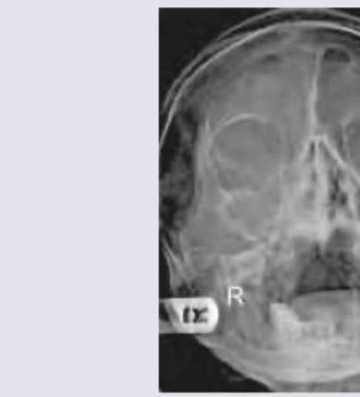
The following X-ray shows:
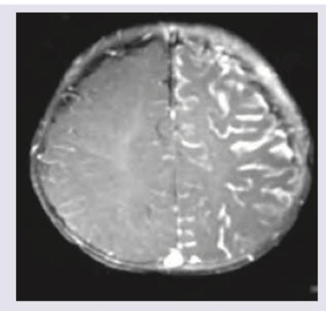
The images show the presence of:
A child with intellectual disability and glaucoma was subjected to NCCT. CT scan findings are?
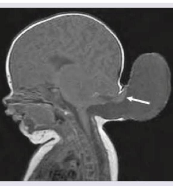
The image shows presence of:
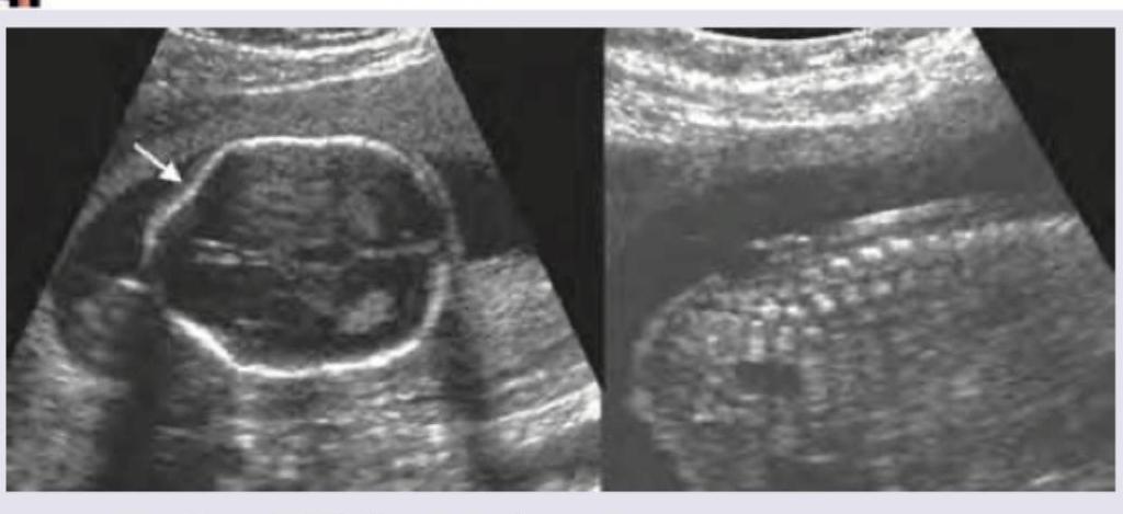
Keyhole sign on fetal ultrasound is seen in:
Practice by Chapter
Neuroanatomy for Radiologists
Practice Questions
Cerebrovascular Diseases
Practice Questions
Intracranial Tumors
Practice Questions
CNS Infections
Practice Questions
Demyelinating and Degenerative Diseases
Practice Questions
Head Trauma Imaging
Practice Questions
Spine Imaging: Trauma and Degenerative Disease
Practice Questions
Spine Tumors and Infections
Practice Questions
Pediatric Neuroradiology
Practice Questions
Congenital CNS Anomalies
Practice Questions
Functional Neuroimaging
Practice Questions
Neurointerventional Procedures
Practice Questions
Want unlimited practice?
Get full access to all questions, explanations, and performance tracking.
Scan to download app