In subarachnoid hemorrhage, where is blood most commonly collected in the skull?
CT scan of the head should be performed before lumbar puncture in all of the following conditions except?
What kind of foreign bodies can be detected by MRI?
A 28-year-old man presented with headache. A CT head shows a low attenuation mass in the left temporoparietal region which has a similar density to CSF and shows no enhancement following contrast administration. Diffusion-weighted MRI shows high signal. Which of the following is the most probable diagnosis?
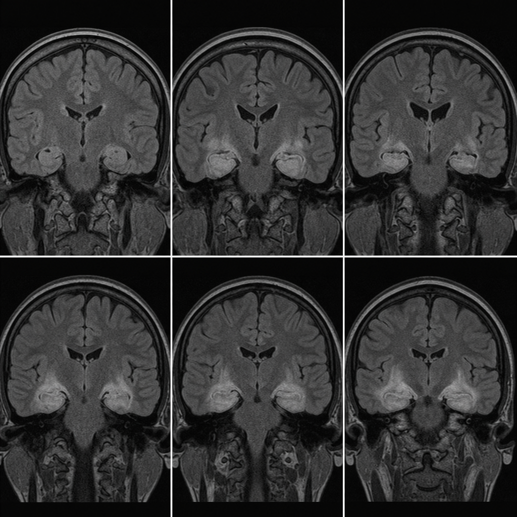
Drop metastases are seen in which of the following tumors?
In idiopathic basal ganglia calcification, which structure is most often calcified?
Characteristic 'tram-line' calcifications in skull radiographs are observed in which of the following conditions?
Basal ganglia calcification is seen in all of the following conditions except:
Kernohan-Woltman sign (notch) is seen in which type of brain herniation?
What is the investigation of choice for SCIWORA?
Explanation: **Explanation:** **Subarachnoid Hemorrhage (SAH)** refers to the presence of blood within the subarachnoid space—the area between the arachnoid mater and the pia mater. This space normally contains cerebrospinal fluid (CSF) and major cerebral blood vessels. **Why Basal Cisterns are the correct answer:** The most common cause of non-traumatic SAH is the rupture of a **saccular (berry) aneurysm**. These aneurysms typically occur at the bifurcations of the arteries forming the **Circle of Willis**, which is anatomically located within the **basal cisterns** (large expansions of the subarachnoid space at the base of the brain). Consequently, when an aneurysm ruptures, blood immediately accumulates in these cisterns (e.g., suprasellar, interpeduncular, and sylvian cisterns). On a non-contrast CT (NCCT), this appears as hyperdense (white) signals replacing the normally dark CSF. **Analysis of Incorrect Options:** * **Around falx cerebri:** While blood can track into the longitudinal fissure, isolated blood here is more characteristic of a subdural hematoma (SDH) or traumatic SAH, rather than the classic presentation of a ruptured aneurysm. * **Near cerebellum:** Blood in the posterior fossa cisterns occurs but is less common as a primary site unless the aneurysm is on the vertebral or basilar arteries. * **Tentorium:** Blood along the tentorium is a classic sign of a subdural hematoma (SDH) tracking along the dural folds. **High-Yield Clinical Pearls for NEET-PG:** * **Gold Standard Investigation:** Non-contrast CT (NCCT) Head is the initial investigation of choice (95-98% sensitive in the first 24 hours). * **Lumbar Puncture:** If CT is negative but clinical suspicion is high, look for **xanthochromia** (yellowish CSF due to bilirubin). * **Classic Sign:** "Star-shaped" hyperdensity in the basal cisterns on CT. * **Most common site for Berry Aneurysm:** Junction of the Anterior Communicating Artery (A-com) and Anterior Cerebral Artery.
Explanation: The primary concern before performing a lumbar puncture (LP) is the risk of **brain herniation** due to a sudden pressure gradient change. A CT scan is indicated prior to LP only when there is clinical suspicion of increased intracranial pressure (ICP) or a space-occupying lesion (SOL). **Explanation of the Correct Answer:** * **A. Hypertension:** Chronic or essential hypertension is not a contraindication to LP. While a hypertensive emergency can cause encephalopathy, hypertension itself does not imply a risk of herniation unless accompanied by focal neurological deficits or papilledema. Therefore, a CT is not routinely required for hypertensive patients before an LP. **Explanation of Incorrect Options:** * **B. Immunocompromised state:** Patients with HIV/AIDS or those on immunosuppressants are at high risk for CNS infections (e.g., Toxoplasmosis, Cryptococcoma) or lymphomas, which act as SOLs. A CT is mandatory to rule out mass effect. * **C. Kernohan-Woltman sign:** This is a "false localizing sign" where a mass (like a subdural hematoma) causes the contralateral cerebral peduncle to be compressed against the tentorial edge, resulting in ipsilateral hemiparesis. It signifies advanced mass effect and impending herniation, making a pre-LP CT vital. * **D. Low GCS score:** A depressed level of consciousness (GCS <12-15) suggests significantly raised ICP or global brain dysfunction, necessitating imaging to ensure safety before CSF drainage. **High-Yield Clinical Pearls for NEET-PG:** * **Indications for CT before LP (IDSA Guidelines):** Age >60, Immunocompromised state, History of CNS disease (stroke, mass), Recent seizure (within 1 week), Papilledema, Focal neurological deficits, and Abnormal level of consciousness. * **Kernohan’s Notch:** Remember it causes **ipsilateral** hemiparesis (paradoxical sign). * **Treatment:** If LP is delayed for CT in suspected meningitis, **always start empirical antibiotics first.**
Explanation: **Explanation:** The detection of foreign bodies (FBs) on MRI depends on the material's physical properties and its interaction with the magnetic field. While MRI is generally contraindicated for certain metallic objects, it is technically capable of "detecting" them through specific imaging characteristics. * **Wooden Foreign Bodies (Option C):** This is a high-yield concept. Wood is non-metallic and often missed on X-rays and CT scans if it is small or chronic. On MRI, dry wood typically appears as a **signal void (hypointense)** on all sequences due to its lack of mobile protons. In chronic cases, as wood becomes hydrated, its signal intensity may change, making MRI a superior modality for detecting organic FBs in soft tissues. * **Magnetic and Iron Foreign Bodies (Options A & B):** These are **ferromagnetic** materials. When placed in an MRI scanner, they cause significant **susceptibility artifacts** (blooming artifacts). These artifacts appear as large areas of signal loss and image distortion, which, while degrading the image quality, clearly signal the presence of a foreign body. **Why "All of the Above" is Correct:** MRI can detect organic materials (like wood) via signal voids and metallic materials (like iron) via susceptibility artifacts. However, in clinical practice, MRI is **strictly contraindicated** if a ferromagnetic foreign body is suspected in a sensitive location (e.g., the orbit or brain) due to the risk of object migration and tissue injury. **NEET-PG High-Yield Pearls:** * **Modality of Choice:** For most suspected foreign bodies (especially glass, metal, or stone), **Non-Contrast CT (NCCT)** is the gold standard. * **Wood Detection:** Ultrasound is excellent for superficial wooden FBs; MRI is used for deep-seated or chronic organic FBs. * **Safety Note:** Always screen with an X-ray or CT before an MRI if a metallic FB is suspected in the eye (e.g., in welders or sheet metal workers). * **Artifact:** Ferromagnetic metals cause "Blooming Artifacts" on Gradient Echo (GRE) sequences.
Explanation: **Explanation:** The clinical presentation and imaging findings are classic for an **Epidermoid Cyst**. **Why Epidermoid Cyst is correct:** * **CT Appearance:** Epidermoid cysts are congenital lesions that contain desquamated keratin. On CT, they appear as well-circumscribed, **low-attenuation masses** (hypodense) that are nearly identical to CSF density. They typically show **no contrast enhancement**. * **MRI (The Key Differentiator):** While they look like CSF on standard sequences (T1/T2), the pathognomonic feature is **restricted diffusion** on Diffusion-Weighted Imaging (DWI), appearing as a **bright/high signal**. This is due to the solid nature of the keratin flakes within the cyst, which restricts the movement of water molecules. **Why other options are incorrect:** * **Arachnoid Cyst:** This is the primary differential on CT as it also follows CSF density. However, on MRI, an arachnoid cyst contains simple fluid and will show **low signal on DWI** (no restricted diffusion), effectively "suppressing" like normal CSF. * **Migraine:** This is a clinical diagnosis of primary headache. It does not present as a space-occupying mass or a low-attenuation lesion on CT. * **Multiple Sclerosis:** MS typically presents with white matter "plaques" that are hyperintense on T2/FLAIR. While acute lesions can show restricted diffusion, they do not appear as large, CSF-density masses on CT. **High-Yield Pearls for NEET-PG:** 1. **"Stealthy" Growth:** Epidermoid cysts tend to grow slowly and "encase" rather than displace adjacent neurovascular structures. 2. **Location:** The most common location is the **Cerebellopontine (CP) angle** (it is the 3rd most common CP angle tumor). 3. **DWI is Gold Standard:** For any "CSF-like" lesion on CT, look for DWI signal. **High DWI = Epidermoid; Low DWI = Arachnoid Cyst.** 4. **Chemical Meningitis:** Rupture of an epidermoid cyst can lead to aseptic (chemical) meningitis due to the release of irritating cholesterol crystals.
Explanation: **Explanation** **1. Why Medulloblastoma is Correct:** Medulloblastoma is a highly malignant primitive neuroectodermal tumor (PNET) arising from the roof of the fourth ventricle. It has a notorious propensity for **leptomeningeal carcinomatosis**, where tumor cells detach and travel via the cerebrospinal fluid (CSF) to seed distant sites. When these cells settle at the base of the spinal cord (theca), they form nodular deposits known as **"drop metastases."** MRI of the entire neuroaxis (brain and spine) is mandatory for staging because up to 30-40% of patients have spinal seeding at the time of diagnosis. **2. Analysis of Incorrect Options:** * **Ependymoma:** While ependymomas (especially the cellular subtype) can occasionally spread via CSF, it is significantly less common than in medulloblastoma. * **Cerebellar Astrocytoma:** These are typically low-grade (Pilocytic Astrocytoma), well-circumscribed, and slow-growing. They do not typically spread via the CSF. * **Neuroblastoma:** This is an extracranial tumor arising from the adrenal medulla or sympathetic chain. While it can metastasize to the bone/orbit (Proptosis), it does not cause "drop metastases" within the CNS. **3. High-Yield Clinical Pearls for NEET-PG:** * **Most common site for drop metastasis:** The lumbosacral region (due to gravity). * **Imaging Gold Standard:** Contrast-enhanced MRI of the spine ("Zuckerguss" or "icing" appearance on the cord). * **Other tumors causing drop metastases:** Germinoma (most common CNS germ cell tumor to do so), Pineoblastoma, and high-grade Gliomas. * **Medulloblastoma Classic Sign:** "Homer-Wright rosettes" on histology and a "hyperdense mass" on non-contrast CT due to high cellularity.
Explanation: **Explanation:** **Idiopathic Basal Ganglia Calcification (IBGC)**, also known as **Fahr’s Disease**, is a rare neurodegenerative disorder characterized by abnormal calcium deposition in the brain parenchyma. **1. Why Globus Pallidus is Correct:** The **globus pallidus** is the most common and typically the earliest site of calcification in this condition. It is highly sensitive to metabolic disturbances and mineral deposition. On CT scans, these calcifications appear as symmetric, high-attenuation areas. While the disease is called "basal ganglia" calcification, the deposition specifically begins or is most prominent in the globus pallidus before involving other structures. **2. Analysis of Incorrect Options:** * **Caudate Nucleus & Putamen:** While these structures (which together form the **Striatum**) are frequently involved as the disease progresses, they are rarely the primary or most heavily calcified site compared to the globus pallidus. * **Striatum:** This is a collective term for the caudate and putamen. While involved in advanced stages, it is not the "most often" or earliest site involved. **3. NEET-PG High-Yield Pearls:** * **Imaging Modality of Choice:** **Non-contrast CT (NCCT) Head** is the gold standard for detecting calcification (appears hyperdense/white). * **Symmetry:** A key feature of Fahr’s is that calcifications are typically **bilateral and symmetric**. * **Other Sites:** Calcification can also be seen in the thalamus, dentate nucleus of the cerebellum, and subcortical white matter. * **Differential Diagnosis:** Always rule out secondary causes of basal ganglia calcification, most notably **Hypoparathyroidism** and **Pseudohypoparathyroidism**, which present with similar radiological findings. * **Clinical Presentation:** Patients may present with movement disorders (parkinsonism, chorea), cognitive decline, or psychiatric symptoms.
Explanation: **Explanation:** **Sturge-Weber Syndrome (SWS)**, also known as encephalotrigeminal angiomatosis, is the correct answer. The characteristic **'tram-line' (or railroad track) calcifications** seen on skull radiographs or CT scans represent gyriform calcifications of the internal granular layers of the cerebral cortex. These occur secondary to chronic ischemia caused by the overlying leptomeningeal angiomatosis (venous malformations). These calcifications typically appear in the parietal and occipital lobes and are usually ipsilateral to the facial port-wine stain. **Analysis of Incorrect Options:** * **Cleidocranial dysostoses:** Characterized by multiple **wormian bones**, delayed closure of fontanelles, and absent/hypoplastic clavicles, but not intracranial calcifications. * **Paget's Disease:** Shows a **'cotton wool' appearance** of the skull due to mixed lytic and sclerotic lesions, along with thickening of the diploic space. * **McCune-Albright syndrome:** Associated with **polyostotic fibrous dysplasia**, which presents as a 'ground-glass' appearance of the bone on imaging, often involving the skull base. **High-Yield Clinical Pearls for NEET-PG:** * **SWS Triad:** Port-wine stain (V1/V2 distribution), leptomeningeal angioma, and glaucoma. * **Imaging Gold Standard:** Contrast-enhanced **MRI** is the most sensitive modality to detect leptomeningeal enhancement. * **Other 'Tram-track' signs in Radiology:** 1. **Orbits:** Optic nerve sheath meningioma. 2. **Chest:** Bronchiectasis (thickened bronchial walls). 3. **Kidney:** Membranoproliferative Glomerulonephritis (MPGN) on basement membrane.
Explanation: **Explanation:** Basal ganglia calcification (BGC) is a common radiological finding caused by the deposition of calcium and other minerals in the basal ganglia, particularly the globus pallidus. **1. Why Acromegaly is the Correct Answer:** Acromegaly is caused by an excess of Growth Hormone (GH) and is associated with bony overgrowth, soft tissue hypertrophy, and visceromegaly. While it affects mineral metabolism (hypercalciuria), it is **not** typically associated with intracranial or basal ganglia calcification. Therefore, it is the "except" option. **2. Analysis of Incorrect Options:** * **Hypoparathyroidism (Option A):** This is the **most common** pathological cause of BGC. Low levels of Parathyroid Hormone (PTH) lead to hyperphosphatemia. The high calcium-phosphate product results in the deposition of calcium in the small vessels of the basal ganglia. * **Hyperparathyroidism (Option C):** Both primary and secondary hyperparathyroidism can lead to metastatic calcification in the brain due to deranged calcium-phosphate homeostasis. * **Hypothyroidism (Option B):** Though less common than parathyroid disorders, hypothyroidism is a recognized metabolic cause of BGC, often associated with altered calcium metabolism or as part of multi-systemic syndromes. **3. NEET-PG High-Yield Pearls:** * **Fahr’s Disease:** A rare, genetic, progressive neurological disorder characterized by idiopathic, symmetrical basal ganglia calcification. * **Physiological Calcification:** Often seen in elderly patients (usually >40 years), typically involving only the globus pallidus and appearing faint/symmetrical. * **Other Causes:** TORCH infections (especially CMV and Toxoplasmosis), Carbon Monoxide poisoning, Lead poisoning, and post-radiotherapy. * **Imaging Modality of Choice:** **CT scan** is significantly more sensitive than MRI for detecting and characterizing intracranial calcification.
Explanation: **Explanation:** The **Kernohan-Woltman notch** is a classic "false localizing sign" seen in cases of severe **Uncal herniation** (Option A). **Pathophysiology:** In uncal herniation, the medial aspect of the temporal lobe (uncus) is pushed over the edge of the tentorium cerebelli. While the uncus typically compresses the ipsilateral (same side) third cranial nerve, in some cases, it displaces the entire midbrain laterally. This causes the **contralateral cerebral peduncle** to be compressed against the sharp, rigid edge of the opposite tentorial notch. Because the motor fibers (corticospinal tract) decussate in the medulla, compression of the *opposite* peduncle results in **ipsilateral hemiparesis** (weakness on the same side as the primary lesion). **Why other options are incorrect:** * **Central herniation (B):** Involves downward displacement of the diencephalon and midbrain through the tentorial notch. It typically presents with bilateral pupillary changes and progressive brainstem dysfunction rather than a focal notch sign. * **Transfalcial (Subfalcine) herniation (C):** The cingulate gyrus is pushed under the falx cerebri. This may compress the anterior cerebral artery but does not involve the tentorial notch or midbrain peduncles. * **Foraminal (Tonsillar) herniation (D):** Cerebellar tonsils are pushed through the foramen magnum, leading to medullary compression and respiratory arrest; it does not produce the Kernohan notch. **High-Yield Clinical Pearls for NEET-PG:** * **Triad of Uncal Herniation:** Ipsilateral dilated pupil (CN III palsy), contralateral hemiparesis, and decreasing level of consciousness. * **Kernohan’s Notch Paradox:** It is a "false localizing sign" because the hemiparesis occurs on the **same side** as the mass lesion. * **Imaging:** Best visualized on MRI as an indentation on the cerebral peduncle contralateral to the herniating uncus.
Explanation: **Explanation:** **SCIWORA** stands for **Spinal Cord Injury Without Radiographic Abnormality**. It is a clinical-radiological syndrome where a patient exhibits signs of post-traumatic myelopathy (spinal cord injury) despite having normal conventional imaging. **Why MRI is the Correct Answer:** The hallmark of SCIWORA is that the injury involves the **soft tissues**—specifically the spinal cord, ligaments, or intervertebral discs—rather than the bony architecture. **MRI** is the investigation of choice because it has superior soft-tissue contrast resolution. It can detect intramedullary pathology such as cord edema, hemorrhage, or contusion, as well as ligamentous disruptions that are invisible on X-rays or CT scans. **Why Other Options are Incorrect:** * **X-ray & CT (Options A & B):** By definition, SCIWORA implies that these modalities are **normal**. They are excellent for identifying fractures, dislocations, or bony instability, but they cannot visualize the internal parenchyma of the spinal cord. If a fracture is seen on CT, the diagnosis is no longer SCIWORA. * **All of the above (Option D):** While X-ray and CT are often the *initial* screening tools in trauma, they are not the definitive investigation for diagnosing the cord injury itself in this specific syndrome. **High-Yield Clinical Pearls for NEET-PG:** * **Demographics:** Most common in the **pediatric population** (especially <8 years) due to increased spinal column elasticity and ligamentous laxity compared to the cord. * **Most Common Site:** Cervical spine. * **Prognostic Indicator:** On MRI, the presence of **cord hemorrhage** signifies a worse prognosis compared to simple edema. * **Management:** Rigid immobilization and avoidance of high-risk activities for several weeks, even if the initial MRI is negative but clinical symptoms persist.
Neuroanatomy for Radiologists
Practice Questions
Cerebrovascular Diseases
Practice Questions
Intracranial Tumors
Practice Questions
CNS Infections
Practice Questions
Demyelinating and Degenerative Diseases
Practice Questions
Head Trauma Imaging
Practice Questions
Spine Imaging: Trauma and Degenerative Disease
Practice Questions
Spine Tumors and Infections
Practice Questions
Pediatric Neuroradiology
Practice Questions
Congenital CNS Anomalies
Practice Questions
Functional Neuroimaging
Practice Questions
Neurointerventional Procedures
Practice Questions
Get full access to all questions, explanations, and performance tracking.
Start For Free