Which of the following symptoms is least likely to be associated with multiple sclerosis?
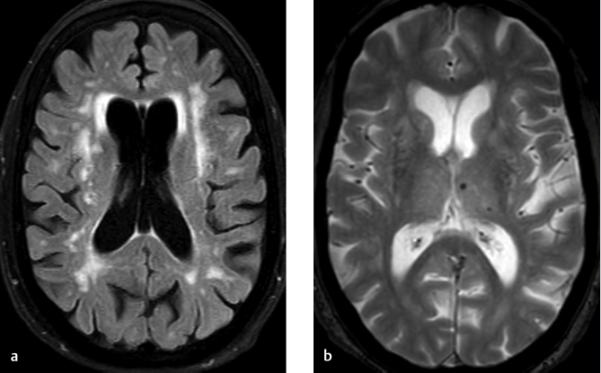
What is the typical MRI finding in multiple sclerosis (MS)?
A 67-year-old male presents with progressive difficulty in walking, frequent falls, and stiffness in the legs. Neurological examination reveals increased muscle tone, brisk deep tendon reflexes, and a positive Babinski sign. MRI of the brain shows multiple white matter lesions. Which of the following is the most likely diagnosis?
Spine MRI shows 'pencil-sharpened' vertebral bodies and 'H-shaped' vertebrae on T1-weighted images. Most likely diagnosis?
Tigroid pattern on MRI is seen in -

Intra-tumoral calcification in the brain is seen in all except?
Which of the following brain tumors is typically hyperdense on CT scan?
A 7-year-old child presents with a posterior fossa mass characterized by cyst formation. On CT, the mass appears hypodense, while on T2-weighted MRI, it is hyperintense. Post-gadolinium administration, a nodular enhancement is observed. What is the most likely diagnosis?
What is the investigation of choice for entrapment neuropathy?
What is the most common tumor of the pineal region?
Explanation: ***Resting tremor*** - A **resting tremor** is much more characteristic of **Parkinson's disease**, resulting from degeneration of **dopaminergic neurons** in the substantia nigra [2]. - While essential tremor and cerebellar tremors (intention tremor) can be seen in MS, a true resting tremor is **uncommon** in multiple sclerosis [1]. *Fatigue* - **Fatigue** is one of the most common and debilitating symptoms in patients with multiple sclerosis, significantly impacting their quality of life. - It often manifests as an overwhelming sense of **physical and mental exhaustion** that is disproportionate to activity and not relieved by rest [1]. *Spasticity* - **Spasticity**, characterized by increased muscle tone and involuntary muscle stiffness or spasms, is a very common symptom in MS due to demyelination in the **corticospinal tracts**. - It can lead to difficulties with movement, pain, and contractures. *Optic neuritis* - **Optic neuritis**, an inflammation of the optic nerve causing sudden vision loss, eye pain, and blurred vision, is a classic and often early symptom of MS. - It is a result of **demyelination** of the optic nerve.
Explanation: ***Periventricular lesions*** - **Demyelinating plaques** in a periventricular distribution are a hallmark MRI finding in **multiple sclerosis**, often appearing as **Dawson's fingers**. - These lesions reflect areas of **demyelination** and inflammation characteristic of the disease. *Basal ganglia calcification* - This finding is more commonly associated with conditions like **Fahr's disease**, **hypoparathyroidism**, or certain infections, not primary to MS. - While calcifications can occur in rare cases of MS, they are not a typical or diagnostic feature. *Diffuse cortical atrophy* - **Cortical atrophy** can be present in later stages of MS, but it is a **non-specific** finding not unique to MS and not a primary diagnostic marker. - It is more commonly seen in neurodegenerative diseases like **Alzheimer's disease** or in the elderly, and less characteristic of early MS. *Subdural hematoma* - A **subdural hematoma** is a collection of blood between the dura mater and arachnoid mater, usually due to trauma. - This is an **acute neurological emergency** and entirely unrelated to the pathology and typical MRI features of multiple sclerosis.
Explanation: ***Multiple sclerosis*** - The combination of **progressive neurological symptoms (walking difficulty, falls, leg stiffness)**, **upper motor neuron signs (increased muscle tone, brisk reflexes, positive Babinski)** and **multiple white matter lesions on MRI** is highly suggestive of **multiple sclerosis (MS)** [1]. - While MS typically presents in younger individuals, presentation in the late 60s, though less common, is possible and referred to as **late-onset MS** [1]. *Amyotrophic lateral sclerosis* - **Amyotrophic lateral sclerosis (ALS)** involves both **upper and lower motor neuron degeneration**, but typically presents with significant **muscle wasting and fasciculations (lower motor neuron signs)** [3]. - The MRI findings of **multiple white matter lesions** are not characteristic of ALS [3]. *Parkinson's disease* - **Parkinson's disease** is primarily characterized by **tremor at rest, bradykinesia, rigidity, and postural instability**, which are **extrapyramidal symptoms** [2]. - While stiffness and walking difficulty can occur, the presence of **brisk deep tendon reflexes** and a **positive Babinski sign (upper motor neuron signs)** are not typical for Parkinson's. *Primary lateral sclerosis* - **Primary lateral sclerosis (PLS)** is a **rare motor neuron disease** characterized by **pure upper motor neuron dysfunction**, leading to progressive **spasticity and weakness**. - While PLS can explain the upper motor neuron signs, **multiple white matter lesions on MRI** are not a defining feature; rather, they are highly indicative of **demyelination seen in MS**.
Explanation: ***Sickle cell disease*** - **Sickle cell disease** can lead to vertebral body changes due to **bone infarction** and **hyperplasia of hematopoietic marrow**, causing central depression and characteristic 'H-shaped' or 'pencil-sharpened' vertebrae. - The abnormal hemoglobin in sickle cell anemia causes red blood cells to stiffen and form a crescent or "sickle" shape, leading to a host of debilitating symptoms and early death. *Thalassemia* - **Thalassemia** can cause widespread marrow expansion leading to generalized osteopenia and widened medullary spaces, but typically does not result in the focal 'H-shaped' vertebral changes seen with infarction. - While it also causes anemia and bone changes, the specific vertebral findings described are not characteristic of thalassemia. *Osteopetrosis* - **Osteopetrosis** is characterized by **increased bone density** and brittle bones, often described as a "stone bone" appearance. - This condition leads to thickened, sclerotic bones and does not produce the 'H-shaped' or 'pencil-sharpened' vertebral deformities. *Paget's disease* - **Paget's disease** is characterized by disorganized bone remodeling, leading to bone enlargement and deformity with a characteristic **"cotton wool" appearance** on imaging. - While it affects vertebrae, it typically results in cortical thickening and coarsened trabeculae, not the specific 'H-shaped' deformity.
Explanation: ***Metachromatic leukodystrophy*** - The **tigroid pattern** on MRI, characterized by **perivascular sparing** within demyelinated white matter, is highly characteristic of metachromatic leukodystrophy. - This pattern results from the accumulation of **sulfatides** in oligodendrocytes and macrophages, leading to central demyelination with spared U-fibers and white matter adjacent to vessels. *Wilson's disease* - Wilson's disease involves **copper accumulation** and typically presents with abnormalities in the **basal ganglia**, thalami, and brainstem. - While it causes neurodegeneration, it does not produce a characteristic tigroid demyelination pattern. *Parkinsonism* - Parkinsonism refers to a group of neurological disorders characterized by motor symptoms like **bradykinesia, rigidity, tremor, and postural instability**. - MRI findings in Parkinsonism often show **nigral degeneration** but do not typically involve a tigroid pattern of leukodystrophy. *GB syndrome* - **Guillain-Barré syndrome (GBS)** is an acute autoimmune peripheral neuropathy affecting the **peripheral nerves and nerve roots**. - It does not involve central nervous system demyelination or display a tigroid pattern on brain MRI.
Explanation: **Explanation:** The correct answer is **Hemangioblastoma**. In neuroradiology, identifying the presence or absence of calcification is a high-yield diagnostic marker for intracranial tumors. **1. Why Hemangioblastoma is the correct answer:** Hemangioblastomas are highly vascular, WHO Grade 1 tumors typically located in the posterior fossa (cerebellum). Characteristically, they present as a **cystic lesion with a highly enhancing mural nodule**. Crucially, hemangioblastomas **do not calcify**. Their primary imaging features are related to vascularity (flow voids on MRI) and associated erythropoietin production, which may lead to polycythemia. **2. Why the other options are incorrect:** * **Oligodendroglioma:** This is the "classic" answer for calcified brain tumors. Calcification is seen in **70–90%** of cases, often described as chunky or ribbon-like. * **Craniopharyngioma:** In the pediatric population (adamantinomatous type), calcification is a hallmark, occurring in approximately **90%** of cases. It follows the "90% rule": 90% are cystic, 90% calcify, and 90% enhance. * **Meningioma:** These are extra-axial tumors that frequently show calcification (about **20–25%**). When the calcification is diffuse and gritty, they are histologically termed "Psammomatous meningiomas." **NEET-PG High-Yield Pearls:** * **Mnemonic for Calcified Brain Tumors (Old Men Are Posh):** **O**ligodendroglioma, **M**eningioma, **A**strocytoma, **P**ineal tumors/ **P**apilloma (Choroid plexus). * **Most common calcified tumor in children:** Craniopharyngioma. * **Most common calcified tumor in adults:** Oligodendroglioma. * **Hemangioblastoma Association:** Frequently associated with **Von Hippel-Lindau (VHL) syndrome**; look for retinal angiomas and renal cell carcinoma in clinical stems.
Explanation: **Explanation:** The density of a tumor on a non-contrast CT (NCCT) scan is primarily determined by its **cellularity** and the **nuclear-to-cytoplasmic (N:C) ratio**. **1. Why Medulloblastoma is correct:** Medulloblastoma is a "Small Round Blue Cell Tumor." These tumors are characterized by extremely high cellular density and very little cytoplasm. Because DNA and cellular proteins attenuate X-rays more than water or lipids, the high concentration of cells makes the tumor appear **hyperdense** relative to the normal brain parenchyma on NCCT. This is a classic radiological hallmark of medulloblastoma, typically seen in the midline (cerebellar vermis) of pediatric patients. **2. Analysis of Incorrect Options:** * **Ependymoma:** Usually appears isodense or heterogeneous on CT. While they often contain calcifications (which are hyperdense), the soft tissue component itself is not typically hyperdense. * **Oligodendroglioma:** These are known for having the highest incidence of **calcification** (up to 90%), which is hyperdense. However, the tumor mass itself is usually hypo-to-isodense. * **Astrocytoma:** Most low-grade astrocytomas are **hypodense** due to high water content and associated edema. High-grade gliomas (GBM) are usually heterogeneous due to necrosis and hemorrhage. **3. High-Yield Clinical Pearls for NEET-PG:** * **Hyperdense tumors on CT (Mnemonic: "M-L-G"):** **M**edulloblastoma, **L**ymphoma (Primary CNS), and **G**erm cell tumors/Meningioma. * **Medulloblastoma:** Most common malignant brain tumor in children; associated with "drop metastases" (seeding via CSF). * **Calcification Mnemonic:** "Old Elephants Can't Dance" (**O**ligodendroglioma, **E**pendymoma, **C**raniopharyngioma, **D**ysembryoplastic Neuroepithelial Tumor).
Explanation: **Explanation:** The clinical presentation and imaging findings are classic for a **Juvenile Pilocytic Astrocytoma (JPA)**, which is the most common brain tumor in children. **1. Why Astrocytoma is correct:** JPA typically arises in the cerebellum (posterior fossa). The pathognomonic radiological appearance is a **large cystic lesion with a brightly enhancing mural nodule**. * **CT:** The cyst is hypodense (fluid-filled). * **MRI T2:** The cyst fluid is hyperintense (similar to CSF). * **Post-Contrast:** The mural nodule shows intense enhancement, while the cyst wall usually does not (unless it contains neoplastic cells). **2. Why other options are incorrect:** * **Medulloblastoma:** This is the most common *malignant* pediatric brain tumor. It typically appears as a **solid, hyperdense mass** on CT (due to high cellularity) arising from the roof of the 4th ventricle (midline/vermis). It rarely shows large cyst formation. * **Ependymoma:** These typically arise from the floor of the 4th ventricle and are characterized by **"plasticity"** (squeezing through the foramina of Luschka and Magendie). Calcification is very common (50%), and they are usually heterogeneous rather than purely cystic with a nodule. * **Cysticercosis:** While cystic, Neurocysticercosis (NCC) usually presents as multiple small parenchymal cysts (vesicular stage) with a visible "scolex" (dot sign) and is not typically a large posterior fossa mass in a child. **3. High-Yield Clinical Pearls for NEET-PG:** * **JPA Location:** Cerebellum is most common; also associated with **Optic Gliomas** in Neurofibromatosis Type 1 (NF1). * **Histology:** Look for **Rosenthal fibers** (corkscrew-shaped eosinophilic bundles) and bipolar cells with long processes (pilocytic). * **Prognosis:** Excellent (WHO Grade I); surgical resection is often curative. * **Key Differentiator:** If a similar "cyst with a nodule" is seen in an **adult**, the diagnosis is likely **Hemangioblastoma** (associated with von Hippel-Lindau syndrome).
Explanation: **Explanation:** **Why MRI is the Correct Answer:** MRI is the investigation of choice for entrapment neuropathy because of its superior **soft-tissue contrast resolution**. It allows for the direct visualization of the nerve itself (MR Neurography). In entrapment syndromes, MRI can detect both **morphological changes** (nerve thickening, flattening, or displacement) and **signal intensity changes** (increased T2/STIR signal indicating intraneural edema or inflammation). Furthermore, MRI is excellent at identifying the extrinsic cause of compression, such as ganglion cysts, tumors, or anatomical variants. **Analysis of Incorrect Options:** * **CT Scan:** While CT is excellent for bone pathology, it has poor soft-tissue resolution and cannot visualize the internal structure of nerves or subtle inflammatory changes. * **Clinical Examination:** While clinical assessment (history and physical exams like Tinel’s or Phalen’s sign) is the first step in diagnosis, it is a **clinical tool**, not an "investigation." It cannot definitively localize the site or cause of compression as accurately as imaging. * **SPECT Scan:** This is a functional nuclear medicine imaging technique used primarily for bone turnover or myocardial perfusion; it has no role in the routine evaluation of peripheral nerves. **High-Yield Facts for NEET-PG:** * **Gold Standard for Function:** While MRI is the investigation of choice for *imaging*, **Electromyography (EMG) and Nerve Conduction Studies (NCS)** remain the gold standard for assessing the *physiological severity* of the nerve injury. * **MR Neurography:** Uses dedicated sequences (like 3D T2-weighted STIR) to suppress fat and vessel signals, making the nerve appear "bright" against a dark background. * **Common Sites:** Carpal Tunnel Syndrome (Median nerve) is the most common entrapment neuropathy, followed by Cubital Tunnel Syndrome (Ulnar nerve).
Explanation: **Explanation:** The pineal region is a complex anatomical area containing the pineal gland, surrounding cisterns, and vascular structures. The most common tumors in this region are **Germ Cell Tumors (GCTs)**, accounting for approximately 50–70% of all pineal masses. Among these, **Germinoma** is the most frequent histological subtype. * **Why Germinoma is correct:** Germinomas are highly radiosensitive tumors that typically affect young males (male-to-female ratio of ~10:1). On imaging, they often present as a solid mass that "engulfs" or surrounds the normal pineal calcification (unlike pineal parenchymal tumors which displace or explode the calcification). * **Why other options are incorrect:** * **Pineocytoma:** This is a slow-growing, well-differentiated Pineal Parenchymal Tumor (WHO Grade I). While it originates from the gland itself, it is significantly less common than Germinomas. * **Pineoblastoma:** A highly aggressive, primitive neuroectodermal tumor (WHO Grade IV) seen primarily in children. It is rare compared to germ cell tumors. * **Secondaries:** Metastasis to the pineal gland is extremely rare and usually occurs in the setting of widespread systemic malignancy (e.g., lung or breast cancer). **High-Yield Clinical Pearls for NEET-PG:** 1. **Parinaud Syndrome:** Pineal tumors often compress the superior colliculus/pretectal area, leading to upward gaze palsy, pupillary light-near dissociation, and convergence-retraction nystagmus. 2. **Hydrocephalus:** Due to its location, these tumors frequently compress the **Aqueduct of Sylvius**, causing obstructive hydrocephalus. 3. **Imaging Sign:** "Exploded" calcification (peripheral displacement) suggests a Pineal Parenchymal Tumor, whereas "Engulfed" calcification suggests a Germinoma. 4. **Tumor Markers:** Always check CSF/Serum for **beta-hCG and AFP** to differentiate between germ cell subtypes (Germinomas may have slightly elevated hCG, but high AFP suggests a Yolk Sac Tumor).
Get full access to all questions, explanations, and performance tracking.
Start For Free