Intra-tumoral calcification in the brain is seen in all except?
Which of the following brain tumors is typically hyperdense on CT scan?
A 7-year-old child presents with a posterior fossa mass characterized by cyst formation. On CT, the mass appears hypodense, while on T2-weighted MRI, it is hyperintense. Post-gadolinium administration, a nodular enhancement is observed. What is the most likely diagnosis?
What is the investigation of choice for entrapment neuropathy?
What is the most common tumor of the pineal region?
Para meningeal Rhabdomyosarcoma is best diagnosed by:
What is the most likely cause of a ring-enhancing lesion in an AIDS patient?
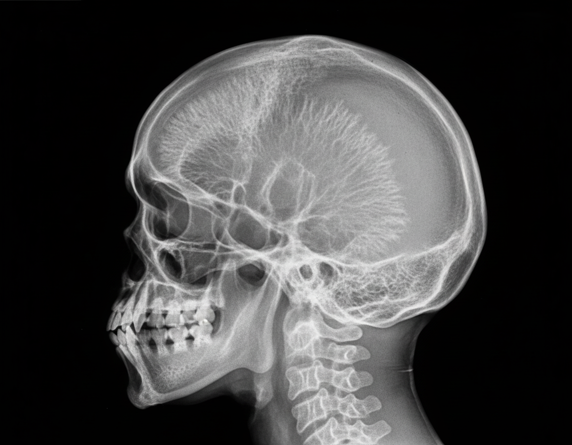
A radiograph of the skull is suggestive of which of the following diagnoses?

All are features of raised intracranial tension in adults except?
Which of the following is a cystic supratentorial tumor?
Explanation: **Explanation:** The correct answer is **Hemangioblastoma**. In neuroradiology, identifying the presence or absence of calcification is a high-yield diagnostic marker for intracranial tumors. **1. Why Hemangioblastoma is the correct answer:** Hemangioblastomas are highly vascular, WHO Grade 1 tumors typically located in the posterior fossa (cerebellum). Characteristically, they present as a **cystic lesion with a highly enhancing mural nodule**. Crucially, hemangioblastomas **do not calcify**. Their primary imaging features are related to vascularity (flow voids on MRI) and associated erythropoietin production, which may lead to polycythemia. **2. Why the other options are incorrect:** * **Oligodendroglioma:** This is the "classic" answer for calcified brain tumors. Calcification is seen in **70–90%** of cases, often described as chunky or ribbon-like. * **Craniopharyngioma:** In the pediatric population (adamantinomatous type), calcification is a hallmark, occurring in approximately **90%** of cases. It follows the "90% rule": 90% are cystic, 90% calcify, and 90% enhance. * **Meningioma:** These are extra-axial tumors that frequently show calcification (about **20–25%**). When the calcification is diffuse and gritty, they are histologically termed "Psammomatous meningiomas." **NEET-PG High-Yield Pearls:** * **Mnemonic for Calcified Brain Tumors (Old Men Are Posh):** **O**ligodendroglioma, **M**eningioma, **A**strocytoma, **P**ineal tumors/ **P**apilloma (Choroid plexus). * **Most common calcified tumor in children:** Craniopharyngioma. * **Most common calcified tumor in adults:** Oligodendroglioma. * **Hemangioblastoma Association:** Frequently associated with **Von Hippel-Lindau (VHL) syndrome**; look for retinal angiomas and renal cell carcinoma in clinical stems.
Explanation: **Explanation:** The density of a tumor on a non-contrast CT (NCCT) scan is primarily determined by its **cellularity** and the **nuclear-to-cytoplasmic (N:C) ratio**. **1. Why Medulloblastoma is correct:** Medulloblastoma is a "Small Round Blue Cell Tumor." These tumors are characterized by extremely high cellular density and very little cytoplasm. Because DNA and cellular proteins attenuate X-rays more than water or lipids, the high concentration of cells makes the tumor appear **hyperdense** relative to the normal brain parenchyma on NCCT. This is a classic radiological hallmark of medulloblastoma, typically seen in the midline (cerebellar vermis) of pediatric patients. **2. Analysis of Incorrect Options:** * **Ependymoma:** Usually appears isodense or heterogeneous on CT. While they often contain calcifications (which are hyperdense), the soft tissue component itself is not typically hyperdense. * **Oligodendroglioma:** These are known for having the highest incidence of **calcification** (up to 90%), which is hyperdense. However, the tumor mass itself is usually hypo-to-isodense. * **Astrocytoma:** Most low-grade astrocytomas are **hypodense** due to high water content and associated edema. High-grade gliomas (GBM) are usually heterogeneous due to necrosis and hemorrhage. **3. High-Yield Clinical Pearls for NEET-PG:** * **Hyperdense tumors on CT (Mnemonic: "M-L-G"):** **M**edulloblastoma, **L**ymphoma (Primary CNS), and **G**erm cell tumors/Meningioma. * **Medulloblastoma:** Most common malignant brain tumor in children; associated with "drop metastases" (seeding via CSF). * **Calcification Mnemonic:** "Old Elephants Can't Dance" (**O**ligodendroglioma, **E**pendymoma, **C**raniopharyngioma, **D**ysembryoplastic Neuroepithelial Tumor).
Explanation: **Explanation:** The clinical presentation and imaging findings are classic for a **Juvenile Pilocytic Astrocytoma (JPA)**, which is the most common brain tumor in children. **1. Why Astrocytoma is correct:** JPA typically arises in the cerebellum (posterior fossa). The pathognomonic radiological appearance is a **large cystic lesion with a brightly enhancing mural nodule**. * **CT:** The cyst is hypodense (fluid-filled). * **MRI T2:** The cyst fluid is hyperintense (similar to CSF). * **Post-Contrast:** The mural nodule shows intense enhancement, while the cyst wall usually does not (unless it contains neoplastic cells). **2. Why other options are incorrect:** * **Medulloblastoma:** This is the most common *malignant* pediatric brain tumor. It typically appears as a **solid, hyperdense mass** on CT (due to high cellularity) arising from the roof of the 4th ventricle (midline/vermis). It rarely shows large cyst formation. * **Ependymoma:** These typically arise from the floor of the 4th ventricle and are characterized by **"plasticity"** (squeezing through the foramina of Luschka and Magendie). Calcification is very common (50%), and they are usually heterogeneous rather than purely cystic with a nodule. * **Cysticercosis:** While cystic, Neurocysticercosis (NCC) usually presents as multiple small parenchymal cysts (vesicular stage) with a visible "scolex" (dot sign) and is not typically a large posterior fossa mass in a child. **3. High-Yield Clinical Pearls for NEET-PG:** * **JPA Location:** Cerebellum is most common; also associated with **Optic Gliomas** in Neurofibromatosis Type 1 (NF1). * **Histology:** Look for **Rosenthal fibers** (corkscrew-shaped eosinophilic bundles) and bipolar cells with long processes (pilocytic). * **Prognosis:** Excellent (WHO Grade I); surgical resection is often curative. * **Key Differentiator:** If a similar "cyst with a nodule" is seen in an **adult**, the diagnosis is likely **Hemangioblastoma** (associated with von Hippel-Lindau syndrome).
Explanation: **Explanation:** **Why MRI is the Correct Answer:** MRI is the investigation of choice for entrapment neuropathy because of its superior **soft-tissue contrast resolution**. It allows for the direct visualization of the nerve itself (MR Neurography). In entrapment syndromes, MRI can detect both **morphological changes** (nerve thickening, flattening, or displacement) and **signal intensity changes** (increased T2/STIR signal indicating intraneural edema or inflammation). Furthermore, MRI is excellent at identifying the extrinsic cause of compression, such as ganglion cysts, tumors, or anatomical variants. **Analysis of Incorrect Options:** * **CT Scan:** While CT is excellent for bone pathology, it has poor soft-tissue resolution and cannot visualize the internal structure of nerves or subtle inflammatory changes. * **Clinical Examination:** While clinical assessment (history and physical exams like Tinel’s or Phalen’s sign) is the first step in diagnosis, it is a **clinical tool**, not an "investigation." It cannot definitively localize the site or cause of compression as accurately as imaging. * **SPECT Scan:** This is a functional nuclear medicine imaging technique used primarily for bone turnover or myocardial perfusion; it has no role in the routine evaluation of peripheral nerves. **High-Yield Facts for NEET-PG:** * **Gold Standard for Function:** While MRI is the investigation of choice for *imaging*, **Electromyography (EMG) and Nerve Conduction Studies (NCS)** remain the gold standard for assessing the *physiological severity* of the nerve injury. * **MR Neurography:** Uses dedicated sequences (like 3D T2-weighted STIR) to suppress fat and vessel signals, making the nerve appear "bright" against a dark background. * **Common Sites:** Carpal Tunnel Syndrome (Median nerve) is the most common entrapment neuropathy, followed by Cubital Tunnel Syndrome (Ulnar nerve).
Explanation: **Explanation:** The pineal region is a complex anatomical area containing the pineal gland, surrounding cisterns, and vascular structures. The most common tumors in this region are **Germ Cell Tumors (GCTs)**, accounting for approximately 50–70% of all pineal masses. Among these, **Germinoma** is the most frequent histological subtype. * **Why Germinoma is correct:** Germinomas are highly radiosensitive tumors that typically affect young males (male-to-female ratio of ~10:1). On imaging, they often present as a solid mass that "engulfs" or surrounds the normal pineal calcification (unlike pineal parenchymal tumors which displace or explode the calcification). * **Why other options are incorrect:** * **Pineocytoma:** This is a slow-growing, well-differentiated Pineal Parenchymal Tumor (WHO Grade I). While it originates from the gland itself, it is significantly less common than Germinomas. * **Pineoblastoma:** A highly aggressive, primitive neuroectodermal tumor (WHO Grade IV) seen primarily in children. It is rare compared to germ cell tumors. * **Secondaries:** Metastasis to the pineal gland is extremely rare and usually occurs in the setting of widespread systemic malignancy (e.g., lung or breast cancer). **High-Yield Clinical Pearls for NEET-PG:** 1. **Parinaud Syndrome:** Pineal tumors often compress the superior colliculus/pretectal area, leading to upward gaze palsy, pupillary light-near dissociation, and convergence-retraction nystagmus. 2. **Hydrocephalus:** Due to its location, these tumors frequently compress the **Aqueduct of Sylvius**, causing obstructive hydrocephalus. 3. **Imaging Sign:** "Exploded" calcification (peripheral displacement) suggests a Pineal Parenchymal Tumor, whereas "Engulfed" calcification suggests a Germinoma. 4. **Tumor Markers:** Always check CSF/Serum for **beta-hCG and AFP** to differentiate between germ cell subtypes (Germinomas may have slightly elevated hCG, but high AFP suggests a Yolk Sac Tumor).
Explanation: **Explanation:** **Parameningeal Rhabdomyosarcoma (RMS)** is a highly aggressive soft tissue sarcoma occurring in sites adjacent to the skull base (e.g., nasopharynx, infratemporal fossa, paranasal sinuses). **Why MRI is the Correct Answer:** MRI is the gold standard for diagnosing and staging parameningeal RMS due to its **superior soft-tissue contrast resolution**. In these locations, the primary clinical concern is **intracranial extension** and **perineural spread**. MRI is far more sensitive than CT in detecting: * Dural enhancement or invasion. * Bone marrow replacement (even before cortical destruction is visible on CT). * Involvement of the cavernous sinus and cranial nerves. * Extension into the orbit or brain parenchyma. **Why Other Options are Incorrect:** * **CT Scan:** While excellent for viewing cortical bone destruction, it lacks the soft-tissue detail required to differentiate tumor from adjacent muscles or to detect early intracranial spread. * **SPECT:** This is a functional imaging modality with low spatial resolution, rarely used in the primary diagnosis of head and neck tumors. * **PET (PET-CT):** While useful for detecting distant metastases (staging) and monitoring recurrence, it is not the primary modality for the initial anatomical diagnosis and local extension assessment of the primary tumor. **High-Yield Clinical Pearls for NEET-PG:** * **Most common site:** The head and neck is the most common site for RMS in children. * **Prognosis:** Parameningeal sites have a **poorer prognosis** compared to orbital sites due to the high risk of meningeal involvement. * **Radiological Sign:** Look for "bone remodeling" or "moth-eaten" bone destruction on imaging. * **Treatment:** Usually involves a combination of chemotherapy, surgery, and radiation (especially if intracranial extension is present).
Explanation: ### Explanation **1. Why Toxoplasmosis is the Correct Answer:** In the context of HIV/AIDS, **Toxoplasmosis** (caused by *Toxoplasma gondii*) is the most common cause of a ring-enhancing lesion. It typically occurs when the CD4 count falls below **100 cells/mm³**. Radiologically, these lesions are often multiple, located in the **basal ganglia** or corticomedullary junction, and exhibit "ring enhancement" due to peripheral inflammation and central necrosis. A pathognomonic (though not always present) sign is the **"Eccentric Target Sign,"** representing an enhancing nodule along the wall of the ring. **2. Why the Other Options are Incorrect:** * **Cryptococcosis:** Usually presents as meningitis. On imaging, it typically shows **"Soap-bubble" appearances** (gelatinous pseudocysts) in the basal ganglia or prominent Virchow-Robin spaces, rather than ring-enhancing masses. * **Hydatid Cyst:** Caused by *Echinococcus granulosus*, these appear as large, well-defined, **unilocular "mother" cysts** with possible internal "daughter" cysts. They do not typically show ring enhancement unless infected. * **Neurocysticercosis (NCC):** While NCC is the most common cause of ring-enhancing lesions in the **general population** (especially in India), it is not specifically associated with the immunocompromised state of AIDS. In NCC, the "scolex" (hole-with-dot appearance) is a key diagnostic feature. **3. NEET-PG High-Yield Pearls:** * **The Great Mimicker:** Primary CNS Lymphoma (PCNSL) is the main differential for Toxoplasmosis in AIDS. * **Toxoplasmosis vs. Lymphoma:** Toxoplasmosis lesions are usually multiple and respond to antitoxoplasmosis therapy (Pyrimethamine + Sulfadiazine). Lymphoma is often solitary, involves the corpus callosum, and shows **increased uptake on Thallium-201 SPECT** or PET scans (Toxoplasmosis is "cold"). * **Prophylaxis:** AIDS patients with CD4 <100 and positive IgG serology should receive **Trimethoprim-Sulfamethoxazole (TMP-SMX)**.
Explanation: ***Sickle cell anemia*** - Classic **"hair-on-end"** or **"crew-cut"** appearance on skull X-ray due to **marrow hyperplasia** and trabecular bone thickening. - Results from chronic **hemolytic anemia** causing compensatory expansion of the **diploic space** between skull tables. *Malignant melanoma* - Skull metastases appear as **non-specific lytic lesions** without the characteristic trabecular pattern. - More commonly presents as **multiple round lesions** rather than diffuse trabecular changes. *Multiple myeloma* - Produces classic **"punched-out" lytic lesions** that are well-demarcated and round without sclerotic borders. - Lacks the **trabecular thickening** and "hair-on-end" pattern seen in hemolytic anemias. *Langerhans cell histiocytosis* - Creates **geographic lytic lesions** with irregular, map-like borders and scalloped edges. - Typically shows **beveled edges** and asymmetric destruction rather than diffuse trabecular changes.
Explanation: **Explanation:** The correct answer is **Sutural diastasis (Option D)**. The fundamental concept here is the difference between the pediatric and adult skull. In **adults**, the cranial sutures are fused (synostosed). Therefore, when intracranial pressure (ICP) rises, the sutures cannot separate. In contrast, **sutural diastasis** (separation of sutures >2mm) is a hallmark sign of raised ICP in **infants and children** (usually up to age 10-12) because their sutures are still patent. **Analysis of Incorrect Options:** * **Erosion of Dorsum Sella (Option A) & Posterior Clinoid Process (Option B):** These are the **earliest and most common** radiologic signs of chronic raised ICP in adults. The pressure from the distended third ventricle (pulsatile CSF) transmits directly onto the sella turcica, leading to rarefaction and erosion of the cortical bone, starting with the posterior clinoid processes and the dorsum sella. * **Inner Table Convolutions (Option C):** Also known as **"Copper Beaten Skull"** or "Fingerprinting," these represent prominent gyral impressions on the inner table of the skull due to chronic pressure from the underlying brain. While it can be a normal variant in children, in adults, it is a classic sign of long-standing raised ICP (e.g., craniosynostosis or obstructive hydrocephalus). **High-Yield Clinical Pearls for NEET-PG:** * **Earliest sign of raised ICP on X-ray:** Erosion of the lamina dura of the polyspastic dorsum sella. * **Sutural Diastasis:** Most commonly involves the **coronal suture** in children. * **J-shaped Sella:** Associated with optic chiasm gliomas, mucopolysaccharidosis (Hurler syndrome), and achondroplasia. * **Empty Sella Syndrome:** Herniation of the subarachnoid space into the sella, often associated with Idiopathic Intracranial Hypertension (Pseudotumor cerebri).
Explanation: **Explanation:** The correct answer is **Craniopharyngioma**. **1. Why Craniopharyngioma is correct:** Craniopharyngiomas are benign (WHO Grade I) tumors derived from the remnants of **Rathke’s pouch**. They are characteristically **supratentorial** (specifically suprasellar) in location. Radiologically, they are known for their heterogeneous appearance, often described by the "90% rule": 90% are cystic, 90% show calcification (especially in the adamantinomatous type), and 90% enhance. The presence of "motor oil" fluid within the cysts is a classic pathological finding. **2. Why the other options are incorrect:** * **Medulloblastoma:** This is a highly malignant (WHO Grade IV) tumor that occurs in the **infratentorial** compartment (posterior fossa). It typically arises from the roof of the 4th ventricle and is solid, not primarily cystic. * **Ependymoma:** While ependymomas can occur supratentorially, they are most commonly found in the **infratentorial** compartment (floor of the 4th ventricle) in children. They often present as solid masses with "plastic" growth (extending through the foramina of Luschka and Magendie). **Clinical Pearls for NEET-PG:** * **Bimodal Age Distribution:** Craniopharyngiomas peak at 5–14 years and 50–75 years. * **Adamantinomatous vs. Papillary:** Adamantinomatous (children) is cystic/calcified; Papillary (adults) is usually solid/mixed and lacks calcification. * **Clinical Triad:** Visual field defects (bitemporal hemianopia), endocrine dysfunction (growth retardation, DI), and increased intracranial pressure. * **Imaging Hallmark:** Suprasellar calcification on CT is the most suggestive feature in a pediatric patient.
Neuroanatomy for Radiologists
Practice Questions
Cerebrovascular Diseases
Practice Questions
Intracranial Tumors
Practice Questions
CNS Infections
Practice Questions
Demyelinating and Degenerative Diseases
Practice Questions
Head Trauma Imaging
Practice Questions
Spine Imaging: Trauma and Degenerative Disease
Practice Questions
Spine Tumors and Infections
Practice Questions
Pediatric Neuroradiology
Practice Questions
Congenital CNS Anomalies
Practice Questions
Functional Neuroimaging
Practice Questions
Neurointerventional Procedures
Practice Questions
Get full access to all questions, explanations, and performance tracking.
Start For Free