Which imaging modality is LEAST useful in the initial diagnosis of stress fractures?
A patient developed paraplegia. On routine examination and X-ray it was found that there are osteoblastic lesions in his spine. What is the MOST probable diagnosis?
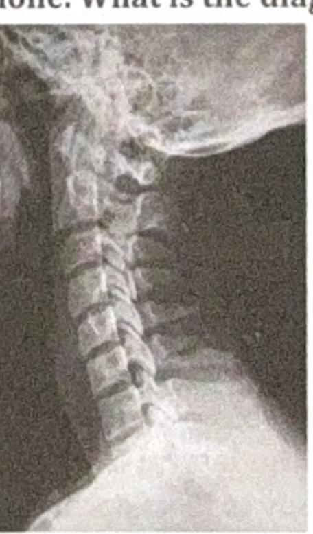
Identify the condition shown in the image:

Jefferson fracture is -
Dennis stability concept is based on which of the following?
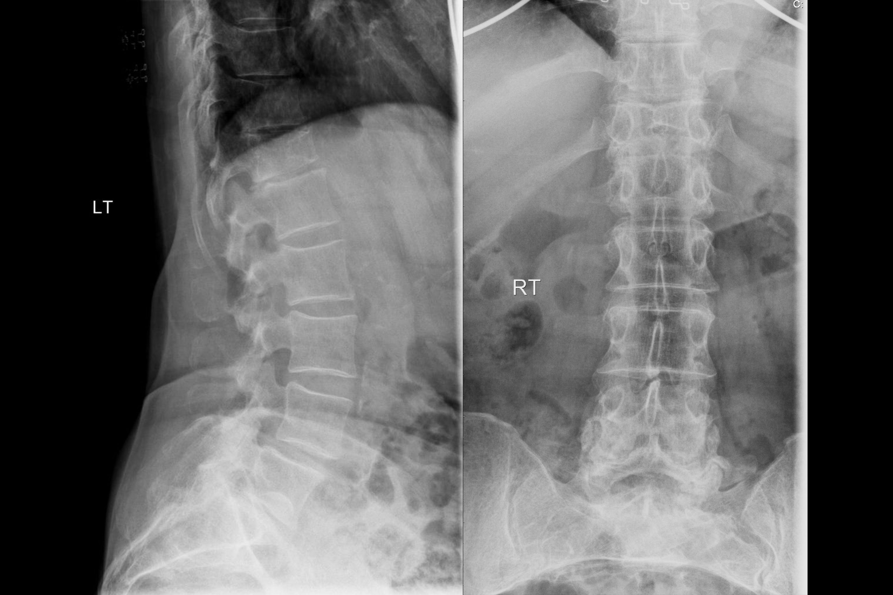
A 75-year-old female has chronic backache. X-ray of the spine is shown. What is the most likely diagnosis?

Following are listed as (SpA) spondyloarthritis features except
Scotty dog sign is seen in:
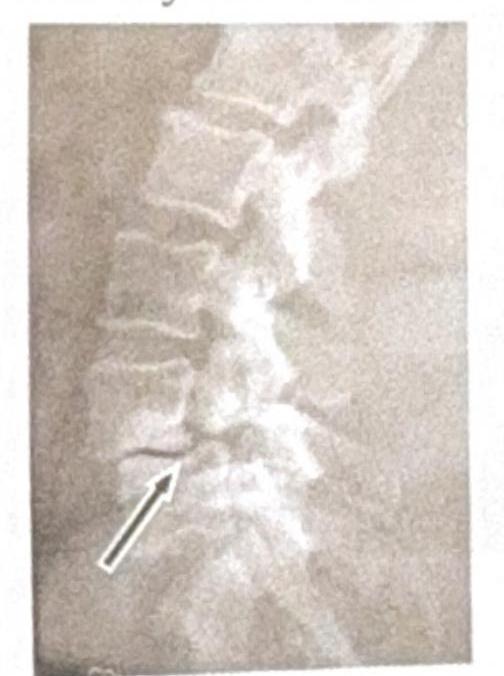
A man presents with back pain following a road traffic accident (RTA). There is no history of neurological deficit. An X-ray of the spine is done. What is the diagnosis based on the image?

Which of the following is least useful for diagnosing spondylolisthesis?
Explanation: ***Correct: CT*** - While **CT** can show bony details, it is **less sensitive** than MRI or bone scan for early stress fractures, which often involve subtle marrow edema or periosteal reaction - Its primary role is usually in defining **fracture geometry** or evaluating **fracture healing**, not initial detection - **CT is rarely used as a first-line imaging modality** for suspected stress fractures *Incorrect: MRI* - **MRI** is highly sensitive for **stress fractures**, detecting early changes such as **marrow edema** and **periosteal reaction** before they are visible on X-ray - It is considered the **gold standard** for confirming a stress fracture when initial X-rays are negative - This is one of the MOST useful modalities, not the least *Incorrect: Bone scan* - **Bone scintigraphy** (bone scan) is very sensitive for **stress fractures**, as it detects increased **osteoblastic activity** at the site of injury - It can identify stress fractures long before they are visible on plain radiographs, showing a focal area of increased radiotracer uptake - This is highly useful for initial diagnosis, not the least useful *Incorrect: X-ray* - **Plain radiographs (X-rays)** are often the **initial imaging modality** for suspected stress fractures due to availability and cost - However, they have **low sensitivity in the early stages** - early stress fractures may appear normal on X-ray - Visible signs such as **sclerosis, periosteal reaction**, or a **fracture line** only develop several weeks after symptom onset - Despite low sensitivity, X-ray is still more commonly used for initial evaluation than CT
Explanation: ***Ca. Prostate*** - **Prostate cancer** has a strong predilection for metastasizing to bone and typically produces **osteoblastic lesions** (new bone formation) in the spine [2]. - The presence of **paraplegia** suggests spinal cord compression due to these metastatic lesions [1]. *Breast Ca.* - While **breast cancer** frequently metastasizes to bone (commonly spine, pelvis, ribs, skull), it typically causes **osteolytic lesions** (bone destruction), though mixed lesions can occur. - Paraplegia can result from breast cancer metastases but the primary lesion type is usually osteolytic. *Carcinoma thyroid* - **Thyroid cancer** metastases to bone are rare and generally lead to **osteolytic lesions**, not osteoblastic. - Although it can cause spinal cord compression, the characteristic osteoblastic appearance is not typical for thyroid cancer. *Pancreatic Ca.* - **Pancreatic cancer** rarely metastasizes to bone, and when it does, the lesions are almost exclusively **osteolytic**. - Therefore, it is highly unlikely to be the cause of osteoblastic spinal lesions and subsequent paraplegia.
Explanation: ***Spondylolysis*** * The image shows a **break in the pars interarticularis** of a vertebra, indicated by the arrow, which is characteristic of spondylolysis. * This condition is a **stress fracture** or defect in the pars interarticularis, a bony segment connecting the superior and inferior articular facets. *Renal osteodystrophy* * Renal osteodystrophy refers to a spectrum of **bone abnormalities** that occur in chronic kidney disease, not a specific vertebral fracture pattern. * It typically involves features such as **osteomalacia**, **osteitis fibrosa cystica**, or **osteoporosis**, which are not directly depicted as a fracture in this image. *Spondylolisthesis* * Spondylolisthesis is the **anterior slippage** of one vertebral body over another, which can be caused by bilateral spondylolysis but is not directly shown as a slip in this specific image. * The image distinctly highlights the **fracture line** itself, rather than the displacement of the vertebral body. *Tuberculosis (TB)* * Spinal tuberculosis (Pott's disease) typically presents with **destruction of vertebral bodies**, disc space narrowing, and often a **paravertebral abscess**. * The image does not show these features; instead, it demonstrates a clear **bony defect** in the pars interarticularis.
Explanation: ***Fracture of atlas*** - A **Jefferson fracture** specifically refers to a **burst fracture** of the **C1 vertebra (atlas)**, caused by a compressive force on the head. - This type of fracture often involves **four separate fractures** within the anterior and posterior arches of the atlas. *Fracture of any cervical vertebra* - This is a too broad a statement; while the atlas is a cervical vertebra, a Jefferson fracture is a specific type affecting only C1. - Cervical spine fractures can involve various vertebrae (C1-C7) and different fracture patterns, not all of which are Jefferson fractures. *Fracture of axis* - The **axis** is the **C2 vertebra**, and its characteristic fractures include a **dens fracture** or a **hangman's fracture**, which involve different mechanisms and anatomical locations than a Jefferson fracture. - Injury to C2 can cause different neurological deficits compared to C1. *Fracture of spinous process of C7* - A fracture of the spinous process of C7 is known as a **clay-shoveler's fracture** and is typically an avulsion injury, not a burst fracture from axial compression. - This type of fracture usually results from forceful neck flexion, leading to avulsion of the spinous process.
Explanation: ***3 columns*** - The **Denis classification** system for spinal stability divides the vertebra into three conceptual columns: **anterior**, **middle**, and **posterior**. - This three-column model helps in assessing the **stability of spinal fractures** and guiding treatment decisions. *4 columns* - The four-column concept is **not standard** for Denis classification; it would overcomplicate the established three-column model. - Adding a fourth column lacks the **clinical utility** and widespread acceptance of the Denis system. *5 columns* - A five-column system is **not recognized** in the standard Denis classification of spinal stability. - Such a detailed breakdown would be **excessive** and not provide additional practical information for assessing stability. *2 columns* - The two-column concept, often seen in older classifications like **Holdsworth classification**, predates Denis's work and was found to be **less comprehensive** for assessing spinal stability. - It does not account for the critical stabilizing role of the **middle column** in spinal fractures.
Explanation: ***Osteoporosis*** - The X-ray shows diffuse **osteopenia** (reduced bone density) and **vertebral compression fractures**, particularly visible in the lateral view, which are characteristic findings in elderly patients with osteoporosis and chronic backache. - The vertebral bodies appear **demineralized** and some exhibit a loss of height, suggesting collapse due to weakened bone structure. *Spondylodiscitis* - This condition involves **inflammation of the vertebral body and adjacent intervertebral disc**, typically showing **erosions** of the vertebral endplates and **narrowing of the disc space** on X-ray, which are not clearly evident here as the primary issue. - While it can cause back pain, the dominant finding on this X-ray is widespread bone density loss and fractures, rather than localized infection-related changes. *Pott's spine* - Pott's spine (**tuberculous spondylitis**) is a form of osteomyelitis that causes **destruction of vertebral bodies** and adjacent discs, often leading to a **gibbus deformity** (sharp posterior angulation of the spine). - The X-ray does not show extensive vertebral destruction, paraspinal abscess formation, or typical kyphotic deformity associated with Pott's spine. *Spondylolisthesis* - Spondylolisthesis is characterized by the **forward slippage of one vertebral body over another**, often due to a defect in the pars interarticularis. - While there may be some degenerative changes, there is no clear evidence of significant anterior translation of a vertebral body on the lateral X-ray that would indicate spondylolisthesis.
Explanation: **Cervical sprain** - A **cervical sprain** is an injury to the ligaments in the neck, typically caused by trauma or sudden movements, and is not a defining characteristic or feature of spondyloarthritis. - Sprains involve the stretching or tearing of soft tissues, whereas spondyloarthritis is a group of **inflammatory conditions** primarily affecting the spine and peripheral joints. *Dactylitis* - **Dactylitis**, or "sausage digits," refers to the **inflammation of an entire digit** (finger or toe) and is a characteristic feature of spondyloarthritis, particularly **psoriatic arthritis** and **reactive arthritis** [1]. - It results from inflammation of both the joints and the tendons within the affected digit [1]. *Enthesitis* - **Enthesitis** is the **inflammation of the entheses**, which are the sites where tendons or ligaments insert into bone [1]. - This is a hallmark feature of spondyloarthritis, commonly seen at sites like the **Achilles tendon insertion** or the plantar fascia [1]. *HLA B27* - **HLA-B27** is a human leukocyte antigen that is strongly associated with spondyloarthritis [1]. - Its presence is a significant genetic marker found in a high percentage of patients with **ankylosing spondylitis** and other forms of spondyloarthritis [1].
Explanation: ***Spondylolysis*** - The **Scotty dog sign** is a classic radiographic finding on an **oblique lumbar spine X-ray** indicating a defect in the pars interarticularis. - This defect, known as **spondylolysis**, causes the "neck" of the Scotty dog to appear broken or wearing a collar. *Fracture femur* - **Femur fractures** are typically identified by discontinuity of the cortical bone and soft tissue swelling, often visualized on **AP and lateral views of the thigh/hip**. - No specific "Scotty dog" appearance is associated with femur fractures, as this sign relates to the **lumbar spine**. *Sarcoidosis* - **Sarcoidosis** is a multi-system inflammatory disease primarily affecting the lungs and lymph nodes, with characteristic **non-caseating granulomas**. - Radiological findings typically include **hilar lymphadenopathy** and pulmonary infiltrates, not a bony defect like the Scotty dog sign. *Osteosarcoma* - **Osteosarcoma** is a primary malignant bone tumor often presenting with a **sunburst pattern** or **Codman triangle** on X-ray. - It primarily affects the metaphysis of long bones and does not produce the specific bony defect seen in the pars interarticularis.
Explanation: ***Spinous process fracture*** - The X-ray image reveals a **fracture of the spinous process** of one of the cervical vertebrae, characterized by a visible discontinuity or separation of this posterior bony projection. - This type of fracture, often caused by direct trauma or forceful hyperextension/hyperflexion, typically presents with localized back pain but often **without neurological deficit** as the spinal canal generally remains intact. *Chance fracture* - A **Chance fracture** (or seatbelt fracture) is a horizontal fracture of a vertebral body, usually in the thoracolumbar region, often caused by distractional forces (e.g., flexion over a seatbelt). - It involves all three columns of the spine (anterior, middle, and posterior) and is not seen in the cervical spine X-ray provided. *Compression fracture* - A **compression fracture** is characterized by the collapse of the vertebral body, often resulting in a wedge shape. - This typically appears as reduced height of the anterior vertebral body on an X-ray, which is not the primary finding in the image. *Fracture of base of vertebrae* - A **fracture at the base of the vertebrae** is a non-specific term; specific vertebral fractures are categorized based on the part of the vertebra affected (e.g., vertebral body, pedicle, lamina, spinous process). - The image distinctly shows a fracture in the **spinous process**, not the main body or base of the vertebra.
Explanation: ***X-ray spine AP view*** - An **AP (Anterior-Posterior) view** of the spine is least useful for diagnosing spondylolisthesis because it does not adequately demonstrate the **forward slippage** of one vertebra over another. - This view primarily visualizes the spine in the **coronal plane**, making it difficult to assess the **sagittal displacement** characteristic of spondylolisthesis. *X-ray spine lateral view* - A **lateral view** of the spine is highly useful as it directly shows the **sagittal alignment** and can clearly demonstrate the **anterior displacement** of a vertebral body. - It is often the **initial imaging modality** for suspecting and classifying spondylolisthesis severity. *MRI* - **MRI** is excellent for evaluating **soft tissue structures**, such as the spinal cord, nerve roots, and intervertebral discs, which can be compressed or damaged by spondylolisthesis. - While it can visualize the slippage, it is usually reserved for assessing **neurological compromise** or if surgical planning requires detailed soft tissue information. *CT scan* - **CT scans** provide detailed **bony anatomy** and are highly effective in visualizing the pars interarticularis defects (spondylolysis) often associated with spondylolisthesis. - It offers superior detail compared to plain X-rays for assessing the **extent of bone displacement** and associated degenerative changes.
Get full access to all questions, explanations, and performance tracking.
Start For Free