What is the first-choice imaging modality for evaluating a shoulder joint?
'Telephone Handle' long bones are seen in which of the following conditions?
Increased heel pad thickness is associated with which of the following conditions?
A person has an injury to the forefinger with glass and it is suspected that there is a retained piece of glass in the finger. What is the first investigation you will perform?
The "floating tooth sign" is seen in which of the following conditions?
A 15-year-old female has an impacted tooth 23, located apical to teeth 22 and 24. How would an oral surgeon determine if the impacted tooth is positioned buccally or palatally to teeth 22 and 24?
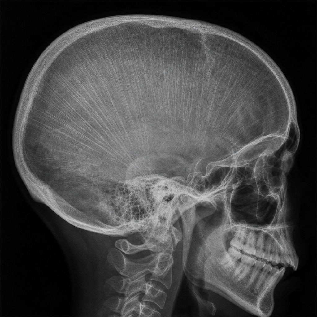
A radiograph of a girl's mandible shows a soap bubble appearance of a lytic lesion. On aspiration, the lesion yields blood. A whirring sound is heard from the lesion, and the girl can feel her heartbeat within it. What is the next recommended investigation?
Which condition is characterized by the presence of ring-shaped epiphyses?
"Bone within bone" appearance is classically seen in?
A young child presents with backache. Imaging reveals a solitary collapsed dorsal vertebra with preserved intervertebral disc spaces and no evidence of a soft tissue mass. What is the most likely diagnosis?
Explanation: **Explanation:** In musculoskeletal radiology, the **Digital X-ray** remains the **initial and first-choice imaging modality** for evaluating the shoulder joint. This is based on the fundamental clinical principle of "starting with the simplest and most cost-effective tool." X-rays provide an immediate overview of bony anatomy, joint alignment, and gross pathology such as fractures, dislocations, or degenerative changes (osteoarthritis). **Why other options are incorrect:** * **CT Scan:** While superior for evaluating complex intra-articular fractures or preoperative planning for glenoid bone loss, it involves high radiation and is considered a second-line investigation for bone detail. * **MRI:** This is the "Gold Standard" for soft tissue evaluation (rotator cuff tears, labral injuries, and marrow edema). However, it is expensive and time-consuming, making it a secondary investigation after X-rays have ruled out bony pathology. * **Ultrasound (USG):** Excellent for dynamic evaluation of the rotator cuff and biceps tendon, but it is operator-dependent and cannot visualize deep intra-articular structures or bony architecture effectively. **High-Yield Clinical Pearls for NEET-PG:** * **Standard Views:** The most common views for the shoulder are Anteroposterior (AP), Internal/External rotation, and the **Axillary view** (best for detecting subtle dislocations). * **Hill-Sachs & Bankart Lesions:** While X-rays may show a "Stryker Notch" or "West Point" view, **MRI/MR Arthrography** is the definitive investigation for these recurrent dislocation sequelae. * **Snowcap Sign:** A classic radiological sign seen on X-ray in **Avascular Necrosis (AVN)** of the humeral head.
Explanation: **Explanation:** The **"Telephone Handle" appearance** of long bones is a classic radiological sign characterized by shortened, bowed femurs with flared metaphyses. This occurs due to defective endochondral ossification, leading to significant rhizomelic (proximal) limb shortening. **1. Why Achondroplasia is correct:** Achondroplasia is the most common cause of disproportionate short-limb dwarfism. It results from a mutation in the **FGFR3 gene**. The long bones (especially the femur) appear short and curved with widened metaphyses, resembling an old-fashioned telephone receiver. Other key radiological features include "squared-off" iliac wings, a narrow interpedicular distance (decreasing caudally), and a "trident hand." **2. Why the other options are incorrect:** * **Thanatophoric Dwarfism:** While this condition also features bowed femurs (often described as "cloverleaf skull" or "French telephone receiver" appearance in Type 1), it is a **lethal** skeletal dysplasia. In the context of standard NEET-PG questions, "Telephone Handle" bones are most classically associated with Achondroplasia, though Thanatophoric dysplasia is the closest differential. * **Mucopolysaccharidosis (MPS):** These conditions (like Hurler or Hunter syndrome) are characterized by **Dysostosis Multiplex**, which includes J-shaped sella, ovoid vertebrae with anterior beaking, and tapering of the proximal metacarpals, rather than telephone-handle femurs. * **Acromegaly:** This involves overgrowth of bone in adults. Key signs include "spade-like" phalangeal tufts, increased heel pad thickness (>25mm), and enlargement of the paranasal sinuses. **High-Yield Clinical Pearls for NEET-PG:** * **Champagne Glass Pelvis:** Seen in Achondroplasia (wide, shallow pelvic inlet). * **Trident Hand:** Persistent space between the 3rd and 4th digits. * **Foramen Magnum Stenosis:** A critical complication in Achondroplasia that can lead to sudden infant death or hydrocephalus. * **Inheritance:** Autosomal Dominant, but 80% of cases are due to *de novo* mutations associated with advanced paternal age.
Explanation: **Explanation:** **Correct Answer: C. Acromegaly** The measurement of heel pad thickness is a classic radiological sign used in the evaluation of **Acromegaly**. Excess Growth Hormone (GH) and Insulin-like Growth Factor-1 (IGF-1) cause soft tissue hypertrophy and overgrowth of connective tissue throughout the body. In the foot, this manifests as thickening of the soft tissue between the calcaneus and the skin surface. * **Diagnostic Threshold:** On a lateral radiograph of the foot, a heel pad thickness **>25 mm in males** and **>23 mm in females** is highly suggestive of acromegaly. **Why other options are incorrect:** * **A. Rickets:** Characterized by defective mineralization of osteoid, leading to radiological signs like cupping, splaying, and fraying of the metaphyses, not soft tissue thickening. * **B. Paget’s Disease:** A disorder of bone remodeling resulting in thickened, disorganized trabeculae and cortical thickening (e.g., "picture frame" vertebra or "cotton wool" skull), but it does not typically cause heel pad hypertrophy. * **D. Fibrous Dysplasia:** A condition where normal bone is replaced by fibrous tissue, showing a characteristic "ground-glass" appearance on X-ray. **High-Yield Clinical Pearls for NEET-PG:** * **Other causes of increased heel pad thickness:** Obesity (most common cause), Myxedema (Hypothyroidism), Acromegaly, and certain drugs (e.g., Phenytoin). * **Radiological signs of Acromegaly:** 1. **Skull:** Enlarged sella turcica, frontal bossing, and prominent external occipital protuberance. 2. **Hands:** "Spade-like" appearance of phalanges and tufting of distal phalanges. 3. **Joints:** Widened joint spaces (due to cartilage hypertrophy) followed by premature osteoarthritis.
Explanation: **Explanation:** The correct answer is **Plain radiograph (X-ray)**. **Why Plain Radiograph is the Correct Choice:** In the context of suspected foreign bodies (FBs) following penetrating trauma, plain radiography is the **initial screening modality of choice**. A common misconception is that glass is only visible if it contains lead; however, **nearly all glass (90%+) is radiopaque** regardless of its lead content, provided it is at least 0.5–1.0 mm in size. X-rays are highly sensitive for detecting radiopaque materials like metal, glass, and gravel, and they are cost-effective and widely available. **Why Other Options are Incorrect:** * **Ultrasound:** While excellent for detecting **radiolucent** foreign bodies (like wood or thorns) and assessing soft tissue complications (abscess/tendon injury), it is operator-dependent and typically performed after a negative X-ray if clinical suspicion remains high. * **CT Scan:** CT is the most sensitive modality for localization but is reserved for complex cases, deep-seated fragments, or when X-ray and Ultrasound are inconclusive. It is not the "first" investigation due to higher radiation and cost. * **MRI:** MRI is generally **contraindicated** as the initial step because if the foreign body is metallic, the magnetic field can cause significant tissue damage by moving the fragment. **High-Yield Clinical Pearls for NEET-PG:** * **Radiopaque FBs:** Glass, metal, stone, and some bones (fish bones vary). * **Radiolucent FBs:** Wood, plastic, and most plant materials (best seen on USG). * **Rule of Thumb:** Always obtain at least **two orthogonal views** (Anteroposterior and Lateral) to accurately localize the depth of a foreign body. * **Lead Content:** The visibility of glass on X-ray does *not* depend on lead content; it depends on the physical density of the glass relative to the surrounding soft tissue.
Explanation: ### Explanation The **"Floating Tooth Sign"** is a classic radiological descriptor where teeth appear to be suspended in mid-air, devoid of bony support. **1. Why Histiocytosis-X (Langerhans Cell Histiocytosis - LCH) is correct:** In LCH, there is an abnormal proliferation of Langerhans cells, leading to focal areas of bone destruction. When this occurs in the mandible or maxilla, the disease causes **extensive alveolar bone resorption**. Because the destruction is so complete around the roots of the teeth, the teeth lose their bony anchorage but often remain in position (as they are not primarily involved by the lesion), creating the appearance of "floating" on a radiograph. **2. Why the other options are incorrect:** * **Ameloblastoma:** Typically presents as a "soap-bubble" or "honeycomb" multilocular radiolucency. While it causes expansion and root resorption, it usually displaces teeth rather than leaving them "floating." * **Odontogenic Myxoma:** Characterized by a "tennis racket" or "stepladder" appearance due to straight, thin bony septa. It is infiltrative but does not typically produce the classic floating tooth sign. * **Odontogenic Fibroma:** A rare benign tumor that presents as a well-defined radiolucency; it lacks the aggressive, diffuse alveolar bone loss seen in LCH. **3. NEET-PG High-Yield Pearls:** * **Differential Diagnosis for Floating Tooth:** Apart from LCH (most common), consider **Aggressive Periodontitis**, **Multiple Myeloma**, **Cherubism**, and **Squamous Cell Carcinoma** of the gingiva. * **LCH Triad (Hand-Schüller-Christian disease):** Exophthalmos, Diabetes Insipidus, and Bone lesions (punched-out skull defects). * **Skull Radiology in LCH:** Look for "punched-out" lytic lesions and the "hole-within-a-hole" sign (due to uneven destruction of the inner and outer tables).
Explanation: ### Explanation The correct answer is **C. Application of the SLOB rule.** The **SLOB rule (Same Lingual, Opposite Buccal)** is a fundamental radiographic principle used to determine the relative buccolingual position of objects within the jaws. It utilizes the concept of **parallax**, where the relative position of two objects changes when the angle of the X-ray beam is altered. * **Mechanism:** Two periapical radiographs are taken at different horizontal or vertical angulations. If the impacted tooth moves in the **same** direction as the X-ray tube head, it is located **Lingually (or Palatally)**. If it moves in the **opposite** direction, it is located **Buccally**. * **Clinical Application:** In this case, comparing a standard periapical view with a second view taken from a more mesial or distal angle allows the surgeon to localize tooth 23 precisely before surgical exposure. #### Why other options are incorrect: * **A. Waters view:** Primarily used to visualize the maxillary sinuses and midfacial fractures; it lacks the resolution and perspective for dental localization. * **B. Panoramic radiograph (OPG):** Provides a 2D overview of the entire dentition but cannot definitively distinguish between buccal and palatal positions due to its tomographic nature. * **D. Lateral head radiograph:** Useful for orthodontic assessment (Cephalometry), but teeth 22, 23, and 24 would be superimposed, making localization impossible. #### High-Yield NEET-PG Pearls: * **Clark’s Rule:** Another name for the SLOB rule (developed by CA Clark in 1910). * **Mnemonic:** **S**ame **L**ingual, **O**pposite **B**uccal. * **Modern Gold Standard:** While SLOB is the traditional radiographic method, **CBCT (Cone Beam Computed Tomography)** is now the clinical gold standard for 3D localization of impacted teeth. * **Tube Shift Technique:** The SLOB rule is also known as the "Tube Shift Technique" or "Buccal Object Rule."
Explanation: **Explanation:** The clinical presentation describes a **Central Arteriovenous Malformation (AVM)** of the mandible. The "soap bubble" appearance is a classic radiological sign of various lytic jaw lesions, but the definitive clues here are the **pulsatile nature** (feeling a heartbeat), the **audible bruit** (whirring sound), and the aspiration of **bright red blood**. 1. **Why CT Angiography is correct:** In the presence of a suspected vascular malformation or high-flow lesion (like an AVM), the primary goal is to map the feeding vessels and assess the extent of vascular involvement. **CT Angiography (CTA)** is the gold standard for visualizing the vascular architecture, planning surgical intervention, or preparing for therapeutic embolization. Performing a biopsy or curettage on such a lesion without prior vascular imaging can lead to life-threatening hemorrhage. 2. **Why other options are incorrect:** * **Non-contrast CT:** While excellent for assessing bone destruction, it cannot characterize the vascular flow or identify feeding arteries. * **CBCT (Cone Beam CT):** Provides high-resolution 3D images of dental structures and bone, but lacks the soft tissue and vascular contrast necessary to diagnose an AVM. * **MRI:** Useful for soft tissue extent, but CTA is generally preferred in the acute diagnostic phase for bony AVMs to specifically delineate the arterial supply for intervention. **Clinical Pearls for NEET-PG:** * **Differential for "Soap Bubble" Jaw Lesions:** Ameloblastoma (most common), Odontogenic Keratocyst (OKC), Giant Cell Granuloma, and AVM. * **High-Yield Sign:** Any lytic lesion of the jaw that exhibits a **bruit or pulsation** is a vascular malformation until proven otherwise. * **Management:** Pre-operative embolization followed by surgical resection is the standard treatment to prevent fatal bleeding.
Explanation: **Explanation:** The correct answer is **Osteogenesis Imperfecta (OI)**. In patients with OI, particularly Type II and III, the epiphyses often appear as thin, radiopaque margins surrounding a lucent center. This radiological sign is known as **"Ring-shaped epiphyses"** or **"Halo epiphyses."** It occurs due to severe osteopenia and defective bone matrix formation, where the peripheral zone of calcification remains visible while the central portion is extremely demineralized. **Analysis of Incorrect Options:** * **Morquio Syndrome (MPS IV):** Characterized by **platyspondyly** (flat vertebrae) with a central anterior beak and irregular, fragmented epiphyses, but not specifically "ring-shaped." * **Zellweger Syndrome:** A peroxisomal disorder classically associated with **stippled epiphyses** (chondrodysplasia punctata), particularly in the patella and long bones. * **Multiple Epiphyseal Dysplasia (MED):** Characterized by delayed and irregular ossification of multiple epiphyses, leading to early-onset osteoarthritis, but the epiphyses are typically small and fragmented rather than ring-shaped. **High-Yield Clinical Pearls for NEET-PG:** * **Osteogenesis Imperfecta (OI):** Look for the triad of **blue sclera**, **fragile bones (multiple fractures)**, and **early deafness**. * **Radiological Signs in OI:** "Zebra-stripe sign" (following cyclical bisphosphonate therapy), "Popcorn calcifications" (metaphyseal/epiphyseal mineralization), and "Codfish vertebrae" (biconcave vertebrae). * **Stippled Epiphyses:** Remember the mnemonic **"CHUNZ"** (Chondrodysplasia punctata, Hypothyroidism, Warfarin embryopathy, Zellweger syndrome).
Explanation: ### Explanation **Correct Answer: B. Osteopetrosis** **Mechanism:** Osteopetrosis (Albers-Schönberg disease or "Marble Bone Disease") is a genetic disorder characterized by **defective osteoclast function**. Because osteoclasts fail to resorb and remodel bone, there is a persistent accumulation of primary spongiosa within the marrow cavity. Radiographically, this creates a "dual-density" effect where a dense, smaller bone contour appears inside the normal bone cortex, classically termed the **"Bone-within-bone" appearance** (Endobone). This is most prominent in the vertebrae, pelvis, and short tubular bones. **Why other options are incorrect:** * **A. Osteomalacia:** Characterized by inadequate mineralization of the osteoid. The hallmark radiographic feature is **Looser’s zones** (pseudofractures), not increased density. * **C. Renal Osteodystrophy:** While it can cause increased bone density (Osteosclerosis), it typically presents as the **"Rugger-Jersey spine"** (increased density at the superior and inferior endplates) rather than a bone-within-bone appearance. * **D. Vitamin C Deficiency (Scurvy):** Presents with osteopenia and specific signs like the **Wimberger sign** (ring epiphysis), **Frankel’s line** (white line of scurvy), and **Pelkan spurs**, but not endobone formation. **High-Yield Clinical Pearls for NEET-PG:** * **Other causes of "Bone-within-bone":** Thorotrast administration, heavy metal poisoning (Lead), Hypervitaminosis D, and normal growth in infants. * **Erlenmeyer Flask Deformity:** Another classic sign of Osteopetrosis (also seen in Gaucher’s disease and Thalassemia). * **Clinical Paradox:** Despite being "dense" on X-ray, the bones in osteopetrosis are brittle and highly prone to fractures. * **Complications:** Bone marrow failure (pancytopenia) and cranial nerve palsies due to narrowing of the neural foramina.
Explanation: ### Explanation The clinical and radiological presentation described is classic for **Vertebra Plana** (Calvé disease), which is most commonly caused by **Langerhans Cell Histiocytosis (LCH)** in children. #### 1. Why Langerhans Cell Histiocytosis (LCH) is Correct LCH is the most common cause of a solitary collapsed vertebra in a child. The hallmark radiological finding is a symmetric, uniform collapse of the vertebral body, resulting in a "coin-on-edge" appearance. * **Preserved Disc Spaces:** Unlike infections (like Pott’s disease), LCH does not cross the disc space. * **No Soft Tissue Mass:** Unlike malignant tumors, LCH typically lacks a significant paraspinal soft tissue component. * **Reversibility:** A unique feature of LCH is that the vertebral height may partially or completely recover over time. #### 2. Why Other Options are Incorrect * **Lymphoma:** Usually presents with multiple levels of involvement, significant paraspinal soft tissue masses, and systemic symptoms (fever, weight loss). * **Ewing’s Sarcoma:** While it can cause vertebral collapse, it is typically associated with an aggressive, large, associated soft tissue mass and significant bone destruction. * **Metastases:** Rare in children (except Neuroblastoma). Metastases usually involve the pedicles (winking owl sign) and often affect multiple levels. #### 3. NEET-PG High-Yield Pearls * **Vertebra Plana Differential (Mnemonic: FEAL):** **F**racture (Eosinophilic Granuloma/LCH), **E**wing’s Sarcoma, **A**neurysmal Bone Cyst, **L**ymphoma/Leukemia. * **Most common site for LCH in spine:** Thoracic (Dorsal) vertebrae. * **Key differentiator from TB Spine:** In Tuberculosis, the intervertebral disc space is **destroyed** early, whereas in LCH, it is **preserved**. * **Skull finding in LCH:** "Punched-out" lytic lesions with beveled edges.
Radiographic Anatomy of Bones and Joints
Practice Questions
Imaging of Fractures and Dislocations
Practice Questions
Arthritides: Inflammatory and Degenerative
Practice Questions
Metabolic Bone Diseases
Practice Questions
Bone and Soft Tissue Tumors
Practice Questions
Congenital Skeletal Anomalies
Practice Questions
Spine Imaging
Practice Questions
Skeletal Infections
Practice Questions
Sports Medicine Imaging
Practice Questions
Imaging of Prostheses and Implants
Practice Questions
Musculoskeletal Ultrasound
Practice Questions
MSK Interventional Procedures
Practice Questions
Get full access to all questions, explanations, and performance tracking.
Start For Free