
Musculoskeletal Radiology — MCQs

On this page
Residual asymmetric deformity of the mandible is seen in which condition?
What radiological feature differentiates myositis ossificans from a bone tumor?
What pathology can be seen on X-ray?
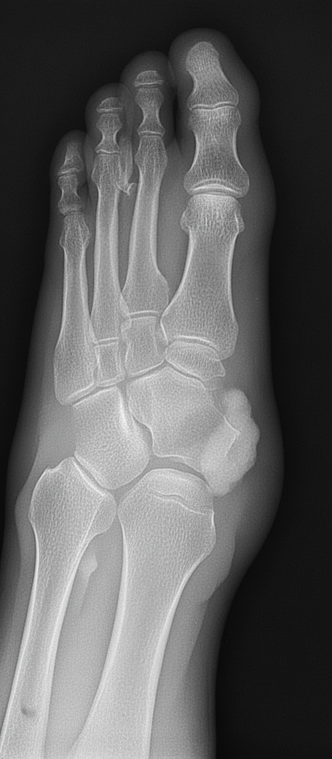
Which disease is shown by the following X-ray of a foot?
Which of the following is NOT a radiological finding seen in rickets?
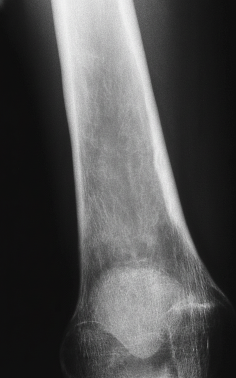
What diagnosis is suggested by the findings on this anteroposterior radiograph of the right tibia?
On an X-ray, what is the appearance of dead bone?
What is the first-choice imaging modality for evaluating a shoulder joint?
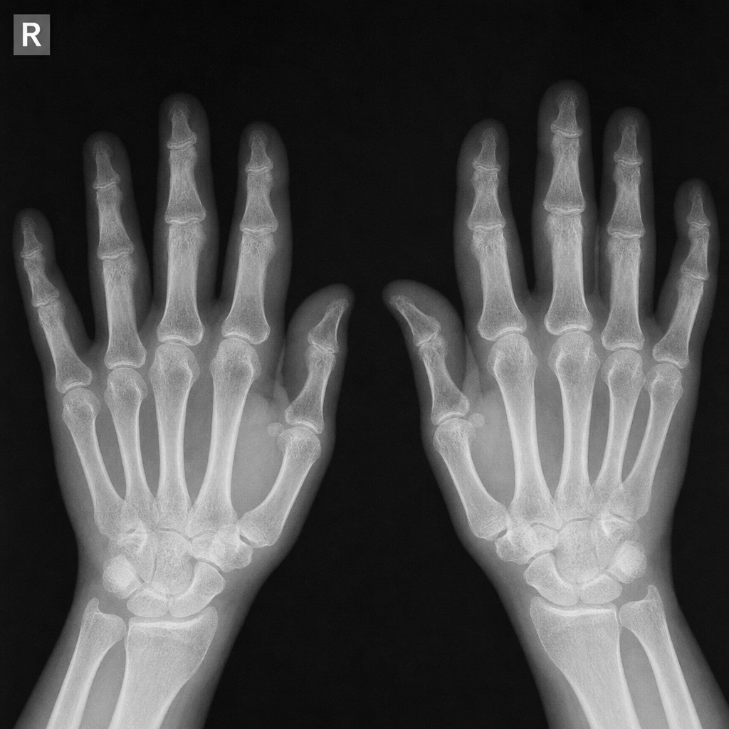
What is the earliest radiological change observed in rheumatoid arthritis?
Increased heel pad thickness is associated with which of the following conditions?
Practice by Chapter
Radiographic Anatomy of Bones and Joints
Practice Questions
Imaging of Fractures and Dislocations
Practice Questions
Arthritides: Inflammatory and Degenerative
Practice Questions
Metabolic Bone Diseases
Practice Questions
Bone and Soft Tissue Tumors
Practice Questions
Congenital Skeletal Anomalies
Practice Questions
Spine Imaging
Practice Questions
Skeletal Infections
Practice Questions
Sports Medicine Imaging
Practice Questions
Imaging of Prostheses and Implants
Practice Questions
Musculoskeletal Ultrasound
Practice Questions
MSK Interventional Procedures
Practice Questions
Want unlimited practice?
Get full access to all questions, explanations, and performance tracking.
Scan to download app