All of the following are radiological features of scleroderma except?
Increased heel pad thickness can be measured on X-ray of the foot. This can be seen in all conditions except?
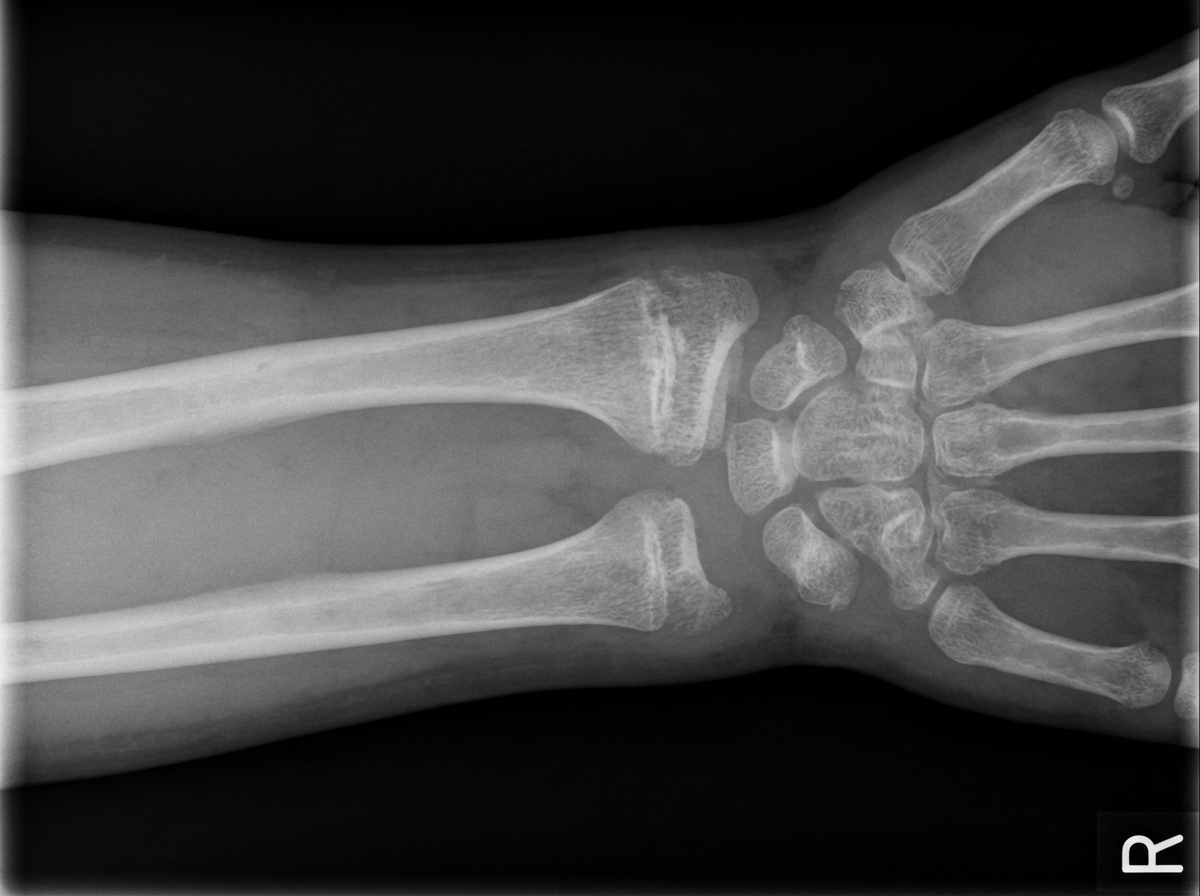
A wrist X-ray is shown. What is the probable diagnosis?

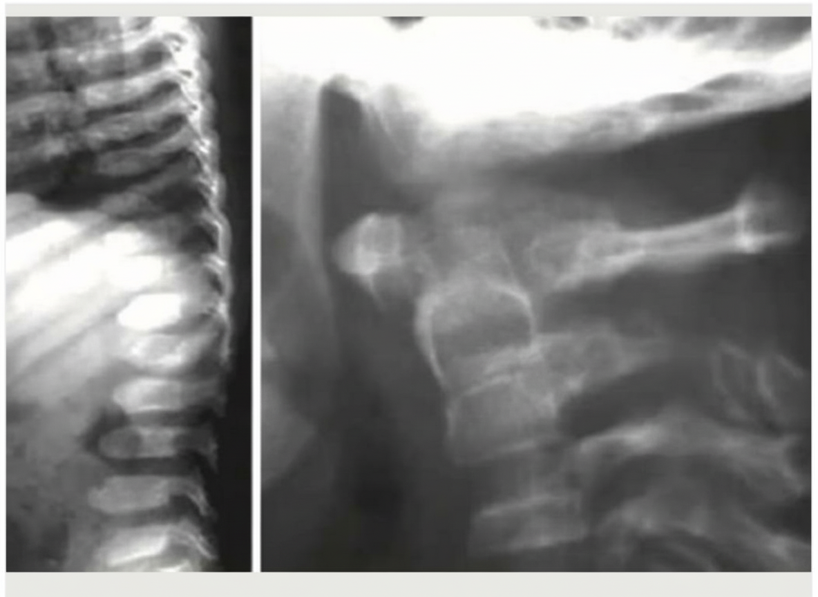
In which of the following diseases are the given X-ray findings typically seen?

Radiographically, how can a bony neoplasm be differentiated from a cyst?
Wormian bones are features of all of the following conditions EXCEPT:
Splaying and cupping of the metaphysis is seen in which condition?
Flaring of the anterior ends of the ribs is characteristically seen in which condition?
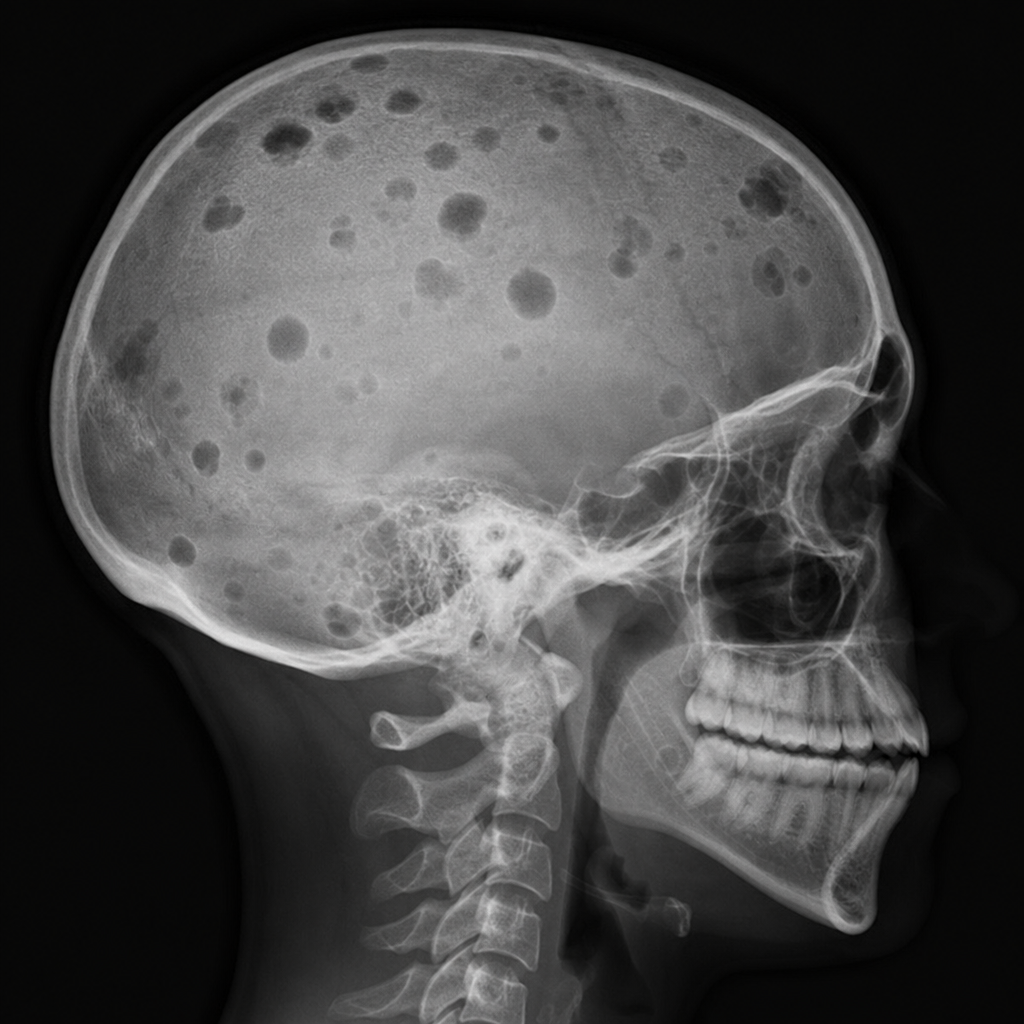
An X-ray of the skull shows multiple lesions. What is the most likely diagnosis?

What is the diagnostic radiological finding in skeletal fluorosis?
Explanation: **Explanation** Scleroderma (Systemic Sclerosis) is a multisystem connective tissue disorder characterized by vascular dysfunction and progressive fibrosis of the skin and internal organs. **1. Why "Diffuse periosteal reaction" is the correct answer:** Scleroderma is primarily a disease of **resorption and fibrosis**, not new bone formation. Diffuse periosteal reaction is typically seen in conditions like Hypertrophic Osteoarthropathy (HOA), thyroid acropachy, or venous stasis. In scleroderma, the musculoskeletal hallmark is **acro-osteolysis** (resorption of the terminal phalanges) rather than periosteal proliferation. **2. Analysis of incorrect options:** * **Oesophageal dysmotility:** This is a classic feature (part of the CREST syndrome). Radiologically, it presents as a "dilated, aperistaltic esophagus" (glass tube esophagus) due to smooth muscle atrophy and fibrosis, leading to gastroesophageal reflux and strictures. * **Erosion of the tip of the phalanges:** Known as **acro-osteolysis**, this is a high-yield radiological sign. It occurs due to digital ischemia (Raynaud’s) and pressure from the tightened skin (sclerodactyly). * **Lung nodules:** While Interstitial Lung Disease (NSIP pattern) is more common, scleroderma patients can develop pulmonary nodules. These may represent rheumatoid nodules (in overlap syndromes), malignancy (increased risk in fibrosis), or organized pneumonia. **Clinical Pearls for NEET-PG:** * **Soft Tissue Calcification:** Calcinosis circumscripta (calcium deposits in periarticular soft tissues) is a hallmark. * **CREST Syndrome:** Calcinosis, Raynaud's, Esophageal dysmotility, Sclerodactyly, Telangiectasia. * **Widening of PDL space:** A characteristic dental radiograph finding in scleroderma is the uniform thickening of the periodontal ligament space. * **Chest HRCT:** Look for the "Ground-glass opacities" or "Honeycombing" (usually at the bases), indicating systemic sclerosis-associated ILD.
Explanation: **Explanation:** The **heel pad thickness** is measured on a lateral radiograph of the foot as the shortest distance between the calcaneus and the skin surface. In a healthy adult, the normal upper limit is typically **21–23 mm in females** and **23–25 mm in males**. An increase in this thickness is a classic radiological sign associated with specific systemic and local conditions. **Why Hyperthyroidism is the Correct Answer:** Hyperthyroidism is **not** associated with increased heel pad thickness. In contrast, **Hypothyroidism (Myxedema)** causes an increase due to the deposition of glycosaminoglycans (mucopolysaccharides) and associated water retention in the soft tissues. **Analysis of Other Options:** * **Granulated GH Cell Adenoma (Acromegaly):** This is the most classic cause. Excess Growth Hormone (GH) leads to soft tissue hypertrophy and hyperplasia, significantly increasing heel pad thickness. It is often used as a screening or monitoring tool for Acromegaly. * **Callus Formation:** Localized mechanical stress leads to hyperkeratosis and thickening of the skin and subcutaneous tissues, directly increasing the measurement on X-ray. * **Hypothyroidism:** As mentioned, the accumulation of interstitial mucopolysaccharides leads to diffuse soft tissue swelling, including the heel pad. **NEET-PG High-Yield Pearls:** * **Mnemonic for Increased Heel Pad Thickness (MAD COAL):** **M**yxedema, **A**cromegaly, **D**ilantin (Phenytoin) use, **C**allus, **O**besity, **A**ndrogen replacement, **L**ocal infection/inflammation. * **Obesity** is the most common non-pathological cause of increased heel pad thickness. * **Acromegaly** should be strongly suspected if the thickness exceeds **25 mm** in a non-obese patient. * Other radiological features of Acromegaly include "spade-like" terminal phalanges and widening of joint spaces.
Explanation: ***Rickets*** - Classic X-ray findings include **widened, cupped, and frayed metaphyses** at the distal radius and ulna, indicating defective mineralization. - **Widened growth plates** and **delayed ossification** are characteristic features of rickets in growing children. *Colles fracture* - Shows a **dorsally angulated fracture** of the distal radius with characteristic **dinner fork deformity**. - Would demonstrate clear **cortical disruption** and **fracture lines**, not the smooth metaphyseal changes seen in rickets. *Scaphoid fracture* - Typically occurs in the **waist of the scaphoid bone** and may not be immediately visible on initial X-rays. - Presents with **tenderness in the anatomical snuffbox** and normal metaphyseal appearance of radius and ulna. *Osteoporosis* - Characterized by **generalized bone demineralization** and **cortical thinning** without specific metaphyseal changes. - Does not cause the distinctive **cupping and fraying** of metaphyses seen in metabolic bone disease.
Explanation: ***Morquio disease*** - Classic **skeletal dysplasia** findings include **platyspondyly** (flattened vertebrae), **odontoid hypoplasia**, and **genu valgum** (knock-knees). - Additional X-ray features show **flared iliac wings**, **epiphyseal dysplasia**, and **short stature** with disproportionate limbs. *Gaucher disease* - Primarily shows **Erlenmeyer flask deformity** of distal femur and **bone infarcts** rather than skeletal dysplasia. - **Hepatosplenomegaly** is prominent, but characteristic skeletal malformations of mucopolysaccharidoses are absent. *Fabry disease* - **X-ray findings are typically normal** as this lysosomal storage disease primarily affects cardiovascular and renal systems. - Clinical features include **angiokeratomas** and **corneal deposits**, not skeletal abnormalities. *Niemann Pick disease* - **Skeletal involvement is minimal** with normal bone architecture on X-rays. - Disease primarily causes **hepatosplenomegaly** and **cherry-red spot** on fundoscopy, not bone dysplasia.
Explanation: In musculoskeletal radiology, the key to differentiating a true neoplasm from a simple bone cyst lies in the **biological response of the host bone**. ### Why "Peripheral Bone Formation" is Correct A **bony neoplasm** (especially malignant or aggressive ones) often triggers a **periosteal reaction** or peripheral bone formation (e.g., Codman’s triangle, sunburst appearance, or onion-skinning). This occurs because the tumor irritates the periosteum or breaks through the cortex, forcing the body to attempt new bone formation. In contrast, a **simple bone cyst** is a benign, fluid-filled cavity that typically causes "expansile" thinning of the cortex without inducing an active periosteal reaction, unless a pathological fracture occurs. ### Why Other Options are Incorrect * **Size:** Both cysts (like Unicameral Bone Cysts) and neoplasms (like Giant Cell Tumors) can vary significantly in size; size alone is not a definitive diagnostic criterion. * **Shape:** While some cysts have classic shapes (e.g., "fallen leaf" sign in fractured cysts), many neoplasms can also appear geographic or multiloculated, making shape an unreliable differentiator. * **Location:** Although certain tumors have "predilection sites" (e.g., Epiphysis for Giant Cell Tumor), both cysts and neoplasms can occur in the metaphysis or diaphysis, leading to significant overlap. ### NEET-PG High-Yield Pearls * **Fallen Leaf Sign:** Pathognomonic for a fractured **Unicameral Bone Cyst (UBC)**; a cortical fragment settles at the bottom of the fluid-filled cavity. * **Periosteal Reactions:** * *Benign/Slow:* Thick, solid, wavy (e.g., Osteoid Osteoma). * *Aggressive/Malignant:* Sunburst, Codman’s triangle, or Spiculated (e.g., Osteosarcoma, Ewing’s Sarcoma). * **Lodwick Classification:** Used to grade the aggressiveness of bone lesions based on the "Zone of Transition." A narrow zone suggests a benign cyst; a wide, ill-defined zone suggests a neoplasm.
Explanation: **Explanation:** **Wormian bones** (sutural bones) are accessory small bones found within the cranial sutures, most commonly in the lambdoid suture. Their presence is often idiopathic but can be a diagnostic marker for several genetic and metabolic bone disorders. **Why Pyle’s Disease is the Correct Answer:** Pyle’s disease (Metaphyseal Dysplasia) is characterized by a failure of remodeling of the long bones, leading to the classic **"Erlenmeyer flask deformity"** of the distal femur and proximal tibia. While it involves significant skeletal dysplasia, it is **not** typically associated with Wormian bones. **Analysis of Incorrect Options:** * **Hypophosphatasia:** A metabolic bone disease caused by low alkaline phosphatase levels, leading to defective mineralization. It is a classic cause of multiple Wormian bones. * **Down’s Syndrome (Trisomy 21):** Children with Down’s syndrome frequently exhibit delayed closure of sutures and the presence of Wormian bones. * **Menke’s Kinky Hair Syndrome:** This is an X-linked recessive disorder of copper metabolism. Radiologically, it presents with metaphyseal spurring and prominent Wormian bones. **NEET-PG High-Yield Pearls:** To remember the causes of Wormian bones, use the mnemonic **"PORK CHOP"**: * **P:** **P**yknodysostosis (also causes osteosclerosis and acro-osteolysis) * **O:** **O**steogenesis Imperfecta (most common pathological cause) * **R:** **R**ickets (healing phase) * **K:** **K**inky Hair Syndrome (Menke’s) * **C:** **C**leidocranial Dysostosis (absent clavicles + Wormian bones) * **H:** **H**ypophosphatasia / **H**ypothyroidism * **O:** **O**ne too many chromosomes (Down’s Syndrome) * **P:** **P**achydermoperiostosis
Explanation: **Explanation:** The correct answer is **Rickets**. This condition is characterized by a failure of osteoid mineralization at the growth plate, leading to an accumulation of uncalcified cartilage. **1. Why Rickets is correct:** In Rickets, the lack of Vitamin D or calcium prevents the provisional zone of calcification from hardening. Under the mechanical stress of weight-bearing and muscle pull, the softened metaphysis becomes distorted. * **Cupping:** The metaphysis assumes a concave shape instead of being flat or convex. * **Splaying (Fraying):** The metaphysis widens and the margins become irregular or "brush-like" due to disorganized osteoid deposition. * **Widening of the Physis:** The radiolucent gap between the epiphysis and metaphysis increases. **2. Why other options are incorrect:** * **Scurvy (Vitamin C deficiency):** Characterized by defective collagen synthesis. Key radiological signs include the **White line of Frankel** (dense zone of provisional calcification), **Wimberger’s ring** (sclerotic margin of epiphysis), and **Pelkan spurs**. * **Paget’s Disease:** A disorder of bone remodeling. It presents with cortical thickening, coarsened trabeculae, and bone enlargement (e.g., **Picture frame vertebra** or **Cotton wool spots** in the skull), but not metaphyseal cupping. * **Lead Poisoning:** Characterized by **Lead lines**, which are dense transverse radiopaque bands at the metaphysis (increased density, not cupping). **Clinical Pearls for NEET-PG:** * **Earliest sign of Rickets:** Loss of the zone of provisional calcification. * **Rachitic Rosary:** Palpable/visible enlargement of costochondral junctions (rounded in Rickets, sharp/angular in Scurvy). * **Healing Rickets:** The first sign of healing on X-ray is the reappearance of the **zone of provisional calcification**.
Explanation: **Explanation:** **Rickets** is the correct answer because it is characterized by a failure of osteoid mineralization at the growth plates. In the thoracic cage, this occurs at the **costochondral junctions**. The accumulation of unmineralized osteoid leads to expansion and bulbous swelling of these junctions, which manifests radiologically as **flaring of the anterior ends of the ribs**. Clinically, this is known as the **"Rachitic Rosary."** Other classic radiological signs of Rickets include cupping, fraying, and splaying of the metaphyses (most prominent at the distal radius and ulna). **Analysis of Incorrect Options:** * **Neurofibromatosis (Type 1):** Characteristically shows **"rib ribboning"** (twisted, thinned appearance) and posterior vertebral scalloping, but not anterior flaring. * **Scurvy:** While it also involves the costochondral junctions (Scorbutic rosary), the swelling is due to subluxation of the cartilaginous plate. Radiologically, Scurvy is defined by the **White line of Frankel**, **Wimberger’s ring**, and **Pelkan spurs**, rather than generalized metaphyseal flaring. * **Hypothyroidism:** Typically presents with delayed bone age, epiphyseal dysgenesis (fragmented epiphyses), and "bullet-shaped" vertebrae, but does not cause anterior rib flaring. **High-Yield Clinical Pearls for NEET-PG:** * **Rachitic Rosary:** Broad and smooth (Rickets). * **Scorbutic Rosary:** Sharp and angular (Scurvy). * **Earliest sign of Rickets on X-ray:** Loss of the sharp zone of provisional calcification. * **Most sensitive site to detect Rickets:** Distal end of the ulna and the knee.
Explanation: ***Multiple myeloma*** - Causes multiple **punched-out lytic lesions** on skull X-ray, creating the classic **"raindrop skull"** or **"pepper-pot skull"** appearance. - These lesions have **well-defined margins** without sclerotic borders and represent plasma cell infiltration causing bone destruction. *Paget's disease* - Skull shows mixed **lytic and sclerotic lesions** creating the **"cotton-wool"** appearance, not purely lytic lesions. - May also present with **osteoporosis circumscripta** (flame-shaped lytic areas) but typically has bone expansion and thickening. *Eosinophilic granuloma* - Usually presents as a **solitary lytic lesion** with **beveled edges** creating a "hole-within-hole" appearance on lateral view. - Part of **Langerhans cell histiocytosis** and rarely causes multiple skull lesions in adults. *Osteomyelitis* - Shows **ill-defined lytic lesions** with **irregular margins** and associated **periosteal reaction**. - Often accompanied by **soft tissue swelling** and **sequestrum formation**, which are absent in purely lytic lesions.
Explanation: ### Explanation **Skeletal Fluorosis** is a chronic metabolic bone disease caused by prolonged ingestion of high amounts of fluoride. The hallmark of the disease is an increase in bone density (osteosclerosis) and pathological calcification of soft tissue structures. **Why Option B is Correct:** The most characteristic and diagnostic radiological feature of skeletal fluorosis is the **ossification of the interosseous membrane**, particularly in the forearm (between the radius and ulna) and the lower leg (between the tibia and fibula). While osteosclerosis occurs in many diseases, the ectopic calcification of ligaments, tendons, and interosseous membranes is highly specific to fluorosis and often serves as a definitive diagnostic clue on X-rays. **Analysis of Incorrect Options:** * **Option A:** Sclerosis of the sacroiliac joint is more characteristic of **Ankylosing Spondylitis** or osteitis condensans ilii. In fluorosis, the joint space is usually preserved despite periarticular sclerosis. * **Option C:** While **osteosclerosis of the vertebral bodies** (chalky white appearance) is a common finding in fluorosis, it is non-specific. It can also be seen in Renal Osteodystrophy (Rugger-jersey spine), Myelofibrosis, and Osteopetrosis. * **Option D:** While ligaments do ossify in fluorosis, the **sacrotuberous and sacrospinous ligaments** are more commonly involved than those of the knee joint. **High-Yield Clinical Pearls for NEET-PG:** * **Earliest Sign:** Osteosclerosis of the pelvis and lumbar spine. * **Classic Triad:** Osteosclerosis, Osteophytosis, and Calcification of ligaments/tendons. * **Dagger Spine:** Can mimic Ankylosing Spondylitis due to ossification of the supraspinous and interspinous ligaments. * **Dental Fluorosis:** Presents as "mottling of enamel" (occurs only if exposure happens during tooth development). * **Safe Limit:** Fluoride in drinking water should ideally be **<1.5 mg/L**.
Radiographic Anatomy of Bones and Joints
Practice Questions
Imaging of Fractures and Dislocations
Practice Questions
Arthritides: Inflammatory and Degenerative
Practice Questions
Metabolic Bone Diseases
Practice Questions
Bone and Soft Tissue Tumors
Practice Questions
Congenital Skeletal Anomalies
Practice Questions
Spine Imaging
Practice Questions
Skeletal Infections
Practice Questions
Sports Medicine Imaging
Practice Questions
Imaging of Prostheses and Implants
Practice Questions
Musculoskeletal Ultrasound
Practice Questions
MSK Interventional Procedures
Practice Questions
Get full access to all questions, explanations, and performance tracking.
Start For Free