
Musculoskeletal Radiology — MCQs

On this page
In early cases, multiple myeloma most commonly presents with which of the following radiographic findings?
Which of the following is the most sensitive investigation for the condition shown in the following X-ray?
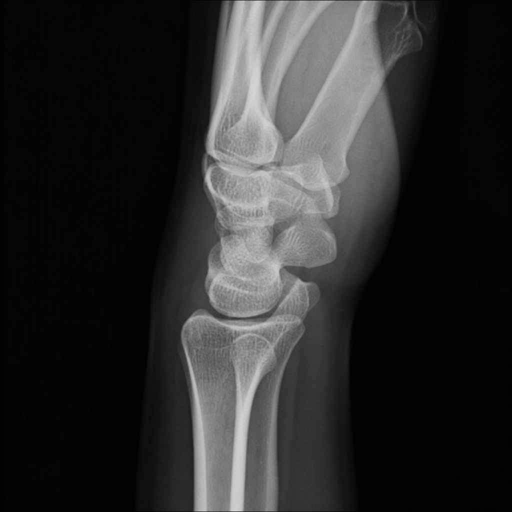
A lateral view X-ray of the wrist shows which of the following findings?
Cotton wool skull is seen in which condition?
Polka dot appearance on CT scan is characteristic of which of the following conditions?
What is the characteristic radiographic appearance of Pindborg's tumor?
What is the most specific but late radiographic feature of scurvy?
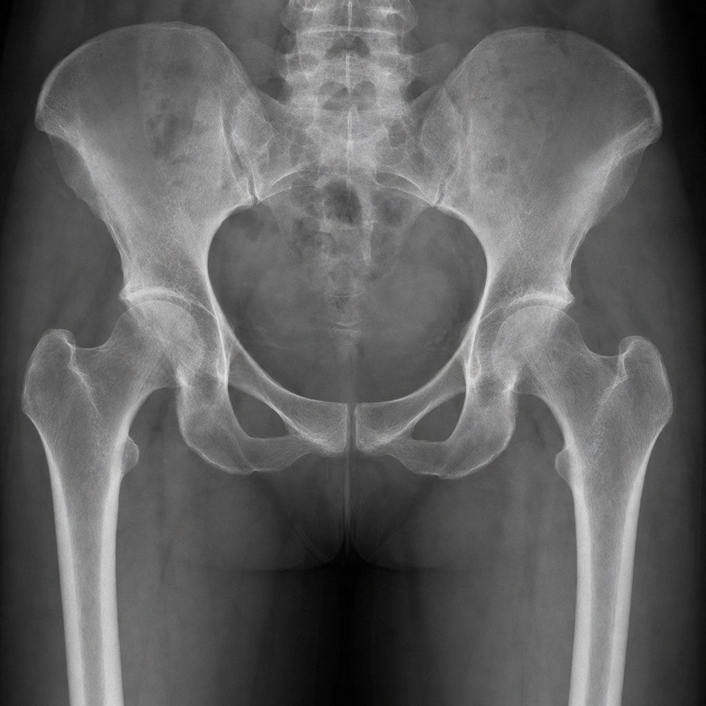
A 40-year-old male patient on long-term steroid therapy presents with recent onset of severe pain in the right hip. What is the imaging modality of choice for this problem?
Expansile type osseous metastases are characteristic of primary malignancy of which organ?
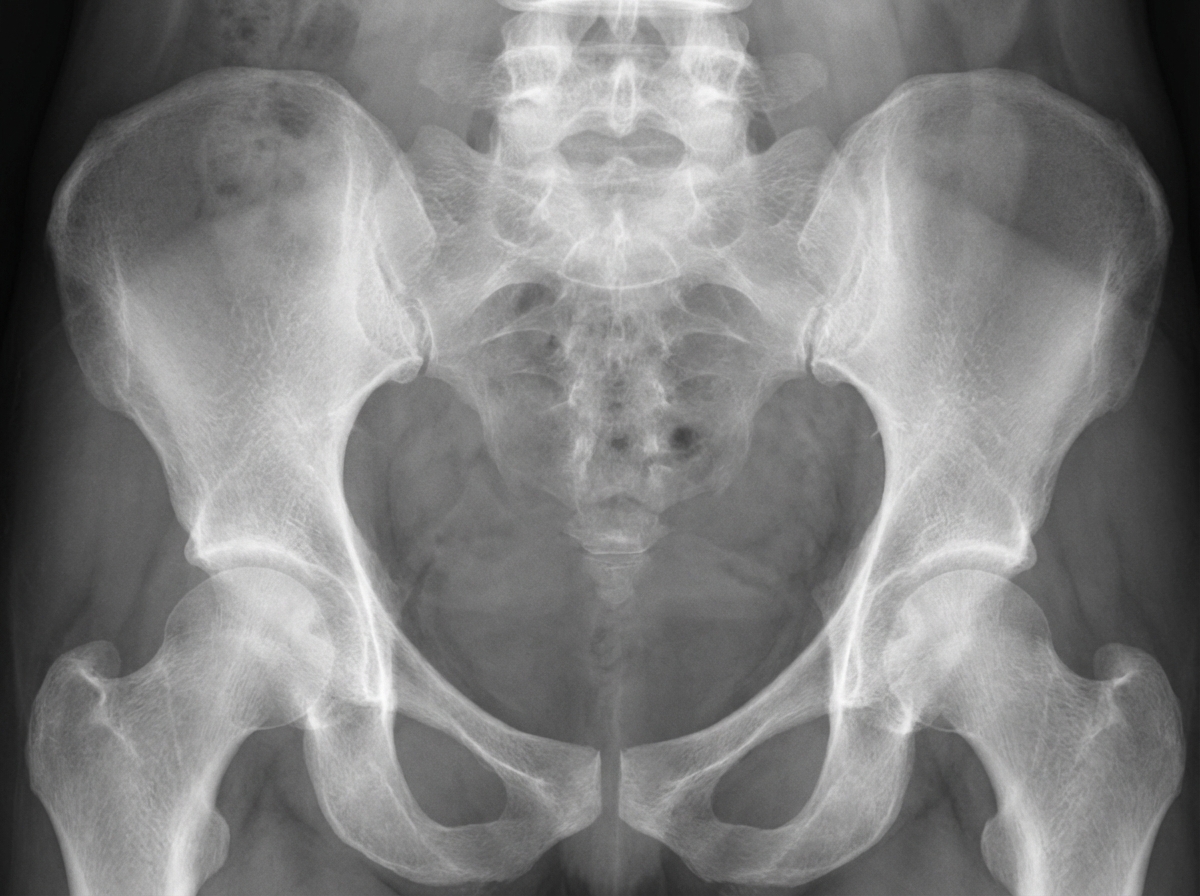
What type of pelvic appearance is this?
Practice by Chapter
Radiographic Anatomy of Bones and Joints
Practice Questions
Imaging of Fractures and Dislocations
Practice Questions
Arthritides: Inflammatory and Degenerative
Practice Questions
Metabolic Bone Diseases
Practice Questions
Bone and Soft Tissue Tumors
Practice Questions
Congenital Skeletal Anomalies
Practice Questions
Spine Imaging
Practice Questions
Skeletal Infections
Practice Questions
Sports Medicine Imaging
Practice Questions
Imaging of Prostheses and Implants
Practice Questions
Musculoskeletal Ultrasound
Practice Questions
MSK Interventional Procedures
Practice Questions
Want unlimited practice?
Get full access to all questions, explanations, and performance tracking.
Scan to download app