
Musculoskeletal Radiology — MCQs

On this page
What causes the sunray appearance in osteosarcoma?
Pepper pot appearance of the skull is seen in which of the following conditions?
Looser's zones are seen in which condition?
The white line of Frenkel is seen in which condition?
Geographic lytic lesions in the vault of the skull with bevelled edges are typically seen with which of the following conditions?
Stress fractures are diagnosed by which imaging modality?
The 'winking owl' sign is characteristic of which of the following conditions?
Sunburst appearance of skull is not seen in which of the following conditions?
The "flowing wax" appearance on the anterior and posterior borders of vertebrae, with normal intervertebral disc spaces, occurring due to ligament calcification, is seen in which condition?
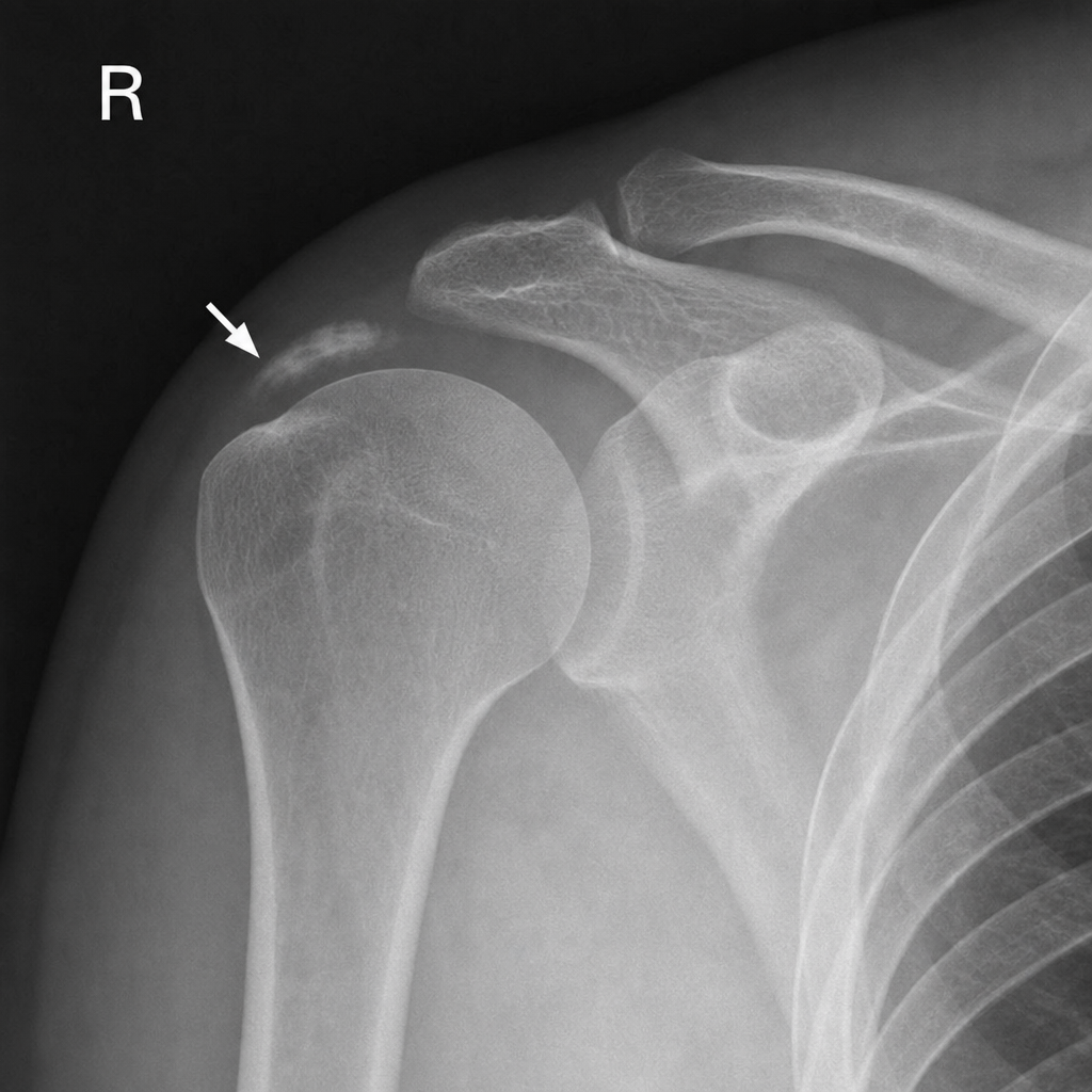
A 40-year-old male gymnast presents with right shoulder pain for 20 days. What does the arrow indicate?
Practice by Chapter
Radiographic Anatomy of Bones and Joints
Practice Questions
Imaging of Fractures and Dislocations
Practice Questions
Arthritides: Inflammatory and Degenerative
Practice Questions
Metabolic Bone Diseases
Practice Questions
Bone and Soft Tissue Tumors
Practice Questions
Congenital Skeletal Anomalies
Practice Questions
Spine Imaging
Practice Questions
Skeletal Infections
Practice Questions
Sports Medicine Imaging
Practice Questions
Imaging of Prostheses and Implants
Practice Questions
Musculoskeletal Ultrasound
Practice Questions
MSK Interventional Procedures
Practice Questions
Want unlimited practice?
Get full access to all questions, explanations, and performance tracking.
Scan to download app