
Musculoskeletal Radiology — MCQs

On this page
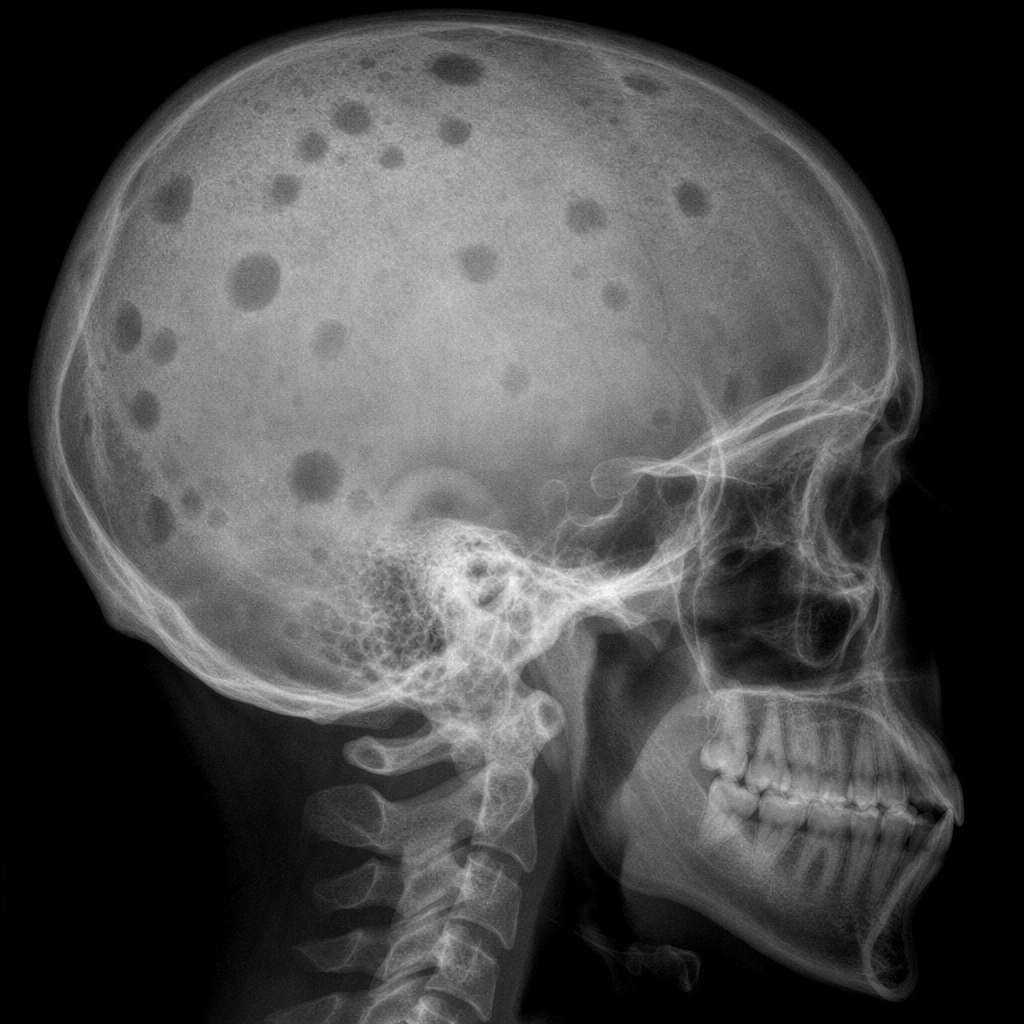
An X-ray of the skull showing multiple lesions is suggestive of what diagnosis?
All of the following are radiological signs of scurvy, except which of the following?
In which condition are Looser's zones typically observed?

Which of the following is NOT a radiological feature of osteoarthritis?
Onion peel appearance is seen with:
Flowing candle wax appearance is seen in:
In which condition is the cleavage plane sign typically observed?
In which of the following conditions is the appearance of H-shaped vertebrae not typically observed?
What is the definitive radiological sign of scurvy observed in an X-ray?
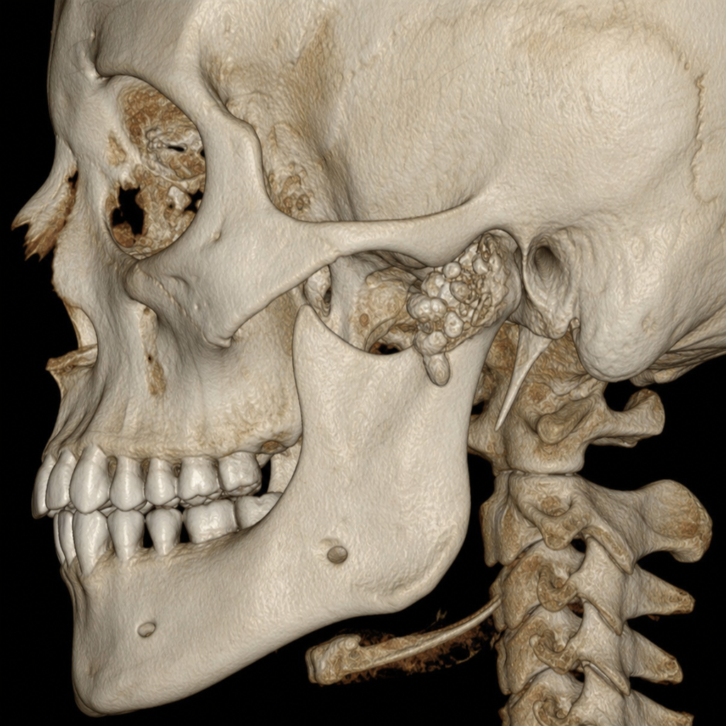
A 3D CT scan of this patient is shown. What is your conclusion?
Practice by Chapter
Radiographic Anatomy of Bones and Joints
Practice Questions
Imaging of Fractures and Dislocations
Practice Questions
Arthritides: Inflammatory and Degenerative
Practice Questions
Metabolic Bone Diseases
Practice Questions
Bone and Soft Tissue Tumors
Practice Questions
Congenital Skeletal Anomalies
Practice Questions
Spine Imaging
Practice Questions
Skeletal Infections
Practice Questions
Sports Medicine Imaging
Practice Questions
Imaging of Prostheses and Implants
Practice Questions
Musculoskeletal Ultrasound
Practice Questions
MSK Interventional Procedures
Practice Questions
Want unlimited practice?
Get full access to all questions, explanations, and performance tracking.
Scan to download app