
Musculoskeletal Radiology — MCQs

On this page
Flowing wax appearance on the anterior and posterior borders of the vertebrae is seen in?
Which of the following X-ray findings is characteristic of fibrous dysplasia?
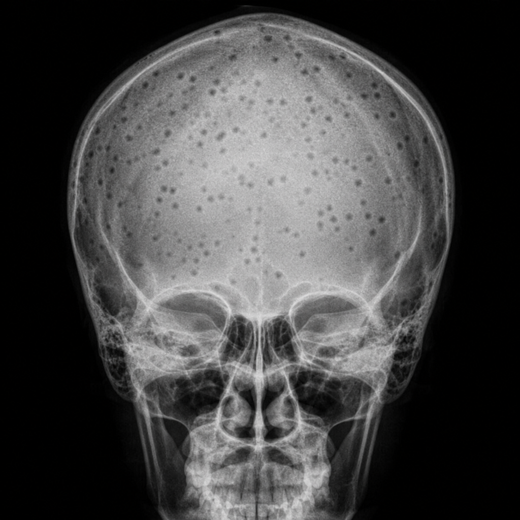
What is depicted in the X-ray of the skull? (Refer to the provided image)
Onion-peel appearance is seen in:
The earliest radiological change to appear in a case of acute osteomyelitis is:
In scurvy, all of the following radiological signs are seen except:
Which of the following is least useful for diagnosing spondylolisthesis?
What is the radiological feature characteristic of osteosarcoma?
Fallen fragment sign is a feature of what?
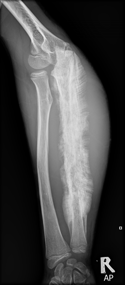
Aggressive bone production with a characteristic "sunray" appearance on x-ray is diagnostic of osteosarcoma in this child because of:
Practice by Chapter
Radiographic Anatomy of Bones and Joints
Practice Questions
Imaging of Fractures and Dislocations
Practice Questions
Arthritides: Inflammatory and Degenerative
Practice Questions
Metabolic Bone Diseases
Practice Questions
Bone and Soft Tissue Tumors
Practice Questions
Congenital Skeletal Anomalies
Practice Questions
Spine Imaging
Practice Questions
Skeletal Infections
Practice Questions
Sports Medicine Imaging
Practice Questions
Imaging of Prostheses and Implants
Practice Questions
Musculoskeletal Ultrasound
Practice Questions
MSK Interventional Procedures
Practice Questions
Want unlimited practice?
Get full access to all questions, explanations, and performance tracking.
Scan to download app