
Musculoskeletal Radiology — MCQs

On this page
What is the main diagnostic feature of osteoarthritis on X-ray imaging?
Which investigation is least effective in diagnosing acute osteomyelitis?
Shenton's line is seen in X-ray of -
Tufting of the distal phalanx is characteristically seen in which of the following conditions?
Which of the following is not a typical radiological finding in rheumatoid arthritis?
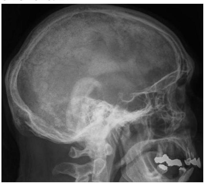
Cotton wool skull is a radiological feature of which condition?
Based on the X-ray image, which condition is indicated by a sunray appearance?
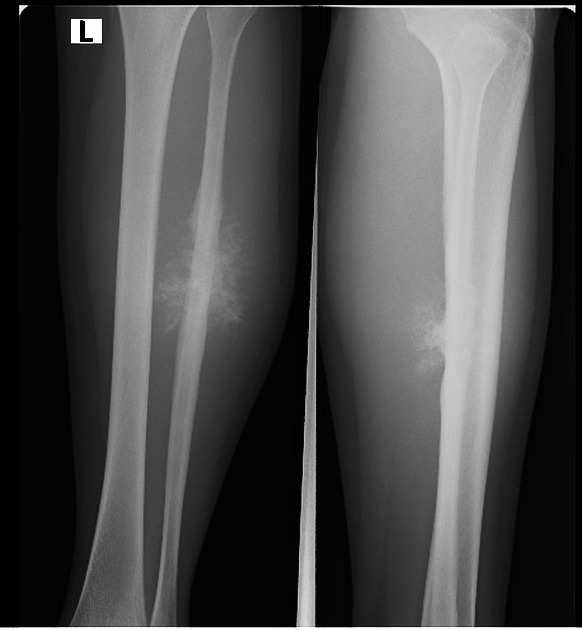
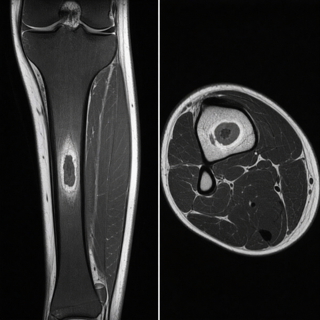
Identify the condition based on the provided image of a bone lesion. Ensure the diagnosis is based on the radiological and clinical features provided.
What type of lesions in the skull bones can be identified on this X-ray?
Which of the following statements about lipoma is radiologically true?
Practice by Chapter
Radiographic Anatomy of Bones and Joints
Practice Questions
Imaging of Fractures and Dislocations
Practice Questions
Arthritides: Inflammatory and Degenerative
Practice Questions
Metabolic Bone Diseases
Practice Questions
Bone and Soft Tissue Tumors
Practice Questions
Congenital Skeletal Anomalies
Practice Questions
Spine Imaging
Practice Questions
Skeletal Infections
Practice Questions
Sports Medicine Imaging
Practice Questions
Imaging of Prostheses and Implants
Practice Questions
Musculoskeletal Ultrasound
Practice Questions
MSK Interventional Procedures
Practice Questions
Want unlimited practice?
Get full access to all questions, explanations, and performance tracking.
Scan to download app