
Musculoskeletal Radiology — MCQs

On this page
In an adult patient, interpret the finding of a vertebral body that has lost almost its entire height anteriorly and posteriorly (pancake vertebra). Which of the following conditions is most likely associated with this finding?
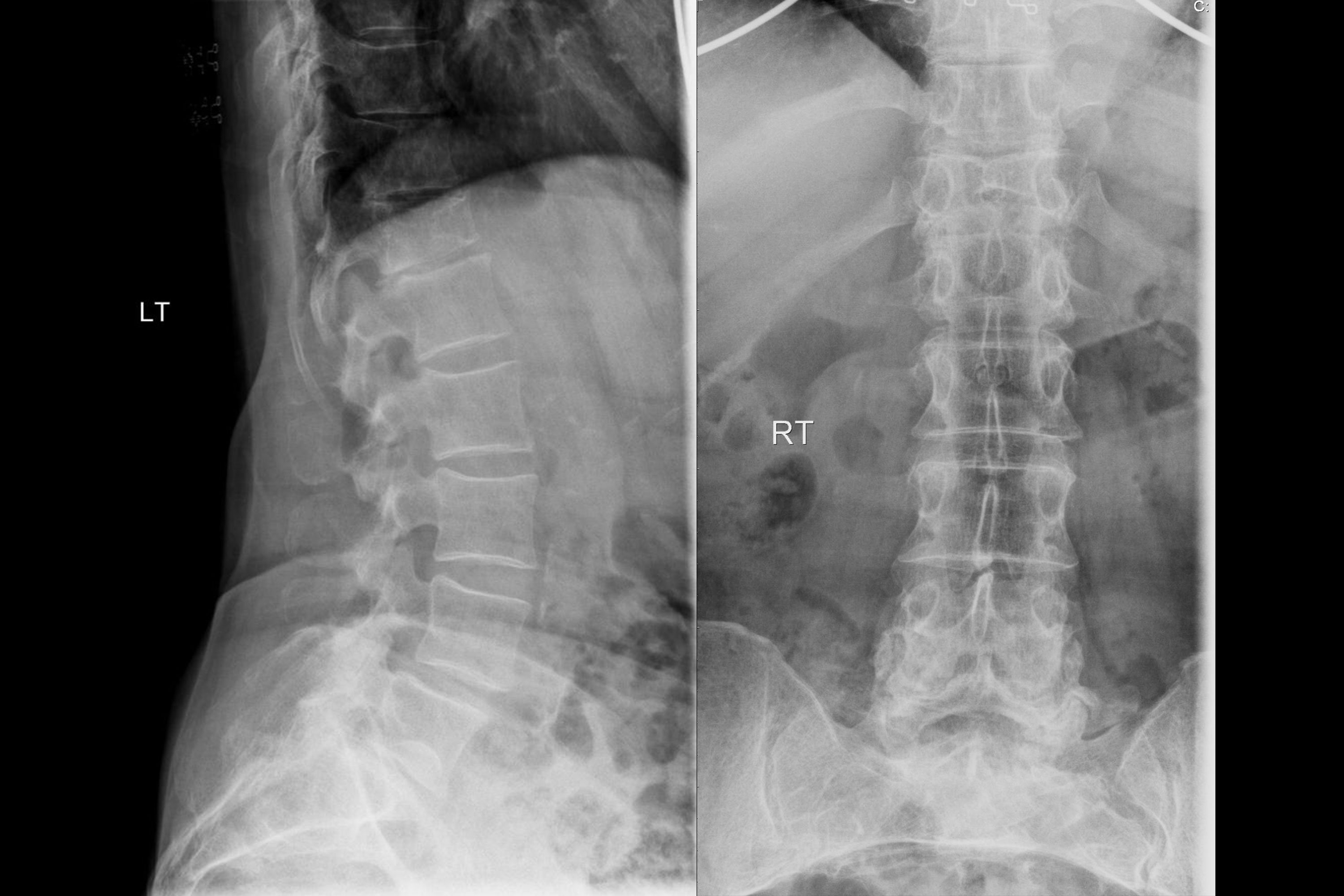
A 75-year-old female has chronic backache. X-ray of the spine is shown. What is the most likely diagnosis?
All the following are seen in Hyperparathyroidism except:
Spine MRI shows 'pencil-sharpened' vertebral bodies and 'H-shaped' vertebrae on T1-weighted images. Most likely diagnosis?
Most sensitive modality for detecting bone metastases
STIR sequence in MRI is most useful for
A child with lymphoma shows 'ivory vertebra' sign. Best imaging modality for evaluation?
Most sensitive imaging modality for detecting early osteomyelitis
What is the most reliable radiological sign of early osteonecrosis of femoral head?
Which MRI finding is suggestive of a torn meniscus in the knee?
Practice by Chapter
Radiographic Anatomy of Bones and Joints
Practice Questions
Imaging of Fractures and Dislocations
Practice Questions
Arthritides: Inflammatory and Degenerative
Practice Questions
Metabolic Bone Diseases
Practice Questions
Bone and Soft Tissue Tumors
Practice Questions
Congenital Skeletal Anomalies
Practice Questions
Spine Imaging
Practice Questions
Skeletal Infections
Practice Questions
Sports Medicine Imaging
Practice Questions
Imaging of Prostheses and Implants
Practice Questions
Musculoskeletal Ultrasound
Practice Questions
MSK Interventional Procedures
Practice Questions
Want unlimited practice?
Get full access to all questions, explanations, and performance tracking.
Scan to download app