
Musculoskeletal Radiology — MCQs

On this page
Judet view is used for fracture of
Punched out lesion in the skull is indicative of:
Which of the following is NOT a radiological finding of scurvy?
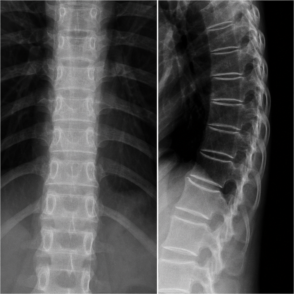
The X-ray findings of the spine shown are most characteristic of
Radiological sign of spondylolysis is
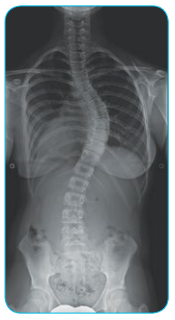
X-ray spine of a child is shown. What is the probable diagnosis?
The PRIMARY mechanisms that cause increased bone density (sclerosis) on X-ray include: a) Increased thickening of trabeculae b) Fracture & Collapse of cancellous bone c) Defective mineralization d) Myositis ossificans
Earliest investigation for diagnosis of Ankylosing spondylitis:
"Hour-glass" shape of the chest and "tri-radiate pelvis" are seen radiologically in -
Which imaging modality is LEAST useful in the initial diagnosis of stress fractures?
Practice by Chapter
Radiographic Anatomy of Bones and Joints
Practice Questions
Imaging of Fractures and Dislocations
Practice Questions
Arthritides: Inflammatory and Degenerative
Practice Questions
Metabolic Bone Diseases
Practice Questions
Bone and Soft Tissue Tumors
Practice Questions
Congenital Skeletal Anomalies
Practice Questions
Spine Imaging
Practice Questions
Skeletal Infections
Practice Questions
Sports Medicine Imaging
Practice Questions
Imaging of Prostheses and Implants
Practice Questions
Musculoskeletal Ultrasound
Practice Questions
MSK Interventional Procedures
Practice Questions
Want unlimited practice?
Get full access to all questions, explanations, and performance tracking.
Scan to download app