
Musculoskeletal Radiology — MCQs

On this page
The 'dot-dash' appearance on imaging is characteristic of which condition?
A dense metaphyseal band is typically seen on which of the following conditions?
What is the normal metacarpal index?
Subperiosteal erosion is seen in which of the following conditions?
All of the following are X-ray findings of Rheumatoid Arthritis except?
Subperiosteal resorption, most apparent on the radial aspect of the middle phalanx of the second and third fingers, is characteristic of which condition?
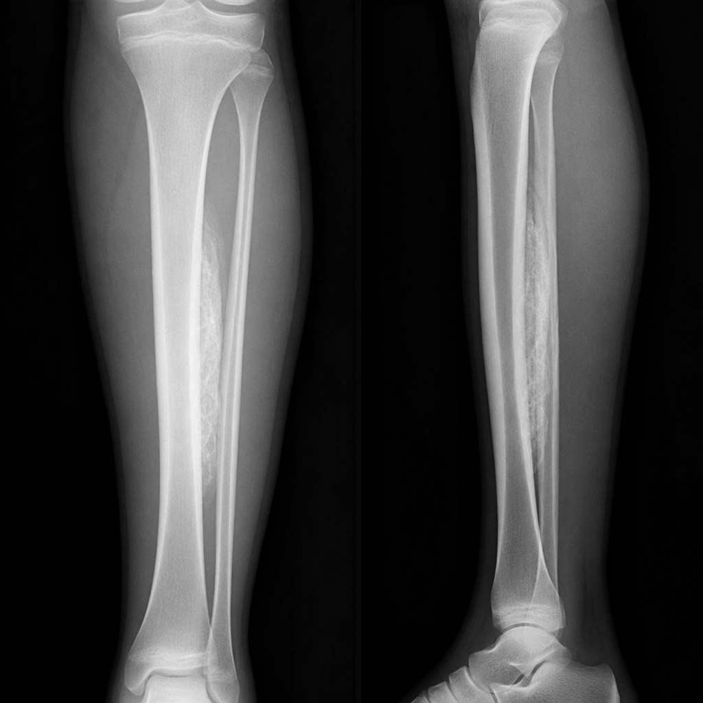
The most likely pathology associated with the following radiograph is:
Anterior scalloping of vertebrae is seen in which of the following conditions?
A 40-year-old female patient on long-term steroid therapy presents with recent onset of severe pain in the right hip. What is the imaging modality of choice for this problem?
A bone bruise or contusion is best identified using:
Practice by Chapter
Radiographic Anatomy of Bones and Joints
Practice Questions
Imaging of Fractures and Dislocations
Practice Questions
Arthritides: Inflammatory and Degenerative
Practice Questions
Metabolic Bone Diseases
Practice Questions
Bone and Soft Tissue Tumors
Practice Questions
Congenital Skeletal Anomalies
Practice Questions
Spine Imaging
Practice Questions
Skeletal Infections
Practice Questions
Sports Medicine Imaging
Practice Questions
Imaging of Prostheses and Implants
Practice Questions
Musculoskeletal Ultrasound
Practice Questions
MSK Interventional Procedures
Practice Questions
Want unlimited practice?
Get full access to all questions, explanations, and performance tracking.
Scan to download app