
Musculoskeletal Radiology — MCQs

On this page
What are the radiological changes observed in Rickets?
Radiological appearance of rickets includes all of the following except?
A "sharpened pencil" appearance of the mandibular condyle on a radiograph indicates which of the following conditions?
Which of the following is NOT a radiological finding of hemophilic arthropathy?
All of the following are features of Achondroplasia except?
Which radiographic view is best for visualizing condylar and ramus fractures?
What are the characteristic radiographic changes seen in the alveolar bone?
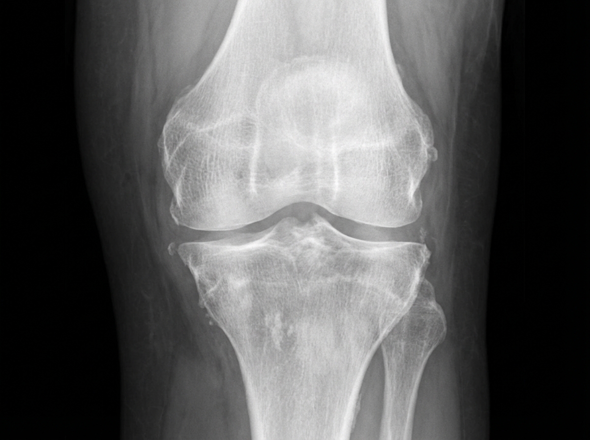
The following X-ray has a feature which is characteristic of which condition?
Which one of the following is a recognized X-ray feature of rheumatoid arthritis?
Phleboliths are typically seen in which of the following locations?
Practice by Chapter
Radiographic Anatomy of Bones and Joints
Practice Questions
Imaging of Fractures and Dislocations
Practice Questions
Arthritides: Inflammatory and Degenerative
Practice Questions
Metabolic Bone Diseases
Practice Questions
Bone and Soft Tissue Tumors
Practice Questions
Congenital Skeletal Anomalies
Practice Questions
Spine Imaging
Practice Questions
Skeletal Infections
Practice Questions
Sports Medicine Imaging
Practice Questions
Imaging of Prostheses and Implants
Practice Questions
Musculoskeletal Ultrasound
Practice Questions
MSK Interventional Procedures
Practice Questions
Want unlimited practice?
Get full access to all questions, explanations, and performance tracking.
Scan to download app