
Musculoskeletal Radiology — MCQs

On this page
Acro-osteolysis is seen in which of the following conditions?
Onion peel appearance on radiograph is not seen in which of the following conditions?
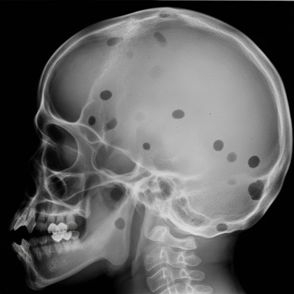
The following radiographs are most likely associated with which condition?
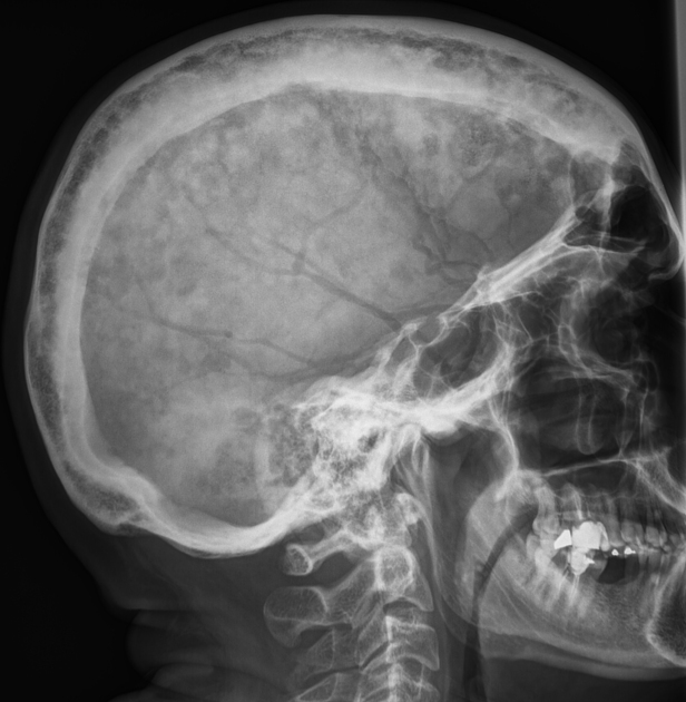
Which of the following conditions is NOT characterized by the radiographic abnormality shown in the X-ray skull?
Which of the following is NOT a radiological change in mucopolysaccharidosis?
Dense calcification is found in which of the following?
What is the earliest radiological feature in rheumatoid arthritis?
The "codfish" vertebra is not commonly seen in which of the following conditions?
An ill-defined lesion margin is described as:
The Insall-Salvati index is used for assessing which anatomical structure?
Practice by Chapter
Radiographic Anatomy of Bones and Joints
Practice Questions
Imaging of Fractures and Dislocations
Practice Questions
Arthritides: Inflammatory and Degenerative
Practice Questions
Metabolic Bone Diseases
Practice Questions
Bone and Soft Tissue Tumors
Practice Questions
Congenital Skeletal Anomalies
Practice Questions
Spine Imaging
Practice Questions
Skeletal Infections
Practice Questions
Sports Medicine Imaging
Practice Questions
Imaging of Prostheses and Implants
Practice Questions
Musculoskeletal Ultrasound
Practice Questions
MSK Interventional Procedures
Practice Questions
Want unlimited practice?
Get full access to all questions, explanations, and performance tracking.
Scan to download app