Subcutaneous calcifications are seen in which of the following conditions?
Which of the following is/are X-ray feature/s of Perthes disease?
Blowout expansile bone metastasis is typically seen with which of the following primary malignancies?
Which imaging view best demonstrates inflammation and temporomandibular joint effusion?
Bohler's angle is reduced in which of the following fractures?
Which sarcoma does not display the radiographic feature of Codman’s triangle?
Which X-ray finding is characteristic of osteomyelitis within 8 days of onset?
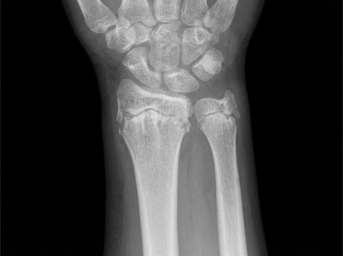
The following X-ray is diagnostic of:

Salt-pepper skull is a characteristic radiographic finding in which of the following conditions?
What are the common causes of vertebral plana?
Explanation: **Explanation:** **Hyperparathyroidism** (specifically secondary and tertiary forms) is a classic cause of **metastatic calcification**. In this condition, an elevated calcium-phosphate product leads to the deposition of calcium hydroxyapatite in soft tissues, including the subcutaneous layers. This is often seen radiologically as amorphous, cloud-like opacities in the periarticular and subcutaneous regions. **Analysis of Options:** * **Hyperparathyroidism (Correct):** Chronic renal failure leads to secondary hyperparathyroidism, which is the most common cause of extensive soft tissue and subcutaneous calcification (often termed *tumoral calcinosis* when large and periarticular). * **Gout (Incorrect):** While gout involves the deposition of monosodium urate crystals (tophi), these are typically **radiolucent** on standard X-rays unless they are very long-standing and have associated secondary calcium deposition. The primary pathology is not subcutaneous calcification but joint destruction and "punched-out" erosions. * **Ochronosis (Incorrect):** Also known as Alkaptonuria, this condition is characterized by the deposition of pigment in connective tissues. Radiologically, its hallmark is **dense disc space calcification** involving multiple levels of the spine and large joint arthropathy, not subcutaneous calcification. **NEET-PG High-Yield Pearls:** * **Differential Diagnosis for Subcutaneous Calcification:** Remember the mnemonic **"DISH-C"**: **D**ermatomyositis/Polymyositis, **I**diopathic, **S**cleroderma (CREST syndrome), **H**yperparathyroidism, and **C**alcinosis cutis. * **Monckeberg’s Sclerosis:** Another form of metastatic calcification seen in diabetics/renal patients, involving the tunica media of arteries. * **Ochronosis Key Sign:** "Vacuum phenomenon" in intervertebral discs followed by wafer-like calcification.
Explanation: **Explanation:** Legg-Calvé-Perthes Disease (LCPD) is an idiopathic avascular necrosis of the capital femoral epiphysis, typically seen in children aged 4–8 years. The radiographic features evolve through stages (Waldenström classification), and the options provided represent various pathological changes seen during these phases. * **Increased medial joint space (Waldenström sign):** This is often the earliest radiographic sign. It occurs due to hypertrophy of the acetabular cartilage, presence of joint effusion, and thickening of the ligamentum teres. * **Metaphyseal cysts and rarefaction:** As the disease progresses, the metaphysis becomes involved. Cysts and radiolucency (rarefaction) occur due to increased vascularity and osteoclastic activity during the repair process. * **Lateral extrusion of the femur head:** During the fragmentation stage, the femoral head becomes flattened (coxa plana) and may subluxate laterally. This "lateralization" is a poor prognostic sign as it leads to abnormal remodeling and joint incongruity. Since all three features are characteristic radiographic findings of Perthes disease, **Option D** is the correct answer. **High-Yield Clinical Pearls for NEET-PG:** * **Gage’s Sign:** A V-shaped lucency on the lateral side of the epiphysis (indicates a poor prognosis). * **Crescent Sign:** A subchondral fracture line, best seen on the frog-leg lateral view. * **Prognosis:** "Head at risk" signs include lateral calcification, Gage’s sign, and lateral subluxation. * **Differential Diagnosis:** Slipped Capital Femoral Epiphysis (SCFE) – usually seen in obese adolescents; Perthes is seen in younger children.
Explanation: **Explanation:** The correct answer is **Renal Cell Carcinoma (RCC)**. **1. Why Renal Cell Carcinoma is correct:** Bone metastases are generally classified as osteolytic, osteoblastic, or mixed. RCC typically produces **purely osteolytic** lesions. The term "blowout" refers to a highly vascular, expansile, and aggressive lytic lesion that causes significant thinning and outward bulging of the bony cortex. This appearance is a hallmark of RCC and **Thyroid carcinoma**. The hypervascular nature of these tumors leads to the rapid destruction of bone and the characteristic "soap-bubble" or "blowout" appearance on X-ray. **2. Why other options are incorrect:** * **Bronchogenic carcinoma:** While lung cancer is a common cause of lytic bone metastases, they are usually "punched-out" or permeative rather than classically expansile/blowout. * **Breast carcinoma:** This is the most common primary to cause **mixed** (both lytic and sclerotic) lesions. While it can be purely lytic, it rarely presents with the classic blowout morphology. * **Prostate carcinoma:** This is the classic cause of **osteoblastic (sclerotic)** metastases, appearing as dense, white spots on a radiograph due to increased bone formation. **3. NEET-PG High-Yield Pearls:** * **Mnemonic for Blowout Metastasis:** "**R**eally **T**errible" (**R**enal cell and **T**hyroid). * **Pulsatile Bone Metastasis:** If a patient presents with a palpable, pulsatile bone mass, think RCC or Thyroid carcinoma (due to high vascularity). * **Most common site for bone mets:** Axial skeleton (spine > pelvis > ribs). * **Investigation of choice:** While Bone Scans are sensitive for most mets, they may be **false negative** in RCC and Multiple Myeloma because these lesions lack osteoblastic activity. MRI or PET-CT is preferred in such cases.
Explanation: **Explanation:** The **Temporomandibular Joint (TMJ)** is best evaluated using Magnetic Resonance Imaging (MRI) because it provides superior soft-tissue contrast for the articular disc and surrounding structures. **Why T2-weighted MRI is the correct answer:** In MRI physics, **T2-weighted images** are highly sensitive to water and fluid. Inflammation in the joint leads to increased capillary permeability and the accumulation of synovial fluid (effusion). On T2 sequences, this fluid appears as a **hyperintense (bright white)** signal. Therefore, T2-weighted MRI is the gold standard for detecting joint effusion, bone marrow edema, and active inflammatory changes in the TMJ. **Analysis of Incorrect Options:** * **B. T1-weighted MR images:** While T1 is excellent for demonstrating anatomical detail and the position of the articular disc (due to high signal from fatty marrow), it is poor at distinguishing fluid from surrounding soft tissues. * **C. Lateral tomogram:** This is a specialized radiographic technique used primarily to visualize bony anatomy and joint space. It cannot detect soft tissue inflammation or minor fluid collections. * **D. Panoramic radiograph (OPG):** This is a screening tool used to view the mandible and dentition. It lacks the resolution to visualize the TMJ disc or internal derangements and cannot detect effusion. **High-Yield Clinical Pearls for NEET-PG:** * **Gold Standard for TMJ Disc:** MRI (specifically T1 for anatomy/position). * **Gold Standard for TMJ Effusion:** T2-weighted MRI. * **Best for Bony Erosions:** Non-contrast CT (NCCT) or Cone Beam CT (CBCT). * **Closed vs. Open Mouth Views:** MRI is typically performed in both positions to assess the mobility and reduction of the articular disc.
Explanation: **Explanation:** **Bohler’s Angle** (also known as the Tuber-joint angle) is a crucial radiographic measurement used to assess the integrity of the **calcaneus**. It is formed by drawing two lines on a lateral foot X-ray: 1. From the highest point of the anterior process to the highest point of the posterior facet. 2. From the highest point of the posterior facet to the highest point of the calcaneal tuberosity. The **normal range is 20° to 40°**. In a **calcaneal fracture** (typically caused by an axial load, such as falling from a height), the calcaneal body is compressed or flattened, causing the angle to **decrease or even become negative**. **Analysis of Incorrect Options:** * **Supracondylar fracture of humerus:** Assessed using the *Baumann angle* or the *Anterior Humeral Line*. * **Fracture neck of femur:** Evaluated using *Garden’s classification* or *Pauwels' classification*; Bohler’s angle has no anatomical relevance here. * **Colles fracture:** This is a distal radius fracture. Radiographic assessment focuses on *radial inclination, radial height,* and *palmar tilt*. **High-Yield Clinical Pearls for NEET-PG:** * **Gissane’s Angle (Crucial Angle):** Also used for calcaneal fractures; it is the angle between the downward slope of the thalami regalis and the upward slope of the anterior process (Normal: 120°–145°). In fractures, this angle **increases**. * **Don Juan Syndrome:** Calcaneal fractures are often associated with compression fractures of the **lumbar spine** (L1-L2) due to the same axial loading mechanism. Always screen the spine in these patients. * **Mondor’s Sign:** Ecchymosis extending to the sole of the foot, pathognomonic for calcaneal fracture.
Explanation: ### Explanation **Codman’s triangle** is a type of aggressive periosteal reaction where the periosteum is lifted off the bone by a rapidly growing lesion (tumor, pus, or hemorrhage). It appears as a triangular area of new subperiosteal bone at the periphery of the lesion. #### Why Chondrosarcoma is the Correct Answer While **Chondrosarcoma** is a malignant bone tumor, it typically presents with a **"popcorn" calcification** or "rings and arcs" pattern within the matrix. It is generally a slower-growing tumor compared to Osteosarcoma or Ewing’s sarcoma. Because the periosteal reaction depends on the *rate* of growth, Chondrosarcoma often causes endosteal scalloping or cortical thickening rather than the classic aggressive Codman’s triangle. #### Analysis of Incorrect Options * **Osteosarcoma (Option B):** This is the most common primary malignant bone tumor to show Codman’s triangle. It is highly aggressive and frequently displays both Codman’s triangle and the "Sunburst" appearance. * **Ewing’s Sarcoma (Option C):** Known for its "Onion-skin" (lamellated) periosteal reaction, Ewing’s sarcoma is extremely aggressive and frequently demonstrates Codman’s triangle as the tumor breaks through the cortex. * **Fibrosarcoma (Option A):** This is a highly aggressive, osteolytic lesion. Due to its rapid growth and destruction of the cortex, it frequently elicits an aggressive periosteal response, including Codman’s triangle. #### NEET-PG High-Yield Pearls * **Codman’s Triangle is NOT pathognomonic for malignancy:** It can be seen in benign but aggressive conditions like **Osteomyelitis**, active Aneurysmal Bone Cysts (ABC), or subperiosteal hematomas. * **Sunburst/Spiculated appearance:** Most characteristic of Osteosarcoma. * **Onion-skin appearance:** Most characteristic of Ewing’s Sarcoma. * **Popcorn Calcification:** Classic for Chondroid lesions (Chondrosarcoma or Enchondroma). * **Ground Glass Matrix:** Classic for Fibrous Dysplasia.
Explanation: **Explanation:** In the context of acute osteomyelitis, X-ray findings lag significantly behind clinical symptoms. During the first **3 to 10 days** of infection, the only radiographic evidence is typically **soft tissue swelling**, characterized by the blurring of normal fat planes and edema in the deep soft tissues adjacent to the bone. * **Why Option B is correct:** Bone destruction (demineralization) only becomes visible on a plain radiograph after **30–50% of the bone mineral content** has been lost, which usually takes 10–14 days. Therefore, within the first 8 days, soft tissue swelling is the earliest and often the only sign. * **Why other options are incorrect:** * **Cystic swelling (A):** This is not a standard radiographic term for early osteomyelitis; it may be confused with Brodie’s abscess (a chronic form). * **New bone formation (C):** Periosteal reaction (new bone) typically appears after 10–14 days as the infection breaches the cortex. * **Sequestrum formation (D):** A sequestrum (piece of dead bone) is a hallmark of **chronic osteomyelitis** and takes weeks to develop. **NEET-PG High-Yield Pearls:** * **Earliest Sign on X-ray:** Soft tissue swelling (3–5 days in children, up to 10 days in adults). * **Earliest Imaging Modality:** **MRI** is the most sensitive and specific early imaging (detects marrow edema within 24–48 hours). * **Nuclear Medicine:** Triple-phase Bone Scan (Technetium-99m) shows increased uptake within 24–48 hours but is less specific than MRI. * **Involucrum:** New bone formation around a sequestrum (Chronic stage). * **Cloaca:** An opening in the involucrum for pus drainage.
Explanation: ***Rickets*** - Classic X-ray findings include **metaphyseal cupping**, **fraying**, and **widening of the growth plate** due to defective mineralization of the growth plate cartilage. - **Coxa vara**, **bowing deformities** of long bones, and **delayed ossification** are characteristic features that make this diagnosis evident on radiographs. *Scurvy* - X-ray shows **Frankel's line** (dense metaphyseal line), **Pelkan spurs** (corner fractures), and **Trümmerfeld zone** (rarefied metaphyseal area). - Caused by **vitamin C deficiency** affecting collagen synthesis, not calcium metabolism like rickets. *Hairline fracture of radius and ulna* - Would appear as **thin lucent lines** through the cortical bone without affecting the growth plates or causing metaphyseal changes. - **Acute trauma history** would be present, and there would be no generalized bone deformities or growth plate abnormalities. *Osteopetrosis* - X-ray shows **increased bone density** with a **marble bone appearance** and **sclerotic changes** throughout the skeleton. - **Erlenmeyer flask deformity** and **bone-in-bone appearance** are characteristic, opposite to the osteopenic changes seen in rickets.
Explanation: **Explanation:** **1. Why Primary Hyperparathyroidism is Correct:** The "Salt-and-Pepper" skull (also known as the granular skull) is a classic radiographic hallmark of **Hyperparathyroidism** (Primary, Secondary, or Tertiary). The underlying pathophysiology involves increased Parathyroid Hormone (PTH) levels, which stimulate osteoclastic activity. This leads to multiple tiny, well-defined areas of **trabecular bone resorption** (the "pepper") interspersed with areas of normal or sclerotic bone (the "salt"), resulting in a diffuse, mottled appearance of the calvarium. **2. Analysis of Incorrect Options:** * **Paget’s Disease:** Characterized by **"Cotton Wool Spots"** on the skull, which are thick, disorganized patches of sclerotic bone. It also features thickening of the diploic space and "Osteoporosis Circumscripta." * **Eosinophilic Granuloma (Langerhans Cell Histiocytosis):** Typically presents with **"Punched-out" lytic lesions** or a "Beveled edge" appearance due to uneven involvement of the inner and outer tables of the skull. * **Multiple Myeloma:** Characterized by multiple, discrete, small, circular **"Raindrop" lytic lesions**. Unlike the granular appearance of hyperparathyroidism, these lesions are sharply demarcated and uniform in size. **3. NEET-PG High-Yield Pearls:** * **Subperiosteal bone resorption** is the *most specific* radiographic sign of hyperparathyroidism, most commonly seen on the radial aspect of the middle phalanges of the 2nd and 3rd fingers. * **Brown Tumors** (Osteoclastomas) are non-neoplastic lytic lesions also associated with this condition. * **Rugger-Jersey Spine** is a classic finding in Secondary Hyperparathyroidism (Renal Osteodystrophy).
Explanation: **Vertebra Plana** refers to the complete, uniform collapse of a vertebral body, resulting in a thin, wafer-like or "pancake" appearance, while the adjacent intervertebral discs remain preserved. ### Explanation of Options: 1. **Tuberculosis (Correct):** While TB of the spine (Pott’s disease) typically involves the disc space, it is a leading cause of vertebral collapse in endemic regions. In the context of this specific question format (often seen in older NEET-PG/AIIMS patterns), TB is highlighted because it can cause significant bone destruction leading to a flattened vertebra, especially in children. 2. **Eosinophilic Granuloma (EG):** This is the **most common cause** of vertebra plana in children (Langerhans Cell Histiocytosis). It typically involves a single level, and remarkably, the bone often reconstitutes over time. 3. **Metastasis:** In adults, malignant infiltration (Metastasis or Multiple Myeloma) is a frequent cause of pathological collapse leading to vertebra plana. **Why "All of the Above" is the logical clinical answer:** In clinical practice, all three conditions are classic differentials for a flattened vertebral body. However, if forced to choose a single primary cause in a competitive exam context, **Eosinophilic Granuloma** is the most classic "textbook" association, while **Tuberculosis** is a high-incidence cause in the Indian subcontinent. ### High-Yield Clinical Pearls for NEET-PG: * **Mnemonic (FEET):** Causes of Vertebra Plana include **F**racture (Trauma), **E**osinophilic Granuloma, **E**wing’s Sarcoma, and **T**uberculosis/Tumor (Metastasis/Myeloma). * **Calvé Disease:** An older term specifically referring to vertebra plana caused by Eosinophilic Granuloma. * **Key Radiological Sign:** The preservation of disc space helps differentiate EG and Metastasis from pyogenic osteomyelitis (where the disc is destroyed early).
Radiographic Anatomy of Bones and Joints
Practice Questions
Imaging of Fractures and Dislocations
Practice Questions
Arthritides: Inflammatory and Degenerative
Practice Questions
Metabolic Bone Diseases
Practice Questions
Bone and Soft Tissue Tumors
Practice Questions
Congenital Skeletal Anomalies
Practice Questions
Spine Imaging
Practice Questions
Skeletal Infections
Practice Questions
Sports Medicine Imaging
Practice Questions
Imaging of Prostheses and Implants
Practice Questions
Musculoskeletal Ultrasound
Practice Questions
MSK Interventional Procedures
Practice Questions
Get full access to all questions, explanations, and performance tracking.
Start For Free