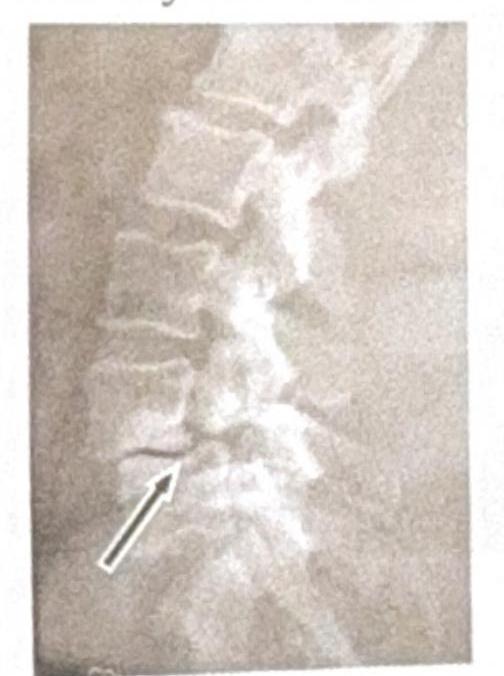
Identify the condition shown in the image:

An athlete sustained an injury around the knee joint, suspecting cartilage damage. Which of the following is the investigation of choice?
Which of the following thyroid carcinomas cannot be definitively diagnosed by fine needle aspiration cytology (FNAC)?
A 10 cm tumor is found on the anterior surface of the thigh. What is the most appropriate procedure to obtain a diagnosis?
A 10-year-old obese boy was referred to the emergency department with a history of hip pain. He was observed to be limping and complained of severe pain. Which of the following investigations is least appropriate for this condition?
In which condition is the cleavage plane sign typically observed?
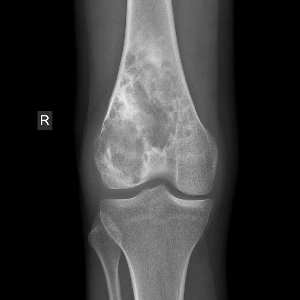
Which imaging modality is best for evaluating retinoblastoma?
Calcification around the joint is seen in which of the following conditions?
A "cold spot" on bone scintigraphy is seen in all the following conditions except?
Superior rib notching is caused by which of the following?
Explanation: ***Spondylolysis*** * The image shows a **break in the pars interarticularis** of a vertebra, indicated by the arrow, which is characteristic of spondylolysis. * This condition is a **stress fracture** or defect in the pars interarticularis, a bony segment connecting the superior and inferior articular facets. *Renal osteodystrophy* * Renal osteodystrophy refers to a spectrum of **bone abnormalities** that occur in chronic kidney disease, not a specific vertebral fracture pattern. * It typically involves features such as **osteomalacia**, **osteitis fibrosa cystica**, or **osteoporosis**, which are not directly depicted as a fracture in this image. *Spondylolisthesis* * Spondylolisthesis is the **anterior slippage** of one vertebral body over another, which can be caused by bilateral spondylolysis but is not directly shown as a slip in this specific image. * The image distinctly highlights the **fracture line** itself, rather than the displacement of the vertebral body. *Tuberculosis (TB)* * Spinal tuberculosis (Pott's disease) typically presents with **destruction of vertebral bodies**, disc space narrowing, and often a **paravertebral abscess**. * The image does not show these features; instead, it demonstrates a clear **bony defect** in the pars interarticularis.
Explanation: ***Arthroscopy*** - **Arthroscopy** is the definitive investigation for **cartilage damage** as it allows for direct visualization of the knee joint's internal structures. - It not only confirms the diagnosis but can also facilitate simultaneous **repair or débridement** of damaged cartilage. *X-ray* - **X-rays** are primarily used to assess **bone structures** and detect fractures or significant joint space narrowing, not soft tissue injuries like cartilage. - They are generally **insufficient** for diagnosing subtle or early cartilage damage. *Clinical examination* - A **clinical examination** is crucial for initial assessment and suspicion of cartilage injury, but it cannot definitively diagnose the extent or type of cartilage damage. - It helps guide further investigations but is **not specific enough** to confirm cartilage integrity. *Arthrotomy* - **Arthrotomy** involves a larger incision to open the joint, which is more **invasive** than arthroscopy and typically reserved for open surgical repairs or complex reconstructions, not as a primary diagnostic tool for cartilage. - It carries a **higher risk of complications**, such as infection and prolonged recovery, compared to arthroscopy.
Explanation: ***Follicular carcinoma of thyroid*** - The definitive diagnosis of **follicular carcinoma** requires the presence of **capsular or vascular invasion**, which cannot be assessed through **fine needle aspiration cytology (FNAC)** alone [1], [5]. - FNA may show features suggestive of follicular neoplasm (e.g., hypercellularity with microfollicles), but differentiation from **follicular adenoma** requires histological examination of the excised specimen [1], [4]. *Anaplastic carcinoma of thyroid* - **Anaplastic carcinoma** is highly aggressive and characterized by **pleomorphic, bizarre cells** that are easily identifiable on FNAC [2], [5]. - The distinctive cytological features, including **spindle cells, giant cells, and rapid cellular atypia**, allow for a relatively straightforward diagnosis via FNAC [2]. *Medullary carcinoma of thyroid* - **Medullary carcinoma** cells have characteristic cytological features, such as **plasmacytoid appearance**, **amyloid deposition**, and **neuroendocrine granules**, which can be identified on FNAC [5]. - Confirmation can be made by **immunohistochemical staining for calcitonin** on the FNA sample [5]. *Papillary carcinoma of thyroid* - **Papillary carcinoma** has distinct cytological features, including **orphan Annie eye nuclei**, **intranuclear grooves**, **pseudoinclusions**, and **papillary structures**, readily identified by FNAC [3]. - These features are highly specific and often allow for a definitive diagnosis of papillary thyroid carcinoma [3]. **References:** [1] Kumar V, Abbas AK, et al.. Robbins and Cotran Pathologic Basis of Disease. 9th ed. The Endocrine System, pp. 1100-1101. [2] Kumar V, Abbas AK, et al.. Robbins and Cotran Pathologic Basis of Disease. 9th ed. The Endocrine System, pp. 1101-1102. [3] Cross SS. Underwood's Pathology: A Clinical Approach. 6th ed. Common Clinical Problems From Liver And Biliary System Disease, pp. 429-430. [4] Cross SS. Underwood's Pathology: A Clinical Approach. 6th ed. Common Clinical Problems From Liver And Biliary System Disease, pp. 428-429. [5] Cross SS. Underwood's Pathology: A Clinical Approach. 6th ed. Common Clinical Problems From Liver And Biliary System Disease, pp. 430-431.
Explanation: ***Incision biopsy*** - An **incision biopsy** is most appropriate for a large tumor (10 cm) to obtain a tissue diagnosis without performing a potentially morbid or disfiguring complete excision upfront. - It involves removing a representative section of the tumor for histopathological analysis, providing adequate tissue for diagnosis, grading, and subtyping. - This allows definitive treatment planning based on confirmed histopathology. *Excision biopsy* - **Excision biopsy** is generally reserved for smaller tumors (typically <3-5 cm) that can be completely resected with acceptable cosmetic and functional outcomes. - Excision of a 10 cm tumor on the thigh would be a significant surgical procedure, potentially causing substantial morbidity, without a prior definitive diagnosis. - Could compromise subsequent definitive surgery if margins are inadequate. *FNAC* - **FNAC (Fine Needle Aspiration Cytology)** provides only cytological diagnosis, which is insufficient for definitive diagnosis, grading, and subtyping of soft tissue tumors, especially sarcomas. - It misses crucial architectural features and tissue patterns needed for accurate classification. - May yield inadequate or non-diagnostic samples from large heterogeneous tumors. *USG* - **USG (Ultrasound)** is an imaging modality, not a tissue diagnosis procedure. - While useful for characterizing mass features (size, location, vascularity, solid vs cystic), it cannot provide histopathological diagnosis. - The question specifically asks for a procedure to "obtain a diagnosis," which requires tissue sampling for microscopic examination.
Explanation: ***USG of hip*** - An **ultrasound (USG)** of the hip is generally not the primary imaging modality for diagnosing conditions like **slipped capital femoral epiphysis (SCFE)**, which is suggested by the patient's presentation. - While USG can detect effusions or synovitis, it provides poor visualization of bony structures and the physeal plate, which are crucial for diagnosing SCFE. *X-ray of the hip* - **X-rays** (AP and frog-leg lateral views) are the **initial and most important imaging study** for diagnosing SCFE. - They effectively visualize the **epiphyseal displacement** relative to the metaphysis and are sufficient for diagnosis in most cases. *MRI of the hip* - **MRI** is highly sensitive for detecting early or subtle SCFE, especially when X-rays are inconclusive. - It can evaluate the **physeal edema**, chondral changes, and avascular necrosis, providing more detailed information than X-rays. *CT scan of hip* - A **CT scan** provides excellent bony detail and can precisely assess the **degree of physeal slip** and femoral head deformity. - It may be used for surgical planning, especially in complex cases or when the slip is difficult to assess with X-rays.
Explanation: ***Parosteal osteosarcoma*** - The **cleavage plane sign** refers to the presence of a fat-filled or fibrous plane separating the tumor from the underlying cortex, which is characteristic of **parosteal osteosarcoma**. - This sign indicates the **juxtacortical (parosteal) growth pattern** of the tumor, which begins on the bone surface and typically grows outwards. *Ewing's sarcoma* - **Ewing's sarcoma** is a highly aggressive malignant bone tumor that originates in the bone marrow, often presenting with an **"onion-skin" periosteal reaction** due to its intramedullary growth. - It does not typically exhibit a clear cleavage plane between the tumor and the cortex as it grows from within the bone. *Chondrosarcoma* - **Chondrosarcoma** is a malignant tumor of cartilage, often showing **popcorn-like calcifications** and endosteal scalloping, indicating its cartilaginous matrix and intramedullary growth. - While it can be juxtacortical, it does not typically present with a distinct fat or fibrous cleavage plane from the underlying bone like parosteal osteosarcoma. *Metastasis* - **Bone metastases** are secondary cancers that have spread to the bone, often presenting as **lytic or blastic lesions** depending on the primary tumor type. - These lesions typically originate within the bone marrow and invade the bone structure, rather than growing from the surface with a distinct cleavage plane.
Explanation: **Explanation:** Retinoblastoma is the most common primary intraocular malignancy in children. The diagnosis and staging require a multi-modal imaging approach, making **CT scan and MRI** the combined gold standard. * **Why CT and MRI are both essential:** * **CT Scan:** It is the most sensitive modality for detecting **intraocular calcification**, which is the hallmark of retinoblastoma (seen in >90% of cases). CT is crucial for confirming the diagnosis when clinical findings are ambiguous. * **MRI:** It is the modality of choice for **staging**. MRI provides superior soft-tissue contrast to evaluate for optic nerve invasion, extraocular extension, and intracranial involvement (such as trilateral retinoblastoma involving the pineal gland). Crucially, MRI avoids ionizing radiation, which is vital in these patients who often have a genetic predisposition to secondary malignancies (RB1 mutation). **Analysis of Incorrect Options:** * **A. Ultrasonography:** While useful for initial screening and detecting calcified masses (showing high reflectivity with acoustic shadowing), it is operator-dependent and cannot assess posterior extension or intracranial spread. * **B & D. CT or MRI alone:** While both are powerful, neither is sufficient on its own for a complete evaluation. CT excels at identifying pathognomonic calcification, while MRI is mandatory for assessing the extent of the disease and planning management. **High-Yield Clinical Pearls for NEET-PG:** * **Classic Presentation:** Leukocoria (white pupillary reflex) in a child under 3 years. * **Trilateral Retinoblastoma:** Bilateral retinoblastoma associated with a pineal gland tumor (Pineoblastoma). * **Imaging Sign:** "Cloud-like" or "clumpy" calcification on CT. * **Management Tip:** Avoid biopsy (fine-needle aspiration) due to the high risk of tumor seeding along the needle track. Diagnosis is based on clinical exam and imaging.
Explanation: **Explanation:** The presence of calcification within or around a joint is a hallmark of **Pseudogout**, also known as **Calcium Pyrophosphate Deposition Disease (CPPD)**. In this condition, calcium pyrophosphate dihydrate crystals deposit in the hyaline cartilage and fibrocartilage, a radiological finding termed **chondrocalcinosis**. It most commonly affects the knees (meniscus), wrists (triangular fibrocartilage complex), and symphysis pubis. **Analysis of Options:** * **Pseudogout (Correct):** Characterized by linear or punctate calcifications within the joint space. Under polarized microscopy, these crystals are weakly positively birefringent and rhomboid-shaped. * **Hyperparathyroidism:** While it can cause metastatic calcification and is associated with an increased incidence of CPPD, the primary radiological features are subperiosteal bone resorption (classically on the radial aspect of middle phalanges) and "salt and pepper" skull. * **Rheumatoid Arthritis:** This is an inflammatory, non-crystal arthropathy. Key radiological features include periarticular osteopenia, uniform joint space narrowing, and marginal erosions. Calcification is not a feature. * **Gout:** Caused by Monosodium Urate (MSU) crystals. These crystals are radiolucent; therefore, gout typically presents with "punched-out" erosions with overhanging edges (Martel’s sign) and soft tissue tophi, but not joint calcification. **High-Yield Clinical Pearls for NEET-PG:** * **Chondrocalcinosis "Big Three" locations:** Knee (Meniscus), Wrist (TFCC), and Pelvis (Symphysis Pubis). * **Milwaukee Shoulder:** A severe destructive arthropathy associated with Basic Calcium Phosphate (Hydroxyapatite) deposition. * **Polarized Microscopy:** * Gout: Needle-shaped, Strongly Negative birefringent. * Pseudogout: Rhomboid-shaped, Weakly Positive birefringent.
Explanation: **Explanation:** Bone scintigraphy (Technetium-99m MDP scan) depends on two factors: **blood flow** and **osteoblastic activity** (bone turnover). A "cold spot" (photopenia) occurs when there is a localized lack of tracer uptake due to impaired blood supply or purely osteolytic processes with no reactive bone formation. **Why Eosinophilic Granuloma (EG) is the correct answer:** Eosinophilic Granuloma (a form of Langerhans Cell Histiocytosis) typically presents as a **"hot spot"** (increased uptake) on bone scans. While it is a lytic lesion, it usually triggers a significant inflammatory response and reactive osteoblastic repair, leading to increased tracer accumulation. Therefore, it does not typically present as a cold spot. **Analysis of Incorrect Options (Causes of Cold Spots):** * **Multiple Myeloma:** This is the classic cause of a "cold spot." Myeloma cells produce Osteoclast Activating Factors (OAFs) that cause purely lytic bone destruction without stimulating osteoblasts. This is why bone scans are often false-negative in myeloma, and skeletal surveys (X-rays) or MRI/PET-CT are preferred. * **Fibrous Cortical Defect:** These are benign, inactive cortical lesions. Due to the lack of active bone turnover or metabolic activity, they often appear as photopenic areas or are "quiet" on scans. * **Pseudarthrosis:** In a "non-union" or false joint, the lack of vascularity and failed mineralized bridge formation at the fracture site can result in a cold spot. **High-Yield Clinical Pearls for NEET-PG:** * **Hot Spots (Increased uptake):** Osteoblastic metastases (Prostate, Breast), Osteomyelitis, Fractures, Osteoid Osteoma, Paget’s disease. * **Cold Spots (Photopenia):** Multiple Myeloma, Early Avascular Necrosis (AVN), Bone Infarction, Renal Cell Carcinoma metastases (sometimes), and Prosthesis interference. * **"Super Scan":** Uniformly increased uptake with absent renal shadows, seen in diffuse metastatic disease (Prostate CA) or Metabolic Bone Disease (Hyperparathyroidism).
Explanation: **Explanation:** Rib notching is a classic radiological sign characterized by cortical erosion along the rib margins. While **inferior rib notching** is most commonly associated with Coarctation of the Aorta (due to dilated intercostal arteries), **superior rib notching** is typically caused by conditions that affect bone resorption, connective tissue integrity, or chronic pressure from muscle atrophy. **Why "All of the Above" is Correct:** 1. **Hyperparathyroidism (Option A):** This is the most common cause of superior rib notching. Increased parathyroid hormone leads to subperiosteal bone resorption, which frequently occurs along the superior borders of the ribs. 2. **Poliomyelitis (Option B):** Chronic paralysis and muscle atrophy lead to a loss of the normal mechanical stress on the bone and decreased vascular supply. The resulting localized pressure and trophic changes cause thinning and notching of the superior rib margins. 3. **Marfan Syndrome (Option C):** This connective tissue disorder results in weakened periosteal attachments and abnormal bone remodeling, leading to superior rib erosions. **High-Yield Clinical Pearls for NEET-PG:** * **Inferior Rib Notching (Roesler’s Sign):** Classically involves the 3rd to 9th ribs. It is a hallmark of **Coarctation of the Aorta** (post-ductal). It is *not* seen in the 1st and 2nd ribs because they are supplied by the costocervical trunk, not the intercostal arteries. * **Other causes of Superior Notching:** Osteogenesis Imperfecta, Rheumatoid Arthritis, and Systemic Lupus Erythematosus (SLE). * **Unilateral Rib Notching:** Suggests a Blalock-Taussig shunt or subclavian artery stenosis.
Get full access to all questions, explanations, and performance tracking.
Start For Free