Which of the following is the preferred graft material for femoropopliteal bypass?
Among the following arteries, which is dissected most frequently during angiography performed via the femoral route?
The procedure of choice for the evaluation of aortic aneurysms is -
A 60-year-old male patient presented to the OPD with complaints of a mass in the epigastric region with no other complaints. On examination, the mass was found to be pulsatile. A USG abdomen and CT abdomen were performed. The doctor then performed a procedure, accessing an artery in the lower limb and opening a sheath to expose the artery. Which of the following structures is enclosed inside that sheath?
Most common complication of cardiac catheterization is:
A 29-year-old woman with a ruptured ectopic pregnancy is admitted to a hospital for culdocentesis. A long needle on the syringe is most efficiently inserted through which of the following structures?
Which of the following is the best management for radiation induced occlusive disease of carotid artery?
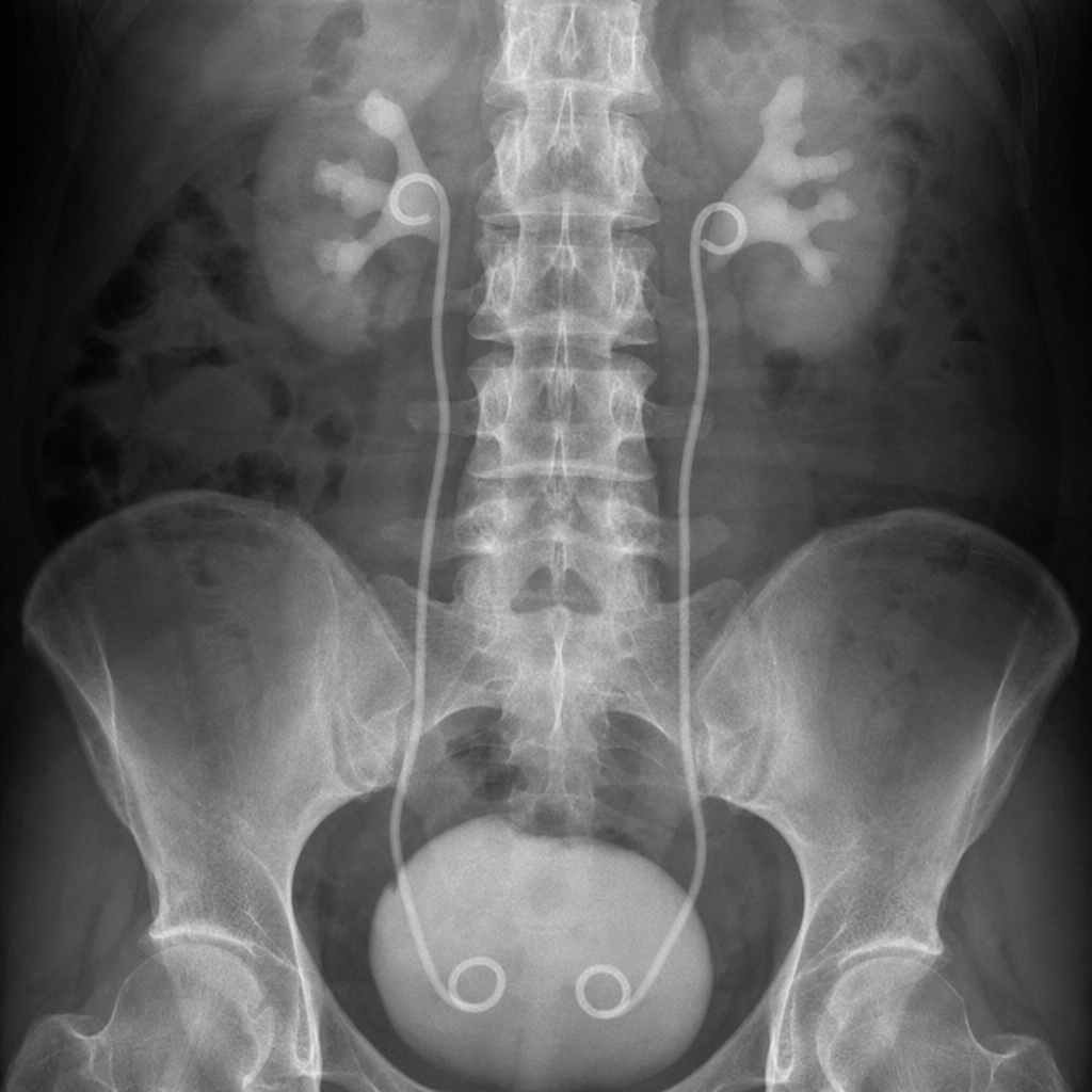
Which condition is most likely associated with specific angiographic findings such as the rosary sign?
What is the investigation of choice in a patient with blunt abdominal trauma with hematuria?
What is the most common complication of TIPS (Transjugular Intrahepatic Portosystemic Shunt) procedure?
Explanation: ***Reversed saphenous*** - The **autologous reversed saphenous vein** is considered the **gold standard** for femoropopliteal bypass due to its superior patency rates and resistance to infection. - The vein's valves are bypassed by reversing its orientation, ensuring unidirectional blood flow. *Dacron* - **Dacron (polyethylene terephthalate)** grafts are synthetic and commonly used for large-diameter arterial bypasses, such as in the aorta, but have **inferior patency in infrainguinal bypasses** compared to autologous vein. - **Higher rates of thrombosis and infection** are observed with Dacron in smaller leg vessels due to compliance mismatch and increased anastomotic intimal hyperplasia. *PTFE* - **Polytetrafluoroethylene (PTFE)** grafts are synthetic and are an option when autologous vein is unavailable, particularly for above-knee femoropopliteal bypasses. - However, PTFE generally has **lower long-term patency rates** and a higher risk of complications like **graft thrombosis and infection** compared to autologous vein grafts. *None of the options* - This option is incorrect because the **reversed saphenous vein** is indeed a preferred and highly effective graft material for femoropopliteal bypass. - The clinical evidence strongly supports its use over synthetic alternatives when available.
Explanation: ***Celiac trunk*** - The **celiac trunk** is the **most frequently dissected artery** during femoral route angiography due to its **challenging anatomical characteristics**. - It arises at a **steep downward angle** (typically 45-90°) from the anterior aorta, requiring significant **catheter manipulation and torque** to engage the orifice. - The acute angulation increases the risk of **subintimal catheter passage** during selective cannulation attempts, leading to iatrogenic dissection. - The need for **repeated attempts** and catheter repositioning when engaging the celiac trunk further elevates dissection risk. *Superior mesenteric artery* - While the **SMA** is frequently cannulated during abdominal angiography, it has a **more favorable anatomy** for catheter engagement. - Its **less acute angle** of origin (typically 30-45° caudally) and more anterior takeoff make it **easier to cannulate** with less manipulation. - Easier cannulation translates to **lower dissection risk** compared to the celiac trunk, despite being commonly studied. *Inferior mesenteric artery* - The **IMA** is a smaller vessel with lower flow, originating from the lower abdominal aorta. - It is **less frequently cannulated** in routine angiography due to its small caliber and distal location. - Lower frequency of selective cannulation means fewer opportunities for dissection. *Gastroduodenal artery* - The **GDA** is a **secondary branch** arising from the hepatic artery (itself a celiac trunk branch). - Due to its **distal location** and smaller size, selective GDA catheterization is uncommon in routine angiography. - Its infrequent selective cannulation makes dissection during femoral angiography extremely rare.
Explanation: ***Computed tomography*** - **Computed tomography (CT)** offers excellent spatial resolution and is the gold standard for diagnosing, staging, and pre-operative planning for aortic aneurysms. - It precisely measures aneurysm size, detects mural thrombus, assesses rupture risk, and evaluates the extent of involvement with surrounding structures. *Ultrasonography* - While useful for initial screening and serial monitoring of known abdominal aortic aneurysms due to its non-invasiveness and cost-effectiveness, its accuracy can be limited by **patient body habitus** and **bowel gas**. - It may not reliably visualize the entire aorta or accurately assess complex anatomy and rupture. *Magnetic resonance imaging* - **Magnetic resonance imaging (MRI)** provides detailed anatomical information and avoids radiation exposure, but it is typically more expensive and time-consuming than CT. - It is often reserved for patients with **renal insufficiency** where iodinated contrast is a concern or when evaluating specific tissue characteristics not well seen on CT. *Arteriography* - **Arteriography** (angiography) is an invasive procedure involving direct contrast injection, carrying risks such as arterial injury and nephrotoxicity. - While it can visualize the aortic lumen, it primarily shows the patent lumen and may **underestimate the true aneurysm size** due to mural thrombus. It is typically used for intervention planning or specific contexts rather than initial diagnosis.
Explanation: ***Femoral canal*** - The description of accessing an artery in the lower limb and opening a sheath to expose it strongly suggests an intervention related to the **femoral artery**, which is part of the structures found in the femoral triangle [1]. - The **femoral sheath** encloses the femoral artery, femoral vein, and the femoral canal (which contains lymphatic vessels and a lymph node called the deep inguinal lymph node of Cloquet). The procedure likely involves accessing one of these [1]. *Cooper's ligament* - **Cooper's ligament** (pectineal ligament) is a fibrous band on the superior aspect of the superior pubic ramus and is involved in the inguinal region but is not part of the femoral sheath or directly accessed for arterial procedures in this context. - It serves as an attachment point for various structures but does not contain major vessels or nerves that would be exposed through this described sheath. *Femoral nerve* - The **femoral nerve** runs lateral to the femoral sheath and is not contained within it. It originates from the lumbar plexus and supplies the anterior thigh muscles. - Accessing the femoral artery for an interventional procedure would typically avoid direct involvement or opening a sheath around the femoral nerve. *Obturator nerve* - The **obturator nerve** is a branch of the lumbar plexus that passes through the obturator foramen to supply the medial compartment of the thigh. - It is anatomically distant from the femoral triangle and the femoral sheath and would not be encountered or enclosed in a sheath during a femoral artery access procedure.
Explanation: ***Vascular Access Site Bleeding*** - This is the **most common complication** of cardiac catheterization, occurring in 2-6% of procedures due to the invasive nature of puncturing an artery or vein for catheter insertion. - Complications can range from a **small hematoma or bruising** to more serious issues like pseudoaneurysm formation or arteriovenous fistula. - Risk factors include larger sheath size, anticoagulation, and femoral access (compared to radial access). *Arrhythmia* - While rhythm disturbances can occur during catheterization, especially when the catheter irritates the myocardium, they are **less frequent** than access site complications. - Most arrhythmias are **transient** and resolve spontaneously without intervention. - Common types include PVCs, NSVT, and rarely sustained ventricular arrhythmias. *Contrast reaction* - Reactions to contrast media can occur, ranging from mild (e.g., rash, itching) to severe (e.g., anaphylaxis). - However, with modern non-ionic, low-osmolar contrast agents and careful patient screening, these are **not the most common complications**. - Incidence of severe reactions is less than 0.1% with modern agents. *Perforation of heart chamber* - This is a **rare but serious complication** (incidence <0.1%) that can lead to cardiac tamponade. - It is typically associated with complex procedures, stiff guidewires, or difficult anatomical features. - Its incidence is significantly lower than access site bleeding.
Explanation: ***Posterior fornix of the vagina*** - Culdocentesis is a procedure where fluid is aspirated from the **cul-de-sac (rectouterine pouch)**. - The **posterior vaginal fornix** is the thinnest and most accessible anatomical landmark for safely accessing the rectouterine pouch. *Anterior wall of the rectum* - Puncturing the **anterior rectal wall** could lead to peritonitis and is not the intended approach for culdocentesis. - The rectum is located posterior to the rectouterine pouch, making it an inappropriate entry point. *Anterior fornix of the vagina* - The **anterior fornix** is anatomically adjacent to the vesicouterine pouch (between the bladder and uterus), not the rectouterine pouch. - Puncturing this area would not access the fluid collection from a ruptured ectopic pregnancy, which accumulates in the rectouterine pouch. *Posterior wall of the uterine body* - Puncturing the **posterior wall of the uterine body** would damage the uterus and is not a route to the cul-de-sac. - The procedure aims to access the space behind the uterus, not the uterine organ itself.
Explanation: ***Carotid angioplasty and stenting*** - **Radiation-induced carotid artery disease** often involves the distal part of the carotid artery, making it less amenable to surgical endarterectomy. - **Angioplasty and stenting** offer a less invasive approach with good technical success in these challenging cases, especially given the increased fragility and fibrosis of radiated tissues. *Carotid endarterectomy* - **Carotid endarterectomy** in previously radiated fields is associated with a significantly higher risk of complications, including **cranial nerve injury**, **wound infection**, and **carotid artery rupture**, due to tissue fibrosis and scarring. - The disease often extends beyond the easily accessible segment for endarterectomy in radiation-induced cases. *Low dose aspirin* - **Low-dose aspirin** is an important component of medical therapy for **atherosclerotic disease** and **stroke prevention**, but it is insufficient as a sole treatment for symptomatic or high-grade occlusive disease of the carotid artery. - It helps manage the underlying **atherosclerotic process** but does not directly address the severe stenosis or occlusion. *Carotid bypass procedure* - **Carotid bypass procedures** are complex surgical interventions usually reserved for cases of **carotid artery occlusion** or **recurrent stenosis** after previous interventions where endarterectomy or stenting is not feasible. - While an option, it is more invasive and technically demanding than angioplasty and stenting, particularly in already radiated tissues with compromised vascular integrity.
Explanation: ***Polyarteritis Nodosa*** - The **rosary sign** on angiography, characterized by alternating areas of stenosis and dilation in medium-sized arteries, is a classic finding in **Polyarteritis Nodosa (PAN)**. - This sign represents **aneurysms and stenoses** resulting from inflammatory destructive lesions in the arterial wall. *Kawasaki disease* - Primarily affects young children and causes **coronary artery aneurysms**, which appear as focal dilations, rather than the "rosary sign" pattern of multiple stenoses and dilations seen in PAN. - While it can involve other medium-sized arteries, the widespread and characteristic "rosary sign" is not typical. *Takayasu arteritis* - This condition primarily affects the **aorta and its major branches**, leading to **stenosis or occlusion** of large arteries, often described as "pulseless disease." - It does not typically present with the **venous beads-like appearance** or rosary sign found in PAN. *Giant cell arteritis* - Characterized by inflammation of **large and medium-sized arteries**, predominantly affecting the temporal arteries. - Angiographic findings often include **long segments of smooth vessel wall thickening, stenosis, or occlusion**, but not the characteristic alternating aneurysms and stenoses of the "rosary sign."
Explanation: ***Correct Answer: CECT*** - **Contrast-enhanced computed tomography (CECT)** is the investigation of choice for evaluating blunt abdominal trauma with hematuria as it accurately assesses the extent of injury to the **kidneys, ureters, bladder**, and surrounding structures. - It provides detailed images for detecting **renal lacerations, hematomas, urine extravasation**, and other abdominal organ injuries. - **Gold standard** in trauma protocols for comprehensive evaluation of renal and abdominal injuries. *Incorrect: USG of the abdomen* - **Ultrasound** can identify gross abnormalities like large hematomas or free fluid but is less sensitive than CECT for subtle renal injuries or collecting system disruptions. - It is often used as an initial screening tool (FAST exam) but not the definitive investigation of choice in this context. *Incorrect: Retrograde urogram* - A **retrograde urogram** primarily evaluates the **lower urinary tract** (ureters and bladder) by injecting contrast directly into the urethra. - It is not suitable for assessing the extent of renal parenchymal injury or other abdominal organ damage in blunt trauma. *Incorrect: IVP* - **Intravenous pyelogram (IVP)** uses intravenous contrast to visualize the kidneys, ureters, and bladder, but it has largely been replaced by CECT due to its lower sensitivity and specificity for traumatic injuries. - It provides less detailed anatomical information about surrounding soft tissues and can miss subtle parenchymal or vascular injuries.
Explanation: **Explanation:** **TIPS (Transjugular Intrahepatic Portosystemic Shunt)** is an artificial channel created between the high-pressure portal vein and the low-pressure hepatic vein to treat complications of portal hypertension. **Why Hepatic Encephalopathy (HE) is the correct answer:** The primary mechanism of TIPS involves bypassing the liver’s filtration system. By creating a shunt, portal blood (rich in ammonia and other neurotoxins derived from the gut) enters the systemic circulation directly without being detoxified by hepatocytes. This leads to **Hepatic Encephalopathy in approximately 25–45% of patients**, making it the most frequent complication post-procedure. **Analysis of Incorrect Options:** * **A. Heart Failure:** While the sudden increase in venous return to the right atrium (preload) can precipitate acute heart failure in patients with underlying cardiac disease, it is far less common than HE. * **C. Thrombosis:** Shunt stenosis or thrombosis was common with bare-metal stents; however, with the modern use of **PTFE-covered stents**, the incidence of thrombosis has significantly decreased. * **D. Recurrent Variceal Bleed:** TIPS is highly effective at decompressing varices. Re-bleeding usually only occurs if the shunt becomes occluded or stenosed, which is a secondary event. **High-Yield Clinical Pearls for NEET-PG:** * **Indications:** Refractory variceal bleeding (most common indication) and refractory ascites. * **Absolute Contraindications:** Severe congestive heart failure (Right-sided), polycystic liver disease, and severe active systemic infection/sepsis. * **Technical Goal:** To reduce the **Portosystemic Pressure Gradient (PSG) to <12 mmHg** to prevent re-bleeding. * **Stent Type:** PTFE-covered stents (e.g., VIATORR) are the gold standard to maintain patency.
Get full access to all questions, explanations, and performance tracking.
Start For Free