Most sensitive investigation for abdominal trauma in a hemodynamically stable patient is-
In a hemodynamically stable patient with abdominal trauma, which imaging modality is considered the gold standard for diagnosing and grading solid organ injuries?
A 45-year-old female presents with the acute onset of a severe headache and vomiting. A non-contrast CT shows a crescent-shaped hyperdense area crossing suture lines. What is the most likely diagnosis?
What is the investigation of choice for diagnosing solid organ injuries in abdominal trauma?
A 75-year-old male with a history of hypertension presents with sudden-onset severe abdominal pain radiating to the back. Which diagnostic tool would be most beneficial in differentiating between a ruptured abdominal aneurysm and acute pancreatitis?
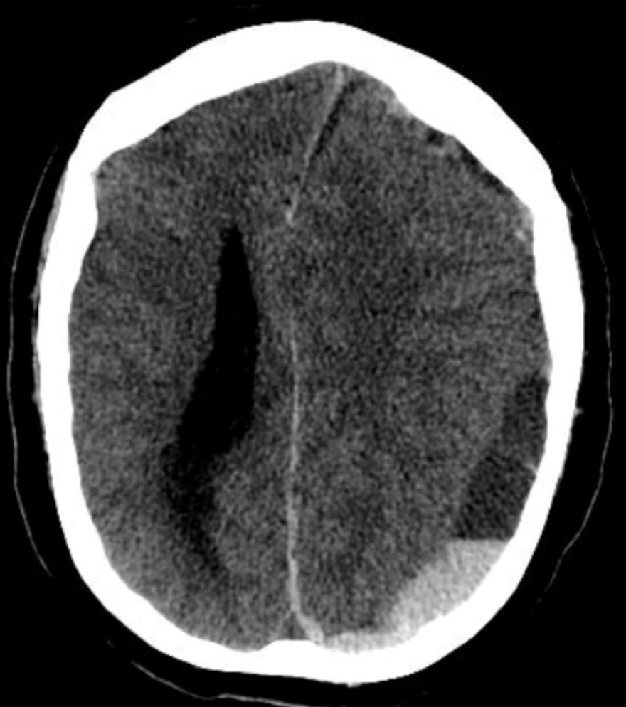
Investigation of choice to evaluate intracranial hemorrhage of less than 48 hours is -
Tear drop sign is seen in?
A polytrauma patient's CT brain shows a crescent-shaped extra-axial collection with a concave inner margin. What is the most likely diagnosis?

The most appropriate investigation to diagnose and determine the extent of renal injury in a 15-year-old boy who presents with hematuria and left-sided abdominal pain 48 hours after sustaining a blunt abdominal injury, with a pulse rate of 96/minute, blood pressure of 110/70 mmHg, hemoglobin of 10.8 gm%, and packed cell volume of 31%, would be-
Thumb sign in lateral X-ray of the neck is seen in?
Explanation: ***CT Scan (Computed Tomography)*** - **CT scans** offer superior anatomical detail and can accurately detect organ damage, hemorrhage, and other injuries in **hemodynamically stable** patients with abdominal trauma. - It is considered the **most sensitive** and specific imaging modality for evaluating blunt and penetrating abdominal trauma when the patient can tolerate the study. *Ultrasonography (FAST)* - While effective for detecting **free fluid** (blood) in specific abdominal areas, **Focused Assessment with Sonography for Trauma (FAST)** has lower sensitivity for solid organ injuries or bowel perforations. - Its primary role is rapid assessment for **hemoperitoneum** to guide immediate management in unstable patients, not detailed injury characterization. *Diagnostic peritoneal lavage (DPL)* - **DPL** is an invasive procedure with high sensitivity for detecting **intraperitoneal bleeding**, but it does not identify specific organ injuries or retroperitoneal hemorrhage. - It is rarely used in hemodynamically stable patients due to its invasiveness and the availability of more detailed imaging techniques. *MRI (Magnetic Resonance Imaging)* - **MRI** provides excellent soft tissue contrast but is typically too **time-consuming** and less accessible in urgent trauma settings compared to CT. - It's generally not the first-line investigation for acute abdominal trauma due to motion artifacts and limited utility in detecting air or bone injuries.
Explanation: ***CT scan (Correct Answer)*** - **Contrast-enhanced CT (CECT)** is the **gold standard** for diagnosing and grading solid organ injuries in hemodynamically stable patients with abdominal trauma - Provides **high sensitivity (95-100%)** and specificity for detecting injuries to liver, spleen, kidneys, and pancreas - Enables accurate **injury grading** using the AAST (American Association for the Surgery of Trauma) classification system - Can detect **active hemorrhage** (contrast extravasation) and guide decisions between operative vs non-operative management - Recommended by **ATLS (Advanced Trauma Life Support)** protocols for stable trauma patients *Ultrasound (Incorrect)* - **FAST (Focused Assessment with Sonography for Trauma)** is excellent for rapid detection of **free intraperitoneal fluid** in unstable patients - However, it has **limited sensitivity (28-50%)** for solid organ parenchymal injuries and cannot grade injury severity - Highly **operator-dependent** with performance affected by body habitus, bowel gas, and subcutaneous emphysema - Best used as a triage tool, not for definitive diagnosis or grading *MRI (Incorrect)* - Offers excellent **soft tissue contrast** but is not suitable for acute trauma evaluation - Major limitations include **long acquisition time** (30-60 minutes), limited emergency availability, and challenges with monitoring critically ill patients - May be used in **specific scenarios** such as pregnant patients where radiation exposure is a concern, or for delayed evaluation of pancreatic/bowel injuries *X-ray (Incorrect)* - **Plain radiographs** have minimal role in evaluating solid organ injuries due to poor soft tissue resolution - Primarily useful for detecting **skeletal fractures**, **pneumoperitoneum** (free air under diaphragm), or **pneumothorax** in trauma patients - Cannot visualize parenchymal injuries, hematomas, or assess organ integrity - Has been largely replaced by CT in modern trauma protocols
Explanation: ***Subdural hematoma*** - A **crescent-shaped hyperdense area** on a non-contrast CT that **crosses suture lines** is the classic imaging finding for a subdural hematoma. - This typically results from the tearing of **bridging veins** and can present acutely or chronically. *Epidural hematoma* - This typically appears as a **lenticular (lens-shaped) hyperdense collection** on CT. - Epidural hematomas characteristically **do not cross suture lines** because the dura is tightly adherent to the skull at these points. *Subarachnoid hemorrhage* - Identified by the presence of blood within the **subarachnoid space**, appearing as **hyperdensity in the sulci and basal cisterns** on CT. - While it causes severe headache, its CT appearance is diffuse and does not form a contained crescent or lenticular shape. *Intracerebral hemorrhage* - Appears as a **focal, hyperdense collection of blood within the brain parenchyma** itself. - It does not conform to the shape of an extra-axial collection like a crescent or lenticular shape.
Explanation: ***CT scan*** - A **CT scan** is the investigation of choice for diagnosing solid organ injuries due to its high sensitivity and specificity in detecting **hemoperitoneum**, organ lacerations, and active bleeding. - It provides detailed cross-sectional images, allowing for precise localization and grading of injuries, crucial for **surgical planning**. *Ultrasound* - While useful for rapid assessment of **free fluid** (FAST scan), it has limited sensitivity for detecting parenchymal organ injuries and cannot reliably assess the extent of damage or active bleeding. - Its utility is primarily in the **initial triage** of hemodynamically unstable patients to identify the presence of significant intra-abdominal bleeding. *MRI* - **MRI** offers excellent soft tissue contrast but is typically too time-consuming and often impractical in the acute trauma setting, especially for **unstable patients**. - Its role is usually reserved for specific indications, such as spinal cord injuries, or when **CT scans** are contraindicated for certain reasons, like **pregnancy**. *X-ray* - **X-rays** are primarily used to detect skeletal injuries, such as fractures, or to visualize air under the diaphragm indicating **hollow viscus perforation**, rather than solid organ damage. - They provide very limited information regarding the **parenchyma of solid organs** and are not sufficient for diagnosing or staging solid organ injuries.
Explanation: ***CT angiography*** - **CT angiography** is the most beneficial tool for differentiating a **ruptured abdominal aortic aneurysm (AAA)** from acute pancreatitis due to its ability to visualize the aorta and detect active extravasation or hematoma [2]. - It clearly identifies the presence, size, and rupture status of an AAA, which is crucial for immediate surgical planning [1]. *MRI abdomen with contrast* - While MRI can provide detailed soft tissue imaging, it is generally **less rapid** and **more expensive** than CT in emergent situations, especially for suspected rupture [2]. - **Motion artifacts** from abdominal breathing can also degrade image quality, and it may not be as readily available in emergency settings as CT. *Endoscopic ultrasound* - **Endoscopic ultrasound (EUS)** is primarily used for detailed imaging of the pancreas, bile ducts, and upper gastrointestinal tract, but it is **invasive** and not suitable for an acute emergency like a suspected AAA rupture. - EUS would not be effective in assessing the aorta for rupture or dissecting an aneurysm. *CT abdomen with oral contrast* - A standard **CT scan of the abdomen with oral contrast** is useful for evaluating the bowel and other intra-abdominal structures, but **oral contrast does not enhance vascular structures sufficiently** to diagnose an AAA rupture. - It would not provide the clear arterial phase imaging necessary to detect active bleeding or the integrity of the aortic wall as effectively as CT angiography [3].
Explanation: ***CT scan*** - **Non-contrast CT** is the most sensitive and rapid imaging modality for detecting acute intracranial hemorrhage, appearing as a **hyperdense** (bright) area within the brain parenchyma or subarachnoid space. - It is readily available in emergency settings and is crucial for immediate diagnosis to guide management, especially within the first **48 hours**. *MRI* - While MRI can detect hemorrhage, its sensitivity for **acute hemorrhage** (especially within the first few hours) is less than CT, and it is more time-consuming. - MRI is superior for detecting older hemorrhage or subtle lesions, but it is not the **first-line investigation** for acute bleeding. *PET* - **Positron Emission Tomography** (PET) scans are primarily used to assess metabolic activity and blood flow in the brain, often for conditions like cancer, epilepsy, or dementia. - It does not directly visualize fresh blood and therefore is not used for the diagnosis of **acute intracranial hemorrhage**. *SPECT* - **Single-Photon Emission Computed Tomography** (SPECT) is used to evaluate cerebral blood flow and neuronal activity, similar to PET but with different tracers and resolution. - It is not indicated for the rapid assessment of **acute intracranial hemorrhage** as it does not directly image blood.
Explanation: ***Blow out fracture*** - The **tear drop sign** on imaging (often CT scan) is characteristic of an **orbital blow-out fracture**, indicating herniation of orbital contents (fat, muscle) into the maxillary sinus. - This fracture typically involves the **orbital floor** or medial wall, often caused by a blunt force trauma to the eye. *Fracture zygomatic arch* - A fracture of the zygomatic arch is often associated with a **flattening of the malar prominence** rather than a "tear drop" sign. - It might lead to restricted jaw movement if the arch impinges on the coronoid process. *Fracture maxilla* - Maxillary fractures (e.g., Le Fort fractures) involve the midface bones and cause **facial deformity**, malocclusion, and mobility of the maxilla. - The tear drop sign is not a primary diagnostic feature of maxillary fractures. *Fracture mandible* - Mandibular fractures present with pain, swelling, and **malocclusion** of the teeth. - Imaging would reveal a break in the mandible, not a tear drop sign associated with orbital contents.
Explanation: ***SDH*** - The image shows a **crescent-shaped collection** of hemorrhage with a concave inner margin, consistent with a **subdural hematoma** (SDH). - SDHs result from the tearing of **bridging veins** and typically conform to the brain's surface, crossing suture lines but not limited by bony sutures. *EDH* - An **epidural hematoma (EDH)** characteristically appears as a **lenticular** or **biconvex** shape on CT, not crescent-shaped. - EDHs are typically caused by arterial bleeding, often from the **middle meningeal artery**, and are limited by cranial sutures. *Contusion* - A **contusion** is brain tissue bruising that appears as **heterogeneous areas** of hemorrhage and edema within the brain parenchyma itself. - It would not manifest as a distinct extra-axial collection with a smooth, concave margin. *Diffuse axonal injury* - **Diffuse axonal injury (DAI)** involves widespread microscopic damage to axons, often at the gray-white matter junction. - It may appear as *punctate hemorrhages* or **small lesions** at these junctions on CT, but often the CT can be normal, and it would not present as a large extra-axial collection.
Explanation: ***Contrast enhanced computed tomography*** - **CT with intravenous contrast** is the gold standard for evaluating **renal trauma**, providing detailed anatomical information on the extent of injury, including lacerations, hematomas, and urinary extravasation, which might be missed by other modalities. - It rapidly assesses the **parenchyma**, **collecting system**, and surrounding structures, allowing for proper staging of the injury and guiding management decisions. *Sonographic evaluation of abdomen* - **Ultrasound** is useful for rapidly detecting **free fluid** (e.g., blood) in the abdomen and assessing major organ integrity, but its ability to characterize renal parenchymal injuries or urinary extravasation is limited. - It is **operator-dependent** and often insufficient for detailed staging of renal trauma compared to CT. *Intravenous pyelography* - **IVP** primarily evaluates the **collecting system** and ureteral patency but has limited sensitivity for assessing renal parenchymal injuries or perinephric hematomas. - It involves radiation exposure and a contrast load, and generally provides **less anatomical detail** than modern CT scans. *MR urography* - **MR urography** provides excellent soft tissue contrast without ionizing radiation, but it is typically **less readily available** in an emergency setting and takes longer to perform than CT. - Its role in acute trauma is usually reserved for cases where **iodinated contrast is contraindicated** (e.g., severe allergy, renal insufficiency) or when specific soft-tissue detail is crucial for follow-up.
Explanation: ***Epiglottitis*** - The **thumb sign** on a lateral neck X-ray is a classic finding in **acute epiglottitis**, caused by the severely swollen epiglottis. - This swelling can lead to severe **airway obstruction** due to its critical location. *Internal hemorrhage* - Internal hemorrhage in the neck might cause soft tissue swelling, but it typically does not produce the specific **"thumb sign" morphology** seen in epiglottitis. - Diagnosis relies more on **clinical signs of bleeding** and potentially imaging like CT scans to localize blood collections. *Saccular cyst* - A **saccular cyst** (or laryngocele) is a benign air-filled or fluid-filled sac - It would appear as a well-defined, often air-filled, or soft-tissue mass, not typically resembling the diffuse, inflamed "thumb" appearance of a swollen epiglottis. *Carcinoma of the epiglottis* - While a **carcinoma of the epiglottis** could cause epiglottic swelling or mass effect, it usually presents as a more irregular or focal mass rather than the uniform, bulbous swelling characteristic of the "thumb sign" in acute epiglottitis. - Carcinoma is also typically a chronic process, unlike the acute, rapidly progressing inflammation of epiglottitis.
Trauma Imaging Protocols
Practice Questions
Head Trauma Imaging
Practice Questions
Spine Trauma Imaging
Practice Questions
Chest Trauma Imaging
Practice Questions
Abdominal Trauma Imaging
Practice Questions
Musculoskeletal Trauma Imaging
Practice Questions
Non-traumatic Neurological Emergencies
Practice Questions
Non-traumatic Thoracic Emergencies
Practice Questions
Non-traumatic Abdominal Emergencies
Practice Questions
Vascular Emergencies
Practice Questions
Pediatric Emergency Imaging
Practice Questions
Imaging of Non-accidental Trauma
Practice Questions
Get full access to all questions, explanations, and performance tracking.
Start For Free