What is the investigation of choice for the evaluation of acute head injury?
Which radiographic view is best for detecting hollow viscus perforation?
The X-ray image provided (img-41.jpeg) shows multiple cylindrical objects in the abdominal area. Which of the following conditions is most likely represented by this image?
A 30 year old apparently healthy man who was carrying laxatives and enema apparatus developed abdominal pain at the airport and an x-ray was done which appears as shown below. Which of the following is the likely diagnosis?
Following a fight between 2 groups, a boy was brought with severe pain in the chest, distended neck veins, dyspnea, and a BP of 80/50 mmHg. X-ray shows the following findings: What is the diagnosis?
Whole-body CT scan protocol for a trauma patient includes all, except
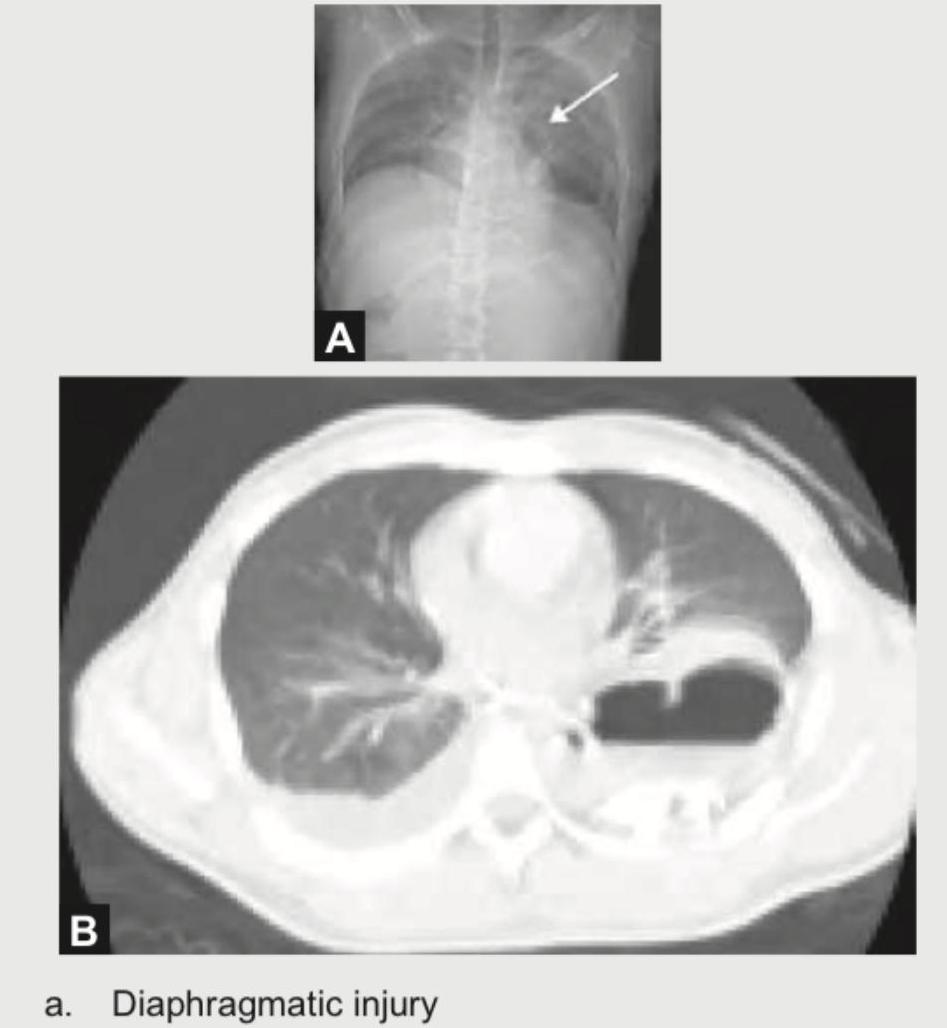
A 25-year-old corporate executive was stabbed in the abdomen in a discotheque. In the emergency room, CXR and CT chest were performed. The radiologist then put an NG tube in the patient, injected contrast and repeated the scan. What is the diagnosis?

A 35-year-old male with history of 4 weeks of immobilization for fracture of femur develops sudden onset breathlessness and blood in sputum. CT angiography shows? (Recent NEET Pattem 2018-19)

Which one of the following statements is correct regarding eFAST in trauma? 1. It is a technique to assess free fluid in abdominal cavity, thoracic cavity, and pericardium. 2. It is a technique to assess free fluid in pelvic cavity. 3. It is a technique to assess free fluid in pleural cavity. Select the correct answer using the code given below:
In an unconscious patient with multiple injuries, what is the best and reliable modality for assessment of cervical spine injury?
Explanation: **Explanation:** **NCCT Head (Non-Contrast Computed Tomography)** is the investigation of choice for acute head injury due to its speed, widespread availability, and superior ability to detect acute intracranial hemorrhage and skull fractures. 1. **Why NCCT Head is Correct:** * **Acute Hemorrhage:** Fresh blood (hyperacute/acute) appears **hyperdense** (bright white) on CT, making it easy to identify Epidural (EDH), Subdural (SDH), and Subarachnoid hemorrhages (SAH). * **Bone Detail:** It is the gold standard for identifying linear, depressed, or basilar skull fractures. * **Speed & Monitoring:** It takes less than a minute, allowing for rapid triage and easier monitoring of unstable trauma patients compared to MRI. 2. **Why Other Options are Incorrect:** * **CECT Head:** Contrast is avoided in acute trauma because intravenous contrast appears white (radio-opaque), which can mask or be confused with acute extravasated blood. * **MRI Brain:** While more sensitive for Diffuse Axonal Injury (DAI) and posterior fossa lesions, it is time-consuming, expensive, and difficult to perform on patients with metallic implants or life-support equipment. * **CT Angiography:** This is indicated only if a vascular injury (e.g., carotid artery dissection or dural venous sinus thrombosis) is suspected, not as a primary screening tool. **Clinical Pearls for NEET-PG:** * **Window of Choice:** For viewing hemorrhage, use the **Brain Window**; for fractures, use the **Bone Window**. * **The "Lucid Interval":** Classically associated with **EDH** (biconvex/lens-shaped), usually due to rupture of the **Middle Meningeal Artery**. * **Crescentic Shape:** Characteristic of **SDH**, usually due to tearing of **bridging veins**. * **Investigation of Choice for DAI:** MRI (specifically GRE or SWI sequences).
Explanation: **Explanation:** The detection of hollow viscus perforation on radiography relies on the visualization of **pneumoperitoneum** (free intraperitoneal air). **1. Why Erect is the Correct Answer:** The **Erect Chest X-ray** (or Erect Abdomen) is the gold standard and most sensitive plain radiographic view. Because air is less dense than abdominal viscera and fluid, it obeys the laws of gravity and rises to the highest point in the peritoneal cavity. In the upright position, this air collects under the **diaphragm**, appearing as a thin, radiolucent crescent (crescent sign) between the liver/stomach and the diaphragmatic leaflets. It can detect as little as 1–2 ml of free air. **2. Analysis of Incorrect Options:** * **Supine:** In this position, air collects anteriorly and spreads out, making it difficult to see. It may show subtle signs like **Rigler’s sign** (gas on both sides of the bowel wall), but it is far less sensitive than erect views. * **Right lateral decubitus:** This is incorrect because air would rise and blend with the gastric bubble or be obscured by the splenic shadow. * **Left lateral decubitus:** This is the **alternative of choice** if the patient cannot stand. Air rises to collect between the **liver and the right lateral abdominal wall**, where it is easily visualized. However, the Erect view remains the primary recommendation. **Clinical Pearls for NEET-PG:** * **Most sensitive imaging:** Non-contrast CT scan is the overall gold standard for detecting pneumoperitoneum. * **Rigler’s Sign:** Also known as the "double wall sign," seen on supine films when large amounts of air are present. * **Cupola Sign:** Refers to air trapped under the central tendon of the diaphragm on a supine film. * **Positioning Tip:** Patients should remain in the erect or decubitus position for at least **5–10 minutes** before the X-ray to allow air to migrate upwards.
Explanation: ***Body packer syndrome*** - The X-ray shows multiple, well-defined, uniformly shaped, dense, cylindrical objects throughout the gastrointestinal tract, a classic radiographic finding for **body packer syndrome**. - These objects are ingested packets containing illicit drugs, often showing a "double-condom sign" (a thin lucent rim of air trapped in the wrapping), confirming their manufactured nature. *Bezoar* - A **bezoar** is a mass of indigestible material (like hair or vegetable fiber) that appears as a single, mottled, intraluminal mass on X-ray, not multiple discrete packets. - The objects in the image have a uniform shape and density, which is inconsistent with the heterogeneous appearance of a bezoar. *Pica due to anaemia* - **Pica** involves the ingestion of non-nutritive substances, which would result in radiopaque foreign bodies of various, irregular shapes (e.g., coins, stones), not uniform cylindrical packets. - While associated with conditions like **iron-deficiency anaemia**, the radiographic findings of pica do not match the organized, manufactured appearance of the objects shown. *Constipation due to fecalith* - A **fecalith** is a hardened mass of stool that appears as a mottled density within the colon, conforming to the haustral pattern, and lacks the smooth, well-defined borders seen here. - While severe **constipation** leads to significant stool burden, it does not present as multiple, encapsulated, geometrically regular objects.
Explanation: ***Body packer syndrome*** - This diagnosis is indicated by the presence of multiple, well-defined, uniformly shaped, and hyperdense foreign bodies within the gastrointestinal tract, as seen on the abdominal X-ray. - The clinical context of being at an airport with laxatives and enema apparatus is highly suggestive of an individual attempting to smuggle illicit drugs by ingesting them in packets. *Bezoar syndrome* - A **bezoar** is a mass of indigestible material (like hair or vegetable fibers) trapped in the GI tract, which appears on X-ray as a mottled, heterogeneous mass, not as multiple discrete, uniform packets. - Bezoars typically conform to the shape of the stomach or bowel lumen and lack the smooth, regular outlines seen in this image. *Pica due to anaemia* - **Pica** is the ingestion of non-nutritive substances and might show foreign objects on an X-ray, but these would typically be of varied shapes and sizes (e.g., coins, dirt, paint chips), not uniform packets. - The clinical presentation does not suggest anaemia, and the scenario points towards illegal activity rather than a compulsive eating disorder. *Constipation due to fecalith* - A **fecalith** is a hardened mass of stool that appears as a mottled density within the colon, consistent with retained feces. - The objects in the X-ray have sharp, smooth borders and a uniform density, which is inconsistent with the appearance of a fecalith.
Explanation: ***Tension pneumothorax*** - The clinical presentation of severe dyspnea, **distended neck veins**, and **hypotension** (BP 80/50 mmHg) after trauma is a classic triad for tension pneumothorax, which is a medical emergency. - The chest X-ray confirms this diagnosis by showing a completely collapsed right lung with a significant **contralateral shift of the mediastinum and trachea** to the left, and flattening of the right hemidiaphragm, indicating high intrapleural pressure. *Primary Spontaneous Pneumothorax* - This type of pneumothorax occurs without any preceding trauma or underlying lung disease, typically in tall, thin young men due to the rupture of **apical blebs**. - The patient's presentation follows a fight, clearly indicating a **traumatic etiology**, which rules out a spontaneous pneumothorax. *Traumatic Pneumothorax* - While the cause is trauma, this is a less specific diagnosis. The term **Tension pneumothorax** is more accurate given the life-threatening signs of **hemodynamic instability** and **mediastinal shift**. - A simple traumatic pneumothorax does not typically involve the one-way valve mechanism that leads to the progressive accumulation of air and the resulting **obstructive shock** seen in this patient. *Bilateral Pneumothorax* - This diagnosis is incorrect as the chest X-ray clearly demonstrates a **unilateral** condition affecting only the right side. - The left lung, although compressed by the shifted mediastinum, remains inflated with visible lung markings extending to the chest wall.
Explanation: ***c.CT Limbs***- Whole-body CT in **polytrauma** focuses on detecting time-critical injuries in the **trunk** and **head** (head, chest, abdomen/pelvis, and spine). - Routine inclusion of **CT Limbs** is not standard unless there is specific clinical suspicion of a major fracture or vascular injury based on physical examination. *a.CT Head* - Essential for rapidly excluding **intracranial hemorrhage**, **subdural/epidural hematomas**, or significant **traumatic brain injury (TBI)**, which are major causes of trauma mortality. - Typically performed first in the whole-body protocol to assess the most immediately life-threatening injuries. *b.CT Cervical spine* - Crucial for identifying potentially unstable **spinal fractures** or **ligamentous injuries** that require immediate management and prevent secondary neurological injury. - High-energy trauma mandates comprehensive assessment of the **cervical spine** as part of the primary survey protocol. *d.CT Abdomen* - Necessary for detecting **solid organ injury** (e.g., liver, spleen lacerations) and **intraperitoneal/retroperitoneal hemorrhage**, which are common sources of **exsanguination** and shock. - The abdominal scan usually extends to include the **pelvis** to assess for **pelvic fractures** and associated bleeding.
Explanation: ***Diaphragmatic injury*** - The chest X-ray (**A**) shows opacification in the left lower lung field with a **raised hemidiaphragm**, and the CT scan (**B**) reveals **herniated bowel loops** and **stomach** (appearing as a gas-filled structure) in the left hemithorax. - The insertion of an **NG tube and contrast injection** to confirm the stomach's presence in the chest is a classical diagnostic approach for **diaphragmatic rupture** following penetrating trauma. *Hemothorax* - Hemothorax would primarily show **fluid collection** in the pleural space, which appears homogeneous on CT, typically without specific organ herniation. - While plausible with penetrating trauma, the images specifically show **organ herniation**, not just blood. *Pneumothorax* - Pneumothorax is characterized by **air in the pleural space**, resulting in lung collapse and a visible pleural line. - The images show solid/fluid-filled structures (bowel, stomach) rather than free air and lung collapse. *Cardiac Tamponade* - Cardiac tamponade involves **fluid accumulation in the pericardial sac**, leading to impaired cardiac filling. - This condition primarily affects heart function and is identified by specific echocardiographic findings, which are not depicted in these chest imaging studies.
Explanation: ***Acute cor-pulmonale*** - **4 weeks of immobilization** is a major risk factor for **deep vein thrombosis (DVT)** leading to **pulmonary embolism (PE)** - **CT pulmonary angiography** is the gold standard investigation for PE, showing filling defects in pulmonary arteries - Massive or submassive PE causes acute **right ventricular strain** = **acute cor-pulmonale** - Clinical presentation of **sudden breathlessness** and **hemoptysis** is classic for pulmonary thromboembolism - The timing (4 weeks post-immobilization) fits thromboembolism, not fat embolism *Fat embolism* - Occurs **acutely within 24-72 hours** after long bone fracture (especially femur/tibia) - The **4-week delay** makes fat embolism extremely unlikely - Presents with **respiratory distress, petechial rash, and neurological symptoms** (Gurd's criteria) - CT findings show diffuse ground-glass opacities, not typical filling defects seen on CT angiography *Pulmonary oedema* - Caused by **left heart failure** or **ARDS**, showing bilateral interstitial and alveolar fluid - Would show diffuse bilateral infiltrates on imaging, not filling defects in pulmonary vessels - **Hemoptysis** is uncommon in cardiogenic pulmonary edema - No clear cardiac history or precipitant in this patient *Aortic dissection* - Involves a tear in the aortic intima with blood dissecting through the aortic wall - Presents with **sudden severe chest/back pain**, not primarily with hemoptysis - CT angiography would show **aortic flap and false lumen**, not pulmonary vascular abnormalities - Unrelated to femur fracture or prolonged immobilization
Explanation: ***1, 2 and 3*** - The **eFAST exam (extended Focused Assessment with Sonography for Trauma)** evaluates multiple areas for **free fluid**, including the abdominal cavity, thoracic cavity (pleural effusions), and pericardium (pericardial effusions). - It also includes specific views for the **pelvic cavity** (pouch of Douglas) to detect free fluid, which is highly relevant in trauma settings. *1 and 2 only* - This option misses the crucial component of assessing the **pleural cavity** for free fluid (hemothorax), which is an integral part of the **eFAST protocol**. - While it correctly includes the abdominal and pelvic cavities, the exclusion of the thoracic cavity (pleural) makes it incomplete. *2 and 3 only* - This option incorrectly omits the assessment of the **abdominal cavity**, which is a primary and essential part of any FAST or eFAST exam to identify intra-abdominal hemorrhage. - The abdominal cavity includes views of the **hepatorenal space (Morison's pouch)**, **splenorenal space**, and **suprapubic window**. *1 and 3 only* - This option incorrectly excludes the specific assessment of the **pelvic cavity**, which is routinely included in the eFAST exam, particularly in trauma, to identify dependent pooling of free fluid. - While it correctly includes the abdominal and thoracic cavities, the absence of the pelvic cavity assessment makes it incomplete.
Explanation: **While doing CT scan of brain take extra cuts at the cervical spine region** - For an unconscious patient, **CT scan** is the most reliable and rapid method for assessing cervical spine injuries, especially in a trauma setting. It is highly sensitive for detecting **fractures** and **misalignments**. - Taking extra cuts during a brain CT is efficient and avoids additional patient movement or delay, providing crucial information for immediate management. *MRI scan* - **MRI** is excellent for soft tissue injuries (ligaments, discs, spinal cord), but it is time-consuming and often not immediately available in acute trauma settings, especially for an unstable patient. - While valuable, it is usually performed after initial stabilization and when neurological deficits are specifically suspected, not as the first-line assessment for bony injury in an acute, unstable trauma patient. *Full AP and lateral radiographs of spine* - **Plain radiographs** have significant limitations in visualizing all cervical spine structures, particularly the **C1-C2 junction** and the **cervicothoracic junction**, which can be obscured. - They have a lower sensitivity for detecting subtle fractures and ligament injuries compared to CT scans, and overlying structures can obscure important details. *Rely only on clinical examination* - In an **unconscious patient**, a reliable clinical examination for cervical spine injury is impossible due to the inability to assess pain, tenderness, or neurological function. - Relying solely on clinical examination in such a patient puts them at **significant risk** for further spinal cord injury if an unstable fracture is present and goes undetected.
Trauma Imaging Protocols
Practice Questions
Head Trauma Imaging
Practice Questions
Spine Trauma Imaging
Practice Questions
Chest Trauma Imaging
Practice Questions
Abdominal Trauma Imaging
Practice Questions
Musculoskeletal Trauma Imaging
Practice Questions
Non-traumatic Neurological Emergencies
Practice Questions
Non-traumatic Thoracic Emergencies
Practice Questions
Non-traumatic Abdominal Emergencies
Practice Questions
Vascular Emergencies
Practice Questions
Pediatric Emergency Imaging
Practice Questions
Imaging of Non-accidental Trauma
Practice Questions
Get full access to all questions, explanations, and performance tracking.
Start For Free