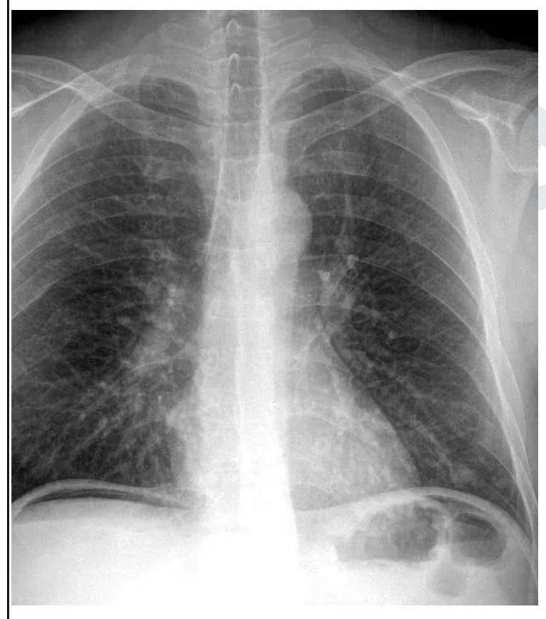
A 26 year old male patient was brought to the emergency department with abdominal pain and obstipation for 3 days. He gives a history of bull gore to the abdomen 3 days back. His chest X-ray is given below. What is the probable diagnosis?

Pulmonary embolism is most commonly produced by which of the following?
IOC for Acute Aortic Dissection in a Clinically Unstable patient is?
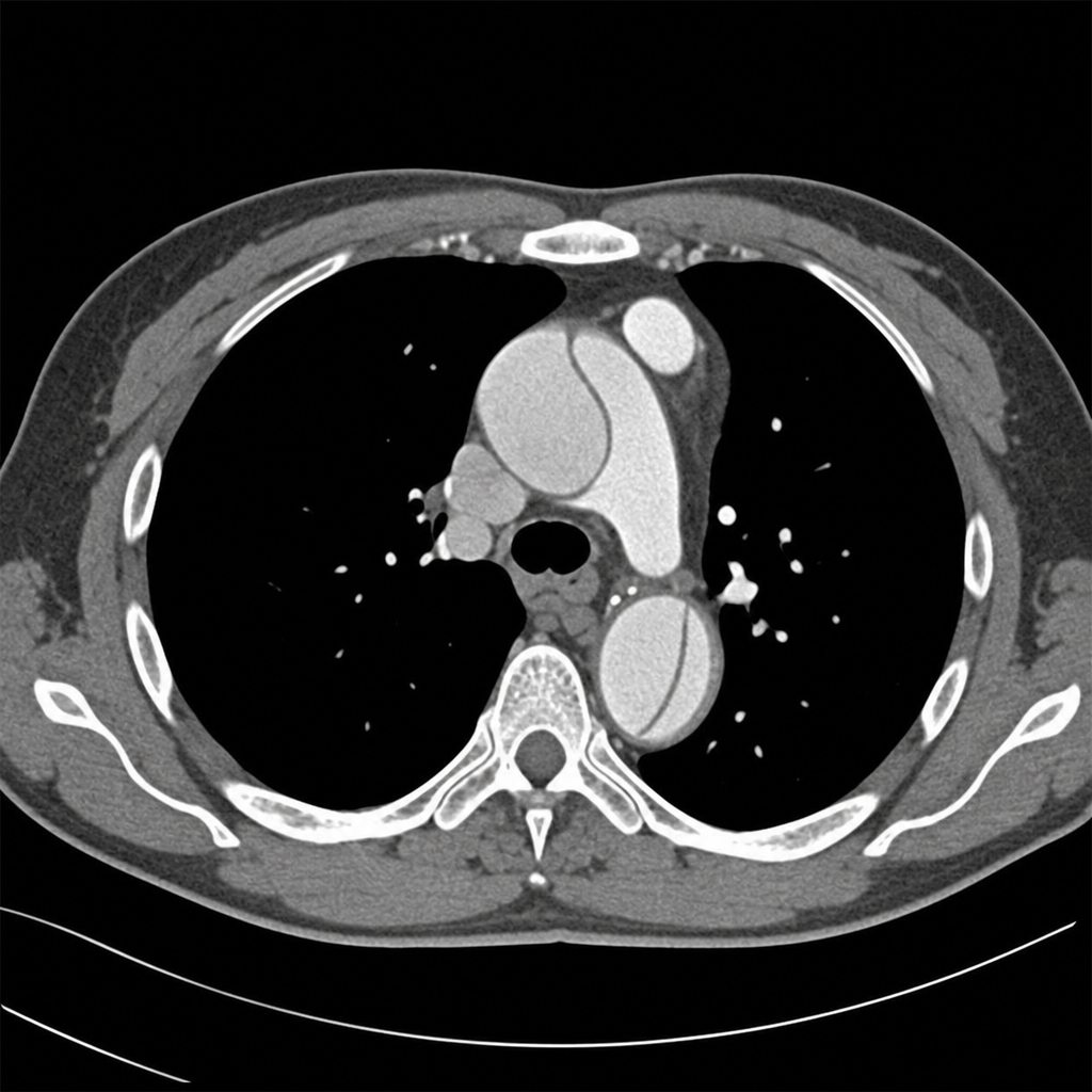
Which of the following statements about pulmonary embolism is false?
A case of spontaneous pneumothorax comes to you. What will be the earliest treatment of choice?
What is a key diagnostic criterion for acute respiratory distress syndrome (ARDS) in a child?
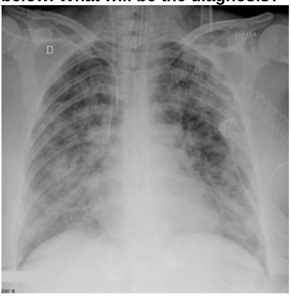
A patient with a known case of acute pancreatitis develops breathlessness and bilateral basal crepitations on day 4. What is the most likely diagnosis based on the chest radiography image?

A Patient presented to emergency with multiple rib fractures. He is conscious speaking single words. On examination, respiratory rate was 40/minute and BP was 90/40 mmHg. What is immediate next step?
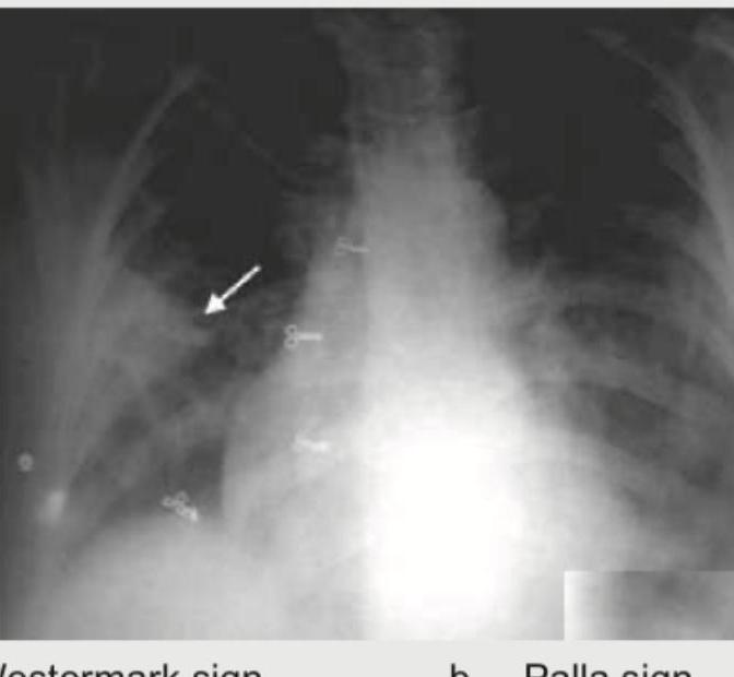
Which of the following is shown in the image below?

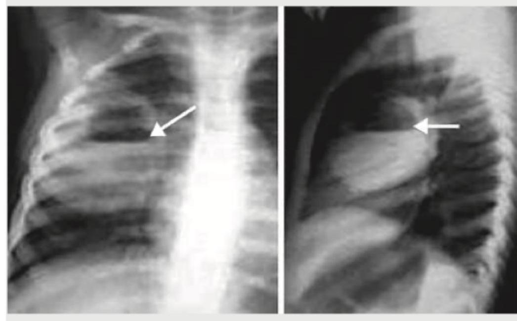
What is the most likely diagnosis based on the chest radiographs shown below?

Explanation: ***Hollow viscus perforation*** - The chest X-ray clearly shows **free air under the diaphragm** (pneumoperitoneum), which is a hallmark sign of a perforated hollow viscus in the abdomen. - The history of **bull gore to the abdomen** and subsequent abdominal pain and obstipation further supports a traumatic perforation of a stomach or intestinal segment. *Hemothorax* - Hemothorax would present as **fluid in the pleural space**, typically seen as blunting of the costophrenic angles or an effusion on X-ray, which is not evident here. - While trauma can cause hemothorax, the prominent finding on this X-ray is intra-abdominal air, not intrathoracic fluid. *Pneumothorax* - Pneumothorax is characterized by the presence of **air in the pleural space**, leading to lung collapse and absence of lung markings in the affected area, which is not observed on this X-ray. - The air seen is clearly **below the diaphragm**, indicating intra-abdominal free air, not air in the chest cavity surrounding the lung. *Intestinal obstruction* - Intestinal obstruction typically presents with **dilated bowel loops** and **air-fluid levels** on an abdominal X-ray, along with abdominal pain and obstipation. - While the patient has obstipation, the primary X-ray finding is free air under the diaphragm, which is not characteristic of an uncomplicated intestinal obstruction.
Explanation: ***Thrombosis of lower limb veins*** - The vast majority of pulmonary emboli originate from **deep vein thromboses (DVTs)** in the lower extremities, particularly the proximal veins (popliteal, femoral, iliac) [1]. - These clots can detach and travel through the right side of the heart to lodge in the **pulmonary arterial system** [1]. - Many patients with suspected PE will have identifiable proximal thrombus in the leg veins [2]. *Trauma* - While severe trauma can increase the risk of DVT due to immobility and venous stasis, **trauma itself is not the direct cause** of the pulmonary embolism. - Trauma is a risk factor for DVT formation, which then leads to PE, rather than directly producing the embolism. *Atherosclerosis* - Atherosclerosis is a disease of arteries involving plaque formation and can lead to conditions like **myocardial infarction** or **stroke**, but it is not a direct source of pulmonary emboli. - While severe atherosclerosis can be a risk factor for DVT in some cases due to systemic inflammation or reduced mobility, it is not the primary mechanism. *No significant cause* - While up to 50% of deep vein thromboses can be asymptomatic, thereby making their "cause" seem insignificant to the patient, PE always has an underlying cause, most commonly **venous thrombosis** [1]. - PE is a serious medical condition with identifiable risk factors and origins, even if the patient is unaware of the initial thrombotic event.
Explanation: ***TEE (Transesophageal Echocardiography)*** - **TEE is the investigation of choice** for acute aortic dissection in **hemodynamically unstable patients** due to its **portability and rapidity**. - Can be performed at the **bedside** without transporting the critically ill patient, minimizing risk. - Provides rapid diagnosis (5-10 minutes) with **>95% sensitivity and specificity** for detecting intimal flap and false lumen. - Simultaneously assesses **complications** such as aortic regurgitation, pericardial effusion/tamponade, and ventricular function. - Particularly excellent for visualizing the **ascending aorta** and aortic root. *CT-Angio* - **CT angiography** is the **investigation of choice** for acute aortic dissection in **hemodynamically STABLE patients**. - Provides excellent anatomical detail of the entire aorta, clearly showing the intimal flap, true and false lumens, and branch vessel involvement. - Requires **patient transport** to the radiology department, which is **unsafe in unstable patients**. - Best for comprehensive surgical planning in stable patients. *MRI* - **MRI** offers the highest anatomical detail and is considered the gold standard for **chronic dissection follow-up**. - Its lengthy acquisition time (30-60 minutes) and incompatibility with monitoring equipment make it **unsuitable for acutely unstable patients**. *NCCT* - **Non-contrast CT** may show indirect signs like the **hyperdense crescent sign** in the aortic wall. - Cannot reliably differentiate true and false lumens or assess the full extent of dissection. - Insufficient for definitive diagnosis or management planning.
Explanation: Most commonly presents within 2 weeks - This statement is **false** because while pulmonary embolism can occur at any time, its onset is not restricted to or "most commonly presents" within a two-week period. - The timing of presentation can vary widely depending on the predisposing factors and the specific event leading to the embolus formation and migration. *Dyspnea is the most common symptom* - **Dyspnea** (shortness of breath) is indeed the **most frequent symptom** reported by patients experiencing pulmonary embolism. - This symptom often occurs acutely and can range from mild to severe, correlating with the size and location of the embolism. *Pulmonary embolism often arises from deep vein thrombosis in the legs.* - The vast majority of **pulmonary emboli (PEs)** originate from **deep vein thromboses (DVTs)**, particularly those located in the large veins of the legs and pelvis [1, 2]. - These clots can detach and travel through the venous system to the heart and then into the pulmonary arteries [2]. *Longer survival time generally indicates a better chance of recovery.* - Patients who survive longer after a pulmonary embolism are more likely to have received timely treatment and may have experienced a less severe embolic event or have better underlying health. - Prolonged survival after the initial acute phase suggests the patient has overcome the immediate life-threatening aspects and is on the path to recovery, potentially with less long-term sequelae.
Explanation: ***Needle aspiration*** - For a spontaneous pneumothorax, especially if it is of moderate size or the patient is symptomatic, **needle aspiration** is often the earliest and least invasive treatment option. - This procedure removes air from the pleural space, allowing the lung to re-expand and relieving symptoms quickly. *ICD* - **Intercostal chest drain (ICD)** insertion is typically reserved for larger pneumothoraces, recurrent cases, or when needle aspiration is unsuccessful [1]. - It is a more invasive procedure than needle aspiration and is generally not the *earliest* treatment of choice for an initial, uncomplicated spontaneous pneumothorax [1]. *Wait and watch* - A "wait and watch" approach is appropriate only for very **small, asymptomatic spontaneous pneumothoraces** (e.g., <2 cm from the chest wall at the level of the hilum) [1]. - The question implies a case that "comes to you," suggesting the need for intervention rather than simple observation. *IPPV* - **Intermittent Positive Pressure Ventilation (IPPV)** is a form of mechanical ventilation used in patients with respiratory failure. - It is not a primary treatment for pneumothorax; rather, pneumothorax could be a complication of IPPV, or IPPV might be required if the pneumothorax leads to severe respiratory compromise, but it is not the initial intervention.
Explanation: ***Within 7 days of known clinical insult*** - A key diagnostic criterion for **Acute Respiratory Distress Syndrome (ARDS)** in children is the onset of respiratory symptoms within **7 days of a known clinical insult**. - This temporal relationship helps distinguish ARDS from other causes of respiratory failure that may have a more chronic or delayed onset. *Respiratory failure not fully explained* - **Respiratory failure not fully explained** by other conditions is a general characteristic but not a specific diagnostic criterion on its own. - ARDS requires the exclusion of cardiac failure as the primary cause of pulmonary edema, indicated by an absence of left atrial hypertension. *Left ventricular dysfunction* - **Left ventricular dysfunction** would suggest **cardiogenic pulmonary edema**, which needs to be excluded for a diagnosis of ARDS. - ARDS is characterized by **non-cardiogenic pulmonary edema**, meaning the fluid in the lungs is not due to heart failure. *All of the options* - This option is incorrect because **left ventricular dysfunction** is an *exclusion criterion* for ARDS, not a diagnostic criterion, as ARDS is defined by **non-cardiogenic pulmonary edema**. - While the other options relate to aspects of ARDS, only one is a key diagnostic criterion as formulated.
Explanation: ***Acute Respiratory Distress Syndrome (ARDS)*** - The chest radiograph shows **bilateral patchy infiltrates** and **diffuse alveolar opacities** consistent with ARDS, especially in the context of **acute pancreatitis** as a known risk factor. - The development of **breathlessness** and **bilateral basal crepitations** (rales) on day 4 further supports ARDS due to fluid accumulation in the lungs. *Bilateral pneumonia* - While pneumonia can cause bilateral infiltrates, the **symmetrical and widespread distribution** seen on this radiograph, combined with the context of acute pancreatitis, makes ARDS a more likely diagnosis. - Pneumonia typically presents with fever, productive cough, and lung consolidation, which are not specifically highlighted as primary symptoms over the breathlessness. *Carcinogenic Pulmonary Embolism* - Pulmonary embolism typically manifests with **sudden onset dyspnea**, pleuritic chest pain, and sometimes hemoptysis, and chest X-rays are often normal or show subtle findings like a **Westermark sign** or Hampton's hump. - The widespread bilateral infiltrates seen in the image are **not characteristic of pulmonary embolism**. *Lung collapse (atelectasis)* - Atelectasis usually appears as a ** localised area of increased opacification**, often with volume loss (e.g., tracheal deviation, elevated hemidiaphragm), and is often unilateral or segmental. - The **diffuse, bilateral, and often fluffy infiltrates** seen in this image are not consistent with typical atelectasis.
Explanation: ***Needle insertion in 2nd intercostal space*** - The patient's presentation with multiple rib fractures, **tachypnea (40/minute)**, **hypotension (90/40 mmHg)**, and speaking only single words suggests **tension pneumothorax**. - Speaking only single words indicates severe **respiratory distress** and inability to complete sentences due to dyspnea. - **Needle decompression** in the 2nd intercostal space at the midclavicular line is the immediate life-saving intervention for tension pneumothorax. - This is a **clinical diagnosis** in an emergency setting and does not require imaging confirmation before intervention. *Urgent IV fluid administration* - While fluid administration may be necessary for shock, the primary issue is likely **impaired ventilation** due to tension pneumothorax, which needs to be addressed first. - Delaying needle decompression to administer fluids could worsen the patient's respiratory and hemodynamic status. *Intubate the patient* - Intubation without addressing the cause of respiratory compromise, especially tension pneumothorax, can worsen the condition by increasing **intrathoracic pressure**. - Positive pressure ventilation in the presence of tension pneumothorax can be **life-threatening**. - **Relief of the tension pneumothorax** is the priority before considering definitive airway management. *Chest X-ray* - A chest X-ray is a diagnostic tool but should **not delay immediate life-saving interventions** in a patient with suspected tension pneumothorax. - Tension pneumothorax is a **clinical diagnosis** based on symptoms and immediate intervention takes precedence over imaging.
Explanation: ***Westermark sign*** - This image displays a **dilated pulmonary artery proximal to an area of oligemia**, specifically noted in the upper right lung field, as indicated by the arrow. This finding is characteristic of the **Westermark sign**, which is suggestive of a **pulmonary embolism**. - The Westermark sign represents **distal collapse of the pulmonary vasculature** due to a reduction in blood flow, making the lung parenchyma appear unusually lucent compared to adjacent normal lung fields. *Palla sign* - The Palla sign refers to a **dilated right descending pulmonary artery** (interlobar artery) on a chest X-ray. While it is also associated with pulmonary embolism, the image prominently shows oligemia, the defining feature of the Westermark sign, not solely an enlarged artery. - This sign indicates **increased pulmonary artery pressure** due to the embolus, but the key feature in the provided image is the reduced vascularity distally, not just the proximal vessel size. *Hampton hump* - A Hampton hump is a **wedge-shaped pleural-based opacity** with a rounded convex border facing the hilum. - It results from a **pulmonary infarction** due to a large pulmonary embolus and is not visible in this image. *Round pneumonia* - Round pneumonia is a **circular or oval-shaped consolidation** that is typically seen in children and appears as a mass-like lesion. - It is an infectious process with consolidation of lung tissue and does not involve vascular abnormalities like oligemia, which is clearly depicted in the image.
Explanation: ***Segmental collapse*** - The frontal image shows a **wedge-shaped opacity** in the right upper lobe, and the lateral view reveals a **triangular area of increased density** consistent with collapsed lung tissue. - This pattern, particularly the triangular density on the lateral view and volume loss indicated by the position of the **minor fissure (white arrow)**, points towards segmental collapse. *Lung abscess* - A lung abscess typically presents as a **cavity with an air-fluid level**, which is not depicted in these images. - The lesion shown is mostly **solid and dense**, unlike the characteristic appearance of an abscess. *Lobar emphysema* - Lobar emphysema involves **overinflation of a lung lobe**, characterized by increased lucency and vascular attenuation, which is the opposite of the findings here. - There is no evidence of **air trapping** or **hyperinflation** in the images provided. *Bronchiectasis* - Bronchiectasis is characterized by **permanent abnormal dilation of the bronchi**, often seen as "tram tracks" or "ring" opacities on imaging. - The images do not show these specific findings; instead, they indicate a loss of lung volume.
Get full access to all questions, explanations, and performance tracking.
Start For Free