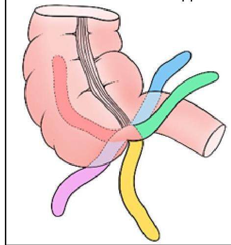
Identify the position of the appendix marked in BLACK in the given image:

Investigation of choice in an unstable patient with suspected intra-abdominal injury is -
Which of the following is the most common cause of acute mesenteric ischemia?
Which of the following is a complication of gallstones?
The coffee bean appearance on imaging is specifically associated with which condition?
Thumb print sign in a plain X-ray is seen in:
A man comes to the emergency department with stab injury to left flank. He has stable vitals. What would be the next step in management?
Most sensitive investigation for abdominal trauma in a hemodynamically stable patient is-
A 29 year old woman presents in emergency ward with amenorrhea of 6 weeks and pain. Urine pregnancy test shows positive. Examination shows diffuse significant lower abdomen tenderness. The pelvic examination is difficult to accomplish due to guarding. Her Beta-hCG level is 4000 mIU/ml. Transvaginal ultrasound shows no pregnancy in the uterus and no adnexal mass but moderate fluid in abdomen. Which of the following is the next best step?
Causes of thickened gallbladder wall on ultrasound examination are all except:
Explanation: ***Retrocecal*** - The **retrocecal** position (represented by the black color in the image) indicates the appendix is located behind the cecum, often a common variant. - This position can make diagnosis of appendicitis challenging as it may cause atypical pain patterns. *Pelvic* - The **pelvic** appendix descends into the true pelvis, which can mimic gynecological or urological conditions. - It usually causes pain that is more generalized in the lower abdomen or suprapubic region. *Subcecal* - The **subcecal** appendix is located directly below the cecum and is a relatively rare position. - While somewhat straightforward in presentation, it is less common than retrocecal or pelvic positions. *Preileal* - The **preileal** position indicates the appendix lies in front of the terminal ileum. - This is a less common anatomical variation, often associated with specific clinical presentations related to its anterior location.
Explanation: ***USG*** - **Focused assessment with sonography for trauma (FAST) exam** is the investigation of choice in an **unstable patient** due to its rapid, non-invasive nature and ability to detect free fluid (blood) in the peritoneal, pericardial, and pleural spaces. - It can be performed at the **bedside** without moving the patient, making it ideal for hemodynamically unstable individuals with suspected intra-abdominal injury. *CT scan* - While a **CT scan** provides detailed anatomical information, it requires the patient to be stable enough for transport to a radiology suite and prolonged scanning time. - It is often difficult to obtain in **unstable patients** who may require continuous resuscitation and monitoring. *X-ray abdomen* - An **X-ray abdomen** has limited utility for detecting intra-abdominal injuries and primarily identifies issues like free air under the diaphragm (suggesting hollow organ perforation) or foreign bodies. - It is **not sensitive** for detecting free fluid (hemoperitoneum) or solid organ injuries, which are critical in trauma. *DPL* - **Diagnostic peritoneal lavage (DPL)** is an invasive procedure that involves inserting a catheter into the peritoneal cavity to detect blood or other fluid. - While sensitive, it is **invasive**, can complicate subsequent imaging, and has largely been replaced by the FAST exam due to the latter's non-invasive nature and comparable diagnostic accuracy for free fluid.
Explanation: ***Embolism*** - **Embolic occlusion** (typically from the heart, e.g., atrial fibrillation) accounts for a significant majority of acute mesenteric ischemia cases. - This typically leads to sudden onset of severe abdominal pain with minimal physical findings initially. *Thrombosis* - **Arterial thrombosis** of the mesenteric vessels is another cause but is less frequent than embolism in acute settings. - It often occurs in the context of pre-existing **atherosclerotic disease** and can present with a more gradual onset of symptoms. *NOMI* - **Nonocclusive Mesenteric Ischemia (NOMI)** is caused by severe vasoconstriction and hypoperfusion, not a physical blockage. - It is often seen in critically ill patients with conditions like **shock**, sepsis, or heart failure. *Venous thrombosis* - **Mesenteric venous thrombosis** is a less common cause of acute mesenteric ischemia compared to arterial causes. - It is often associated with hypercoagulable states and can present with more insidious abdominal pain and bowel wall edema.
Explanation: ***Cholangitis*** [2] - **Cholangitis** refers to an infection of the **biliary tree**, most commonly caused by obstruction of the bile ducts by gallstones, leading to bacterial overgrowth. [2] - The obstruction (often due to choledocholithiasis) allows bacteria from the duodenum to ascend into the biliary system, causing inflammation and infection. *Hemobilia* - **Hemobilia** is bleeding into the **biliary tract**, typically caused by trauma, iatrogenic injury (e.g., biopsy), or vascular anomalies, not directly from gallstones. - While gallstones can cause inflammation, they do not typically lead to the direct arterial or venous bleeding characteristic of hemobilia. *Acute pancreatitis* [1] - **Acute pancreatitis** can be caused by gallstones if a stone temporarily obstructs the **ampulla of Vater**, blocking both the common bile duct and the pancreatic duct. [1] - However, it's considered a complication of **choledocholithiasis** (gallstones in the common bile duct), not a direct complication of gallstones themselves. *Biliary enteric fistula* [1] - **Biliary enteric fistula** is an abnormal connection between the biliary tree and the gastrointestinal tract, usually caused by chronic inflammation and erosion by a gallstone (e.g., a **gallstone ileus**). [1] - While a direct complication of gallstones, the question asks for *a* complication, and cholangitis is a more immediate and common infectious complication directly arising from biliary obstruction.
Explanation: ***Sigmoid volvulus*** - The **coffee bean sign** on an abdominal X-ray is a classic finding in **sigmoid volvulus**, representing the hugely dilated, gas-filled loop of bowel. - This characteristic appearance is due to the **mesentery twisting** on itself, creating a closed-loop obstruction. *Testicular torsion* - This condition involves the **twisting of the spermatic cord**, leading to interrupted blood supply to the testis. - Imaging focuses on the **scrotum** (e.g., ultrasound) and does not produce a "coffee bean" sign on abdominal films. *Meconium ileus* - This is a form of **small bowel obstruction** in newborns caused by abnormally thick and sticky meconium. - Imaging typically shows **dilated loops of small bowel** and a **"soap bubble" appearance** due to trapped gas in meconium, not a coffee bean shape. *Ileal atresia* - This condition involves a **congenital blockage of the ileum**, leading to proximal bowel dilation. - While it causes bowel obstruction, the characteristic imaging features are **dilated bowel loops** with **air-fluid levels**, not the specific coffee bean shape seen in sigmoid volvulus.
Explanation: ***Ischemic colitis*** - The **thumbprint sign** on a plain X-ray or CT scan is characteristic of ischemic colitis, resulting from submucosal edema and hemorrhage. - This appearance is due to the thickened, edematous **haustral folds** projecting into the colonic lumen. *Ulcerative colitis* - While it affects the colon, classic imaging findings for ulcerative colitis include **loss of haustral folds** (lead pipe sign) and pseudopolyps, not the thumbprint sign. - **Toxic megacolon** is a severe complication, identifiable by colonic dilation and wall thickening, distinct from thumbprint sign. *Pseudomembranous colitis* - This condition is caused by *Clostridioides difficile* infection and typically manifests with **thickened, nodular colonic walls** or inflammatory pseudomembranes on imaging. - It does not typically present with the classic "thumbprint" appearance indicative of ischemic changes. *Appendicitis* - Appendicitis is an inflammation of the appendix, diagnosed usually by findings like a **dilated appendix** with surrounding fat stranding on imaging. - The imaging findings are localized to the right lower quadrant and do not involve diffuse colonic changes like the "thumbprint sign."
Explanation: ***CECT*** - A **Contrast-Enhanced Computed Tomography (CECT)** scan is the preferred initial diagnostic step for a hemodynamically stable patient with a stab wound to the flank. - It effectively assesses the **depth of penetration** and identifies potential internal organ injuries in the abdomen or retroperitoneum, guiding further management. *Diagnostic peritoneal lavage* - **Diagnostic peritoneal lavage (DPL)** is less commonly used for stab wounds in hemodynamically stable patients due to its **invasiveness** and lower specificity compared to CT scans. - While it can detect peritoneal penetration or significant hemorrhage, it often leads to **unnecessary laparotomies** and is not as precise in identifying specific organ injuries. *Laparotomy* - **Laparotomy** (surgical exploration) is indicated for **hemodynamically unstable** patients or those with definitive signs of peritonitis or evisceration. - Since the patient has **stable vitals**, immediate laparotomy is not the next step, as diagnostic imaging is needed first. *Laparoscopy* - **Laparoscopy** is a minimally invasive surgical procedure that can be used diagnostically or therapeutically in stable patients. - However, in the initial assessment of a flank stab wound, a **CECT scan** is typically performed first to get a comprehensive view of potential organ damage before considering a more invasive procedure like laparoscopy.
Explanation: ***CT Scan (Computed Tomography)*** - **CT scans** offer superior anatomical detail and can accurately detect organ damage, hemorrhage, and other injuries in **hemodynamically stable** patients with abdominal trauma. - It is considered the **most sensitive** and specific imaging modality for evaluating blunt and penetrating abdominal trauma when the patient can tolerate the study. *Ultrasonography (FAST)* - While effective for detecting **free fluid** (blood) in specific abdominal areas, **Focused Assessment with Sonography for Trauma (FAST)** has lower sensitivity for solid organ injuries or bowel perforations. - Its primary role is rapid assessment for **hemoperitoneum** to guide immediate management in unstable patients, not detailed injury characterization. *Diagnostic peritoneal lavage (DPL)* - **DPL** is an invasive procedure with high sensitivity for detecting **intraperitoneal bleeding**, but it does not identify specific organ injuries or retroperitoneal hemorrhage. - It is rarely used in hemodynamically stable patients due to its invasiveness and the availability of more detailed imaging techniques. *MRI (Magnetic Resonance Imaging)* - **MRI** provides excellent soft tissue contrast but is typically too **time-consuming** and less accessible in urgent trauma settings compared to CT. - It's generally not the first-line investigation for acute abdominal trauma due to motion artifacts and limited utility in detecting air or bone injuries.
Explanation: ***Emergency laparotomy*** - The patient presents with **amenorrhea**, **positive pregnancy test**, significant lower **abdominal tenderness**, and **free fluid in the abdomen** without an intrauterine pregnancy on ultrasound, strongly suggesting a **ruptured ectopic pregnancy**, which is a life-threatening emergency requiring immediate surgical intervention. - The high **Beta-hCG level of 4000 mIU/ml** with no intrauterine pregnancy on ultrasound, combined with acute abdominal pain and tenderness, points to a rapidly progressing ectopic pregnancy that may have already ruptured, necessitating **emergency laparotomy** for hemorrhage control and removal of the ectopic gestation. *Repeat Beta-hCG level in 48 hours* - While serial Beta-hCG measurements are used to monitor early pregnancies, this patient's acute symptoms of severe abdominal pain, tenderness, and fluid in the abdomen, along with a high Beta-hCG and no intrauterine pregnancy, indicate an **urgent condition** that cannot wait 48 hours. - Waiting for repeat hCG levels would delay critical intervention for a potentially ruptured ectopic pregnancy, which could lead to **hemorrhagic shock** and death. *Institution of methotrexate* - **Methotrexate** is typically considered for **unruptured, stable ectopic pregnancies** with lower Beta-hCG levels and no signs of acute abdominal distress or rupture. - This patient's presentation with acute pain, tenderness, and free fluid strongly suggests rupture, making **methotrexate inappropriate** and dangerous as it would not address the active bleeding and could worsen her condition. *Wait and watch* - A "wait and watch" approach is inappropriate and extremely dangerous given the patient's acute abdominal pain, tenderness, and evidence of free fluid in the abdomen, which are all signs of a **ruptured ectopic pregnancy**. - Delaying intervention in cases of potential ruptured ectopic pregnancy can lead to **massive hemorrhage**, shock, and maternal death.
Explanation: ***Kawasaki disease*** (Correct Answer) - While Kawasaki disease can cause **gallbladder hydrops** (distension with bile), the primary ultrasound finding is an **enlarged, distended gallbladder** rather than isolated wall thickening. - When gallbladder involvement occurs in Kawasaki disease, it manifests as **acalculous cholecystitis** with hydrops, but this is **not a typical or common presentation** compared to the other causes listed. - The hallmark features of Kawasaki disease are **coronary artery aneurysms** and systemic vasculitis, not primary gallbladder pathology. - In clinical practice, gallbladder wall thickening would **not be attributed to Kawasaki disease** as a primary differential diagnosis. *Incorrect: Congestive cardiac failure* - **Systemic fluid overload** and venous congestion in CHF leads to gallbladder wall thickening due to **transudative edema**. - This is a **common cause** of non-inflammatory gallbladder wall thickening (>3mm). - The wall appears thickened, hypoechoic, and **edematous** without pericholecystic fluid. *Incorrect: Postprandial state* - After eating, the gallbladder **contracts to release bile**, causing the wall to appear thicker on ultrasound due to **accordion-like folding** of the mucosa. - This is a **normal physiological finding** and typically resolves within 1-2 hours. - Scanning should ideally be done after **6-8 hours of fasting** to avoid this pseudo-thickening. *Incorrect: Cholecystitis* - **Acute cholecystitis** is the **classic cause** of gallbladder wall thickening (>3mm, often >5mm). - Associated findings include **gallstones, pericholecystic fluid, positive sonographic Murphy's sign**, and wall edema. - The wall shows **layering** (subserosal edema) and hyperemia on Doppler imaging.
Get full access to all questions, explanations, and performance tracking.
Start For Free