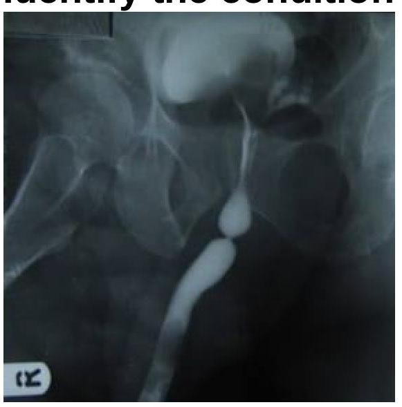
Vesicoureteric reflux is diagnosed by which of the following methods?
Which imaging modality is preferred for diagnosing placenta accreta in a 25-year-old pregnant woman at 20 weeks with a history of a previous cesarean delivery?
The Barium Swallow examination shows a filling defect in the esophagus. What is the most probable diagnosis?

Barium meal follow through is helpful in diagnosing -
Early and late suspected instrumental perforation of the oesophagus should first be assessed using?
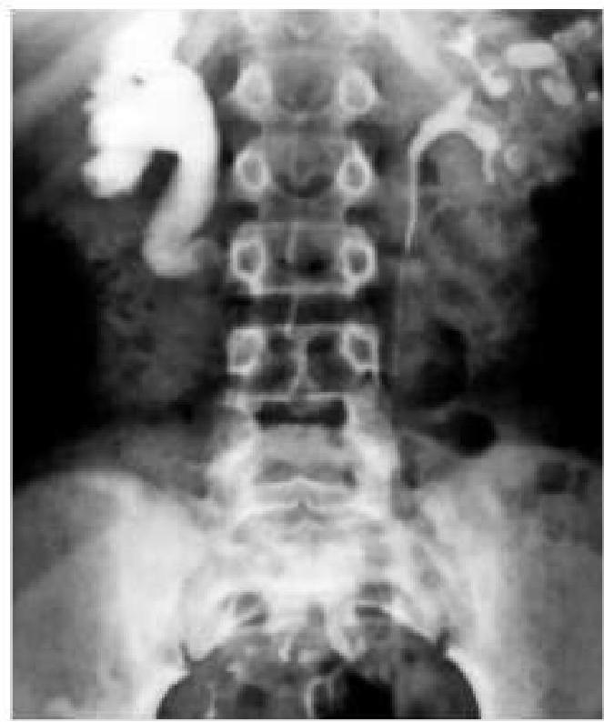
Percentage of renal stones which are radio-opaque:
Which of the following is NOT an indication for a barium meal X-ray?
What is the primary purpose of the Caldwell view in radiology?
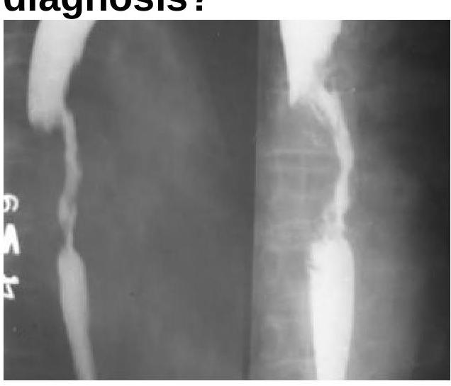
A delayed intravenous urogram of a patient is given below. What is the likely diagnosis?

Identify the radiological procedure shown in the image?

Explanation: ***Micturating cystography*** - **Micturating cystography** (also known as voiding cystourethrography or VCUG) is the gold standard for diagnosing and grading **vesicoureteric reflux (VUR)**. - It involves filling the bladder with contrast medium via a catheter and taking X-ray images during bladder filling and **voiding** to visualize the retrograde flow of urine into the ureters. *Ultrasound bladder* - While ultrasound can assess bladder shape, emptying, and the presence of hydronephrosis (dilation of the renal pelvis and calyces due to urine obstruction), it **cannot directly visualize urine reflux** from the bladder to the ureters. - It is often used as a screening tool or to assess for complications of VUR but not for diagnosis of VUR itself. *Intravenous pyelogram* - **Intravenous pyelogram (IVP)**, also known as intravenous urography (IVU), involves injecting contrast material into a vein and taking X-rays as it's filtered by the kidneys and passes through the urinary tract. - While it can assess kidney structure, collecting systems, and patency of ureters, it is **not ideal for demonstrating VUR** because it primarily visualizes antegrade flow and is not performed during the critical voiding phase. *Magnetic resonance imaging* - **Magnetic resonance imaging (MRI)** provides detailed anatomical information and can be used to evaluate renal parenchyma or complex urinary tract anomalies. - However, MRI is **not the preferred or standard method for diagnosing VUR** due to its limitations in visualizing dynamic reflux during voiding and its higher cost and longer imaging time compared to micturating cystography.
Explanation: ***Transabdominal ultrasound*** - This is the **first-line and preferred imaging modality** for diagnosing placenta accreta spectrum disorders, with **sensitivity of 77-87%** and **specificity of 96-98%**. - Key ultrasound findings include **loss of retroplacental clear space**, **placental lacunae** (irregular vascular spaces giving a "Swiss cheese" appearance), **thinning or loss of the hypoechoic retroplacental myometrial zone**, and **abnormal color Doppler showing turbulent flow**. - In a patient with **previous cesarean delivery** (major risk factor), targeted ultrasound examination of the anterior lower uterine segment at 18-20 weeks can effectively identify accreta, allowing timely multidisciplinary planning. - **Gray-scale ultrasound with color Doppler** provides excellent real-time assessment and is readily available, non-invasive, and does not involve radiation. *MRI* - MRI serves as a valuable **second-line or adjunct imaging modality** when ultrasound findings are **equivocal** or when there is **posterior placentation** limiting ultrasound visualization. - It offers superior soft tissue resolution for assessing the **depth of myometrial invasion** and evaluating extension into adjacent structures like the **bladder** or **parametrium**, which is particularly useful for **surgical planning**. - However, MRI is more expensive, less readily available, time-consuming, and its superiority over ultrasound for routine diagnosis has not been definitively established in most cases. *Transvaginal ultrasound* - While transvaginal ultrasound can complement transabdominal imaging by providing better visualization of the **lower uterine segment** and **cervical region**, it has a **limited field of view**. - It is primarily useful for assessing **cervical length**, **placental location relative to the internal os**, and ruling out **vasa previa**, but is not the preferred modality for comprehensive placenta accreta assessment. - The overall extent of placental invasion, particularly into the anterior wall or beyond, may not be fully evaluated. *CT scan* - CT scanning involves **ionizing radiation** which poses **teratogenic risks to the fetus** and is therefore **contraindicated** during pregnancy except in life-threatening maternal emergencies. - It provides **inferior soft tissue contrast resolution** compared to both ultrasound and MRI for placental evaluation, offering no diagnostic advantages for placenta accreta.
Explanation: ***Esophageal Carcinoma*** - A filling defect on a barium swallow study, especially with irregular borders and luminal narrowing, is highly suggestive of an **esophageal carcinoma**. - The image appears to show an **irregular, obstructing lesion** that displaces the barium column, characteristic of a mass. *Esophageal Ring* - An esophageal ring, such as a **Schatzki ring**, typically presents as a thin, circumferential narrowing of the distal esophagus, forming a smooth, shelf-like indentation, which is not seen here. - Esophageal rings usually cause **intermittent dysphagia** to solids but do not present as a large, irregular filling defect. *Esophageal Tear* - An esophageal tear (e.g., **Mallory-Weiss tear**) is a mucosal laceration that would present with **hematemesis** and would typically appear as a linear defect or streak on a barium swallow if visible, not a filling defect. - A tear is not usually associated with a persistent mass effect or irregular luminal obstruction seen in the image. *Achalasia Cardia* - **Achalasia** is characterized by the failure of the lower esophageal sphincter to relax and **absent peristalsis** in the esophageal body, leading to a classic "bird's beak" or "rat tail" appearance on barium swallow due to distal narrowing and proximal dilation. - While it causes luminal narrowing, it does not typically present as an irregular filling defect within the lumen, but rather as a smooth tapering of the distal esophagus.
Explanation: ***Ileal stricture*** - A **barium meal follow-through** visualizes the small intestine, including the ileum, making it ideal for detecting **strictures** in this region. - The barium contrast can highlight narrowings or areas of abnormal transit time indicative of a stricture. *Colonic obstruction* - **Colonic obstruction** is better diagnosed with a **barium enema** (lower GI series) or a **CT scan**, as barium meal follow-through primarily evaluates the upper GI tract and small intestine. - Barium would typically not reach the colon in a timely or sufficient manner to definitively diagnose an obstruction originating there. *Rectal obstruction* - Similar to colonic obstruction, **rectal obstruction** is best assessed by a **barium enema** or **sigmoidoscopy/colonoscopy**, which directly visualize the rectum and distal colon. - A barium meal follow-through is unlikely to provide adequate detail for a definitive diagnosis in the rectum. *Esophageal obstruction* - **Esophageal obstruction** is primarily diagnosed with an **esophagram** (barium swallow), which specifically focuses on the esophagus. - While a barium meal often starts with an esophagram component, the "follow-through" aspect is for the small intestine, and dedicated esophageal imaging is more appropriate for obstruction.
Explanation: ***Water soluble contrast swallow*** - This is the **traditional first-line investigation** for suspected oesophageal perforation and remains the standard answer for most examinations. - **Water-soluble contrast agents** (e.g., Gastrografin) are less irritating to tissues than barium if they extravasate, making them safer for detecting perforations. - Has good sensitivity (50-75%) and can directly visualize the site of leak. - **Modern practice note:** CT with oral contrast is increasingly preferred in many centers due to higher sensitivity (>90%) and ability to assess complications simultaneously, but water-soluble contrast swallow remains the established first-line investigation in standard protocols. *CT Scan* - A CT scan (particularly with oral contrast) has **higher sensitivity** (>90%) than contrast swallow and can identify fluid collections, air in the mediastinum, and other complications. - In modern practice, CT is often performed early or even as the initial investigation, but traditionally it is considered **second-line** after contrast swallow. - For examination purposes, water-soluble contrast swallow is typically considered the first-line investigation. *Dilute barium swallow* - **Barium** can cause a severe inflammatory reaction (mediastinitis or pleuritis) if it leaks into the mediastinum or pleural cavity in the presence of a perforation. - Its use is **contraindicated** as the initial study if perforation is suspected due to the risk of complications. - May be used as a **second-line investigation** if water-soluble contrast study is negative but clinical suspicion remains high, as barium has better mucosal coating and sensitivity. *MRI* - MRI is generally not used for acute oesophageal perforation due to its **longer acquisition time** and limited ability to effectively visualize active leaks from the oesophageal lumen. - It offers less utility than CT or contrast studies for this specific acute condition and is not readily available in emergency settings.
Explanation: ***80*** - Approximately **80%** of renal stones are radio-opaque, making them visible on X-rays and CT scans [1]. - This high percentage is primarily due to the composition of most stones, such as **calcium oxalate** and **calcium phosphate** [1]. *20* - Only **20%** of renal stones are typically radio-opaque, which is significantly lower than the actual statistic. - This option fails to account for the majority of stones that are indeed visible on radiographic imaging. *40* - A **40%** radio-opacity would indicate a much larger proportion of stones being less visible on imaging, which is inaccurate. - The range for radio-opaque stones is considerably higher, primarily due to common stone types. *60* - While **60%** might suggest a considerable portion of stones are radio-opaque, it still underestimates the actual prevalence. - The more accurate figure reflects that at least **80%** of stones show radio-opacity which aids in diagnosis. **References:** [1] Cross SS. Underwood's Pathology: A Clinical Approach. 6th ed. Common Clinical Problems From Diseases Of The Urinary And Male Genital Tracts, pp. 491-492.
Explanation: ***Ischemic Colitis*** - Barium studies are **contraindicated** in suspected ischemic colitis due to the risk of **perforation** and the poor resolution of mucosal changes. - **CT angiography** or colonoscopy are preferred for diagnosing ischemic colitis. *Duodenal ulcer* - A barium meal X-ray can demonstrate the presence of a **duodenal ulcer** as a niche or crater, and also identify associated deformities like a **cloverleaf sign**. - While endoscopy is more definitive, barium studies were historically important for initial evaluation. *Carcinoma stomach* - Barium meal can reveal filling defects, **mucosal irregularities**, and **loss of distensibility** in the stomach, suggesting gastric carcinoma. - However, **endoscopy with biopsy** is the gold standard for definitive diagnosis. *Carcinoma head of pancreas* - Barium meal can indirectly show signs of a pancreatic head mass, such as a **widened duodenal C-loop** or a **"reversed 3" sign** (Frohberg's sign) due to extrinsic compression. - This is an indirect sign, and cross-sectional imaging like **CT or MRI** is superior for direct visualization and characterization of pancreatic masses.
Explanation: ***Correct: To visualize the frontal sinus*** - The Caldwell view, also known as the **occipitomental view with 15-23 degree caudal angulation**, is specifically designed to best demonstrate the **frontal sinuses and anterior ethmoid air cells**. - This projection minimizes superimposition of the **petrous ridges** over the frontal sinuses by projecting them below the level of the frontal sinuses, allowing for clear visualization. - The Caldwell view is a **PA (posteroanterior) projection** of the skull used primarily in sinus imaging. *Incorrect: To visualize the sphenoid sinus* - The sphenoid sinus is best visualized with the **lateral view** or the **submentovertex (SMV) view**. - The Caldwell view provides poor projection of the sphenoid sinus due to overlying bony structures. *Incorrect: To visualize the maxillary sinus* - The **Waters view** (occipitomental projection at 37-45°) is the primary projection for visualizing the maxillary sinuses, as it projects the petrous ridges below them. - While the maxillary sinuses are visible on a Caldwell view, they are often partially obscured by the petrous ridges. *Incorrect: To visualize the ethmoid sinus* - The ethmoid sinuses (particularly anterior ethmoid cells) are visible on the Caldwell view but are typically best evaluated with both **Caldwell and lateral views** together. - However, the **primary purpose** of the Caldwell view is frontal sinus visualization, not ethmoid assessment.
Explanation: ***Pelviureteric junction obstruction*** - The image shows marked **dilatation of the renal pelvis and calyces** on the right side, with a relatively abrupt narrowing at the junction of the pelvis and ureter. - The delayed nature of the urogram suggests **impaired drainage** of contrast from the renal pelvis, accumulating proximal to the obstruction. *Putty kidney* - A "putty kidney" (or **autonecrotic kidney**) refers to a chronic, severely diseased kidney, often seen in end-stage **renal tuberculosis**, that has become calcified and non-functional. - This image demonstrates active contrast excretion and pelvicalyceal dilatation, not a calcified, non-functional organ. *Staghorn calculus* - A staghorn calculus is a **large, branched kidney stone** that occupies a significant portion of the renal collecting system. - While it can cause hydronephrosis, the image does not show a dense, radiopaque calculus filling the collecting system. *Cystic kidney* - **Cystic kidneys**, such as in polycystic kidney disease, are characterized by multiple fluid-filled sacs within the kidney parenchyma. - The image depicts dilatation of the collecting system, not diffuse cystic changes throughout the renal parenchyma.
Explanation: ***Barium meal follow through*** - The image shows opacification of the stomach and the entire small bowel loops via oral uptake of a contrast agent, which is characteristic of a **barium meal follow-through**. - This procedure tracks the passage of **barium** from the esophagus, stomach, and duodenum, through the jejunum and ileum, to assess the **small intestine's morphology and function**. *Barium enema* - A **barium enema** involves introducing contrast material rectally to visualize the **colon and rectum**, which is not depicted in this image. - The primary structures opacified in a barium enema are the **large intestine**, not the stomach and small bowel as seen here. *Enteroclysis* - **Enteroclysis** is a specialized study of the small bowel where contrast is directly instilled into the **duodenum** or **proximal jejunum** via a nasoenteric tube. - While it visualizes the small bowel, the image shows oral contrast progression from the stomach, not direct jejunal intubation. *Proctography* - **Proctography**, also known as defecography, is a dynamic study focused specifically on the **rectum and anal canal** during defecation. - This procedure is highly specific to the distal gastrointestinal tract for assessing anorectal function and does not visualize the stomach or extensive small bowel loops.
Iodinated Contrast Media
Practice Questions
MRI Contrast Agents
Practice Questions
Ultrasound Contrast Agents
Practice Questions
Adverse Reactions to Contrast Media
Practice Questions
Management of Contrast Reactions
Practice Questions
Contrast-Induced Nephropathy
Practice Questions
Barium Studies
Practice Questions
Intravenous Urography
Practice Questions
Angiography Techniques
Practice Questions
Lymphangiography
Practice Questions
Contrast Administration Protocols
Practice Questions
Pretesting and Premedication
Practice Questions
Get full access to all questions, explanations, and performance tracking.
Start For Free