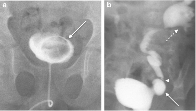
Micturating cystourethrogram shows filling defect in the urinary bladder. Likely diagnosis is________

Which of the following is considered an absolute contraindication for MRI in most clinical scenarios?
A young adult presents with proptosis and pain in eye after 4 days of trauma to eye. Chemosis, conjunctival congestion and extraocular muscle palsy with inability to move eye are seen.Investigation of choice -
All of the following are contrast radiographs except:
All of the following are indications for cystogram, EXCEPT:
Gas shadow in the heart and great vessels on chest imaging most commonly appears in association with-
Bead cystogram is used for the diagnosis of:
Which of the following is the most commonly used MR sequence for non-contrast angiography?
The procedure of choice for the evaluation of an aneurysm is:
Excretory urography should be cautiously performed in
Explanation: ***Ureterocele*** - The image shows a **filling defect** within the urinary bladder, which is characteristic of a **ureterocele**, an abnormal ballooning of the distal ureter into the bladder lumen. This appears as a round or oval lucency surrounded by contrast medium. - The presence of the arrow pointing to this lucency within the contrast-filled bladder strongly supports a ureterocele. *Duplication of ureter* - Ureteral duplication involves two ureters draining a single kidney, which would not typically present as a filling defect in the **bladder lumen** itself, but rather as two separate ureteral orifices. - While it can be associated with other anomalies like ureterocele, the primary finding described and visualized is the **filling defect**, not the duplication of the ureter. *Sacrococcygeal teratoma* - A sacrococcygeal teratoma is a **tumor** located at the base of the spine (sacrum and coccyx), which would not be seen as a **filling defect within the urinary bladder** on a micturating cystourethrogram (MCU). - Its effects on the urinary tract would typically be due to **external compression** or displacement, not an intrinsic bladder lumen anomaly. *Vesicoureteric reflux grade II* - Vesicoureteric reflux (VUR) is the **backward flow of urine** from the bladder into the ureters and potentially kidneys, which would be visualized as contrast filling the ureters during bladder opacification or voiding, not as a filling defect within the bladder itself. - Grade II VUR specifically means reflux into the ureter and collecting system **without dilation**, which is a functional issue, not a structural filling defect within the bladder lumen.
Explanation: ***Pacemaker*** - The strong magnetic fields and radiofrequency pulses of **MRI** can interfere with the function of a **pacemaker**, potentially causing device malfunction, dislodgement, or heating of leads, which can be life-threatening. - While some newer pacemakers are MRI-conditional, the presence of an older or non-MRI-conditional pacemaker is an **absolute contraindication** to MRI. *Claustrophobia* - **Claustrophobia** is a relative contraindication or a challenge for MRI, often managed with sedation or open MRI scanners, but it does not pose an immediate physical threat. - It affects patient comfort and compliance but is not an **absolute contraindication** in terms of safety. *Penile prosthesis* - Most modern **penile prostheses** are made of MRI-compatible materials and are generally safe for MRI. - While some older devices might cause artifacts, they do not typically pose a significant safety risk during an **MRI** scan. *Joint replacement* - **Joint replacements**, especially newer ones, are often made of non-ferromagnetic materials and are increasingly becoming MRI-safe. - While older or certain types of metallic implants can cause **image artifacts**, they are not an **absolute contraindication** for MRI unless the material is known to be ferromagnetic and prone to movement or heating.
Explanation: ***Digital subtraction angiography*** - The combination of **proptosis**, **pain**, **chemosis**, **conjunctival congestion**, and **extraocular muscle palsy** following trauma strongly suggests a **carotid-cavernous fistula (CCF)**. - **Digital subtraction angiography (DSA)** is the **gold standard** for diagnosing and characterizing CCFs, providing detailed visualization of arterial and venous flow. *MR angiography* - While MRA can provide information about vascular structures, it is less sensitive and specific than DSA for detecting and characterizing subtle shunts in **carotid-cavernous fistulas**. - It might miss smaller fistulas or provide insufficient detail for therapeutic planning. *CT* - **Computed tomography (CT)** is useful for assessing orbital bony structures, but it offers limited information regarding the dynamic blood flow and shunt characteristics crucial for diagnosing **carotid-cavernous fistulas**. - **CT angiography** can provide some vascular detail, but it is generally less comprehensive than DSA for this specific condition. *MRI* - **Magnetic Resonance Imaging (MRI)** can show orbital soft tissue changes and identify potential vascular abnormalities, but it lacks the real-time, high-resolution vascular detail of DSA, especially for depicting the exact location and flow dynamics of an **arteriovenous shunt**. - It is often used as a preliminary imaging modality but is not the definitive diagnostic tool for **carotid-cavernous fistulas**.
Explanation: ***Tomography*** - **Tomography** (e.g., CT scan) involves imaging structures in "slices" using X-rays but does not inherently require contrast medium for basic imaging. - While contrast material can be used in tomography to enhance specific structures, the technique itself is not exclusively a **contrast radiograph**. *Arthrography* - **Arthrography** is a type of contrast radiograph where a contrast medium is injected directly into a joint space to visualize soft tissue structures like ligaments, menisci, and articular cartilage. - The use of contrast is fundamental to distinguish these structures, which are not visible on plain X-rays. *Arthrotomography* - **Arthrotomography** combines arthrography with tomography, meaning a contrast agent is injected into a joint, and then tomographic slices are obtained. - This technique provides detailed cross-sectional images of the joint with enhanced contrast from the injected medium. *Sialography* - **Sialography** is a contrast radiograph procedure used to visualize the salivary ducts and glands. - A contrast medium is injected into the ductal system to identify obstructions, strictures, or inflammation within the salivary glands.
Explanation: ***Polycystic kidney disease*** - A cystogram evaluates the bladder and urethra, not the kidneys. **Polycystic kidney disease** primarily affects the **kidneys**, causing cysts to form within them, and is typically diagnosed with ultrasound, CT, or MRI. - A cystogram would not provide relevant information for the diagnosis or management of polycystic kidney disease. *Bladder rupture* - A cystogram is a primary diagnostic tool for **bladder rupture**, enabling visualization of contrast extravasation outside the bladder. - It helps determine the location and extent of the rupture, guiding subsequent management. *Stress incontinence* - A cystogram, particularly a **voiding cystourethrogram (VCUG)**, can be used to assess bladder and urethral function during voiding, which is relevant for evaluating **stress incontinence**. - It can help identify anatomical abnormalities or dynamic changes contributing to urine leakage. *Ca bladder* - A cystogram can reveal bladder wall irregularities, filling defects, or masses suggestive of **bladder carcinoma (Ca bladder)**. - While cystoscopy with biopsy is definitive, a cystogram can be a useful initial imaging study to delineate tumor extent or identify other suspicious areas.
Explanation: ***Post-procedural complications*** - **Most common cause** of gas shadow in the heart and great vessels on chest imaging in modern clinical practice - Iatrogenic air introduction during **central venous catheter insertion**, cardiac catheterization, hemodialysis, or cardiopulmonary bypass - Air can enter through catheter placement, line flushing, or during interventional procedures - Also seen with mechanical ventilation causing barotrauma leading to systemic air embolism - Typically appears as **discrete gas bubbles** or linear lucencies within cardiac chambers or great vessels *Cardiac trauma* - While penetrating or severe blunt chest trauma can introduce air into the heart, it is **less common** than iatrogenic causes in current practice - Would require direct communication between air-containing spaces (lung, atmosphere) and cardiac chambers - Usually associated with other traumatic findings like hemopericardium or pneumothorax *Gas embolism* - Can occur with decompression illness (diving), surgical procedures, or pulmonary barotrauma - May show intracardiac gas, but is **less common** as a primary presentation on chest imaging - Often presents with neurological or systemic symptoms rather than as an incidental imaging finding *Pneumopericardium* - Refers to air in the **pericardial sac** surrounding the heart, not within the cardiac chambers or great vessels themselves - Creates a different radiological appearance with air outlining the heart border - Does not produce gas shadows **within** the heart chambers as described in the question
Explanation: ***Stress incontinence*** - A **bead cystogram** is a diagnostic test specifically designed to evaluate the **urethral vesical angle** and assess for bladder neck descent during stress maneuvers, which are characteristic of **stress incontinence**. - The procedure involves placing a metal bead chain within the urethra and bladder, and then taking X-rays to visualize the relative positions of the bladder neck and urethra during changes in intra-abdominal pressure. *Thimble bladder* - A **thimble bladder** is a small, contracted bladder, often seen in conditions like **tuberculosis** or interstitial cystitis. - While cystography might reveal a small bladder, a bead cystogram is not specifically used to diagnose the size or capacity of the bladder. *Diverticulum* - A **diverticulum** is an outpouching of the bladder wall, which would be visualized as an abnormal projection during a standard cystogram. - A bead cystogram's primary purpose is to assess urethral and bladder neck morphology, not to identify diverticula. *Ca bladder* - **Bladder cancer (Ca bladder)** is typically diagnosed through **cystoscopy with biopsy**, urine cytology, or imaging techniques like CT scans or MRI, which look for masses or wall thickening. - A bead cystogram is not a primary diagnostic tool for bladder cancer and does not provide information about malignant lesions.
Explanation: ***Time-of-flight (TOF)*** - This sequence is widely used for non-contrast angiography because it relies on the **inflow of unsaturated spins** into the imaging plane to generate signal from flowing blood. - Due to its ability to visualize blood vessels without contrast, it is particularly valuable for detecting **aneurysms** and **arteriovenous malformations**. *Phase contrast* - Phase contrast MR angiography measures the **phase shift** induced by blood flow within a magnetic field gradient. - While it can quantify flow velocity, it is generally **less sensitive** for visualizing small vessels and complex vascular pathologies compared to TOF. *TRUFI* - TRUFI (True Fast Imaging with Steady-State Precession) is a **balanced steady-state free precession (bSSFP)** sequence known for its high signal-to-noise ratio and bright fluid signal. - While TRUFI can show vessels, it is not primarily an angiographic sequence but rather used for general imaging where **good tissue contrast** and **fluid visualization** are desired. *Arterial spin labeling* - Arterial spin labeling (ASL) is a technique that uses **arterial blood water as an endogenous tracer** to measure cerebral blood flow **quantitatively**. - It is not an angiographic sequence for visualizing vessel anatomy but rather for **perfusion imaging**, assessing tissue blood supply.
Explanation: ***Computed tomography*** **Computed tomography (CT)**, particularly **CT angiography (CTA)**, is widely considered the procedure of choice for evaluating aneurysms due to its **rapid acquisition**, **high spatial resolution**, and ability to visualize the vessel lumen and surrounding structures. **Key advantages:** - Particularly useful for assessing aneurysm size, morphology, thrombus formation, and rupture - Excellent for both emergent and elective settings - Widely available and fast imaging acquisition - Provides comprehensive anatomical detail *Ultrasonography* **Ultrasonography** is an excellent and cost-effective **screening tool for abdominal aortic aneurysms (AAA)** because it is non-invasive and does not involve radiation. However, its utility is limited for: - Complex aneurysms requiring detailed anatomical information - Less accessible locations (e.g., thoracic, cerebral aneurysms) - **Operator dependence** and **limited field of view** restrict its use as a definitive diagnostic tool *Magnetic resonance imaging* **Magnetic resonance imaging (MRI)** and **magnetic resonance angiography (MRA)** provide excellent soft tissue contrast without ionizing radiation and can accurately evaluate aneurysm morphology and flow characteristics. However, MRI is: - More time-consuming and expensive - May be contraindicated in patients with metallic implants or claustrophobia - Less suitable for initial acute evaluation compared to CT *Angiography* **Angiography**, traditionally a catheter-based invasive procedure, provides detailed images of the vessel lumen and is excellent for evaluating precise anatomy and planning endovascular repair. While it offers highly detailed images, its: - Invasiveness - Exposure to radiation and contrast agents - Potential for complications These factors typically reserve it for **interventional planning** or when non-invasive methods are inconclusive, rather than as the primary diagnostic tool.
Explanation: ***Multiple myeloma*** - Excretory urography (intravenous pyelography or IVP) involves the administration of **iodinated contrast media**, which can precipitate **Bence Jones proteins** in the renal tubules, leading to or worsening **acute kidney injury** in patients with multiple myeloma. - Patients with multiple myeloma often have **pre-existing renal dysfunction** (myeloma kidney) due to light chain deposition, making them highly susceptible to contrast-induced nephropathy. *Bone metastases* - While bone metastases can be painful and may require imaging, they do not directly contraindicate excretory urography; the primary concern with IVP is renal function. - The presence of bone lesions itself does not increase the risk of **contrast-induced nephropathy** in the same way that proteinuria from multiple myeloma does. *Neuroblastoma* - Neuroblastoma is a **childhood cancer** affecting the adrenal glands or sympathetic nervous system, and it is not typically associated with a specific risk for contrast-induced nephropathy from excretory urography. - The primary diagnostic imaging for neuroblastoma often involves ultrasound, CT, or MRI, and while contrast may be used, the specific renal risk seen in multiple myeloma is not present. *Leukemia* - While some forms of leukemia can affect the kidneys, particularly through infiltration, it does not typically pose the same specific risk for **contrast-induced nephropathy** as multiple myeloma. - The renal manifestations in leukemia are generally different from the **light chain proteinuria** seen in multiple myeloma, which directly interacts with iodinated contrast.
Iodinated Contrast Media
Practice Questions
MRI Contrast Agents
Practice Questions
Ultrasound Contrast Agents
Practice Questions
Adverse Reactions to Contrast Media
Practice Questions
Management of Contrast Reactions
Practice Questions
Contrast-Induced Nephropathy
Practice Questions
Barium Studies
Practice Questions
Intravenous Urography
Practice Questions
Angiography Techniques
Practice Questions
Lymphangiography
Practice Questions
Contrast Administration Protocols
Practice Questions
Pretesting and Premedication
Practice Questions
Get full access to all questions, explanations, and performance tracking.
Start For Free